Review
. 2008 Sep;98(1):85-106.
doi: 10.1016/j.pbiomolbio.2008.05.007. Epub 2008 Jun 20.

Intrinsic disorder in scaffold proteins: getting more from less

Affiliations
Review

Intrinsic disorder in scaffold proteins: getting more from less

Marc S Cortese et al. Prog Biophys Mol Biol. 2008 Sep.

Abstract

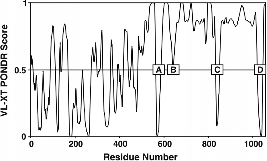
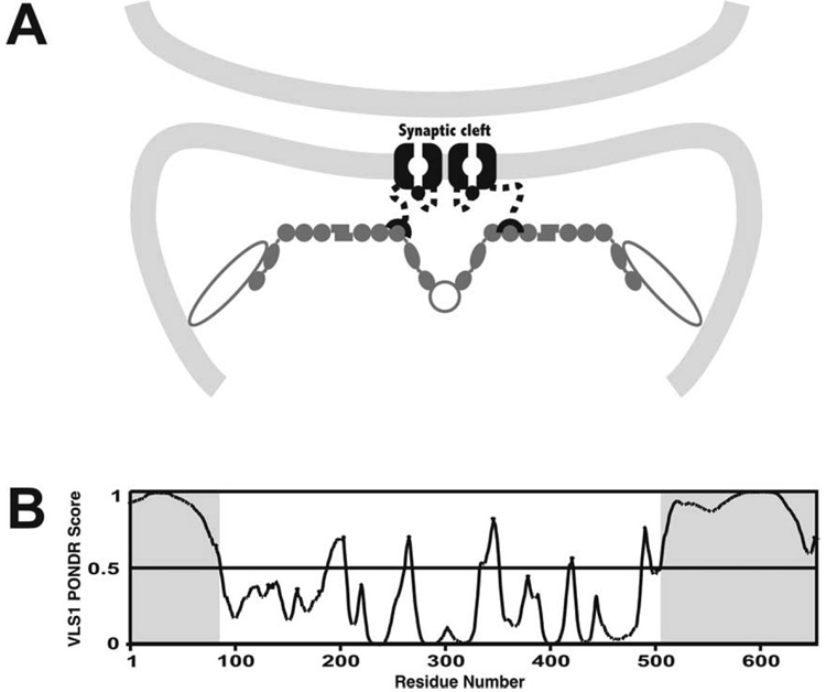
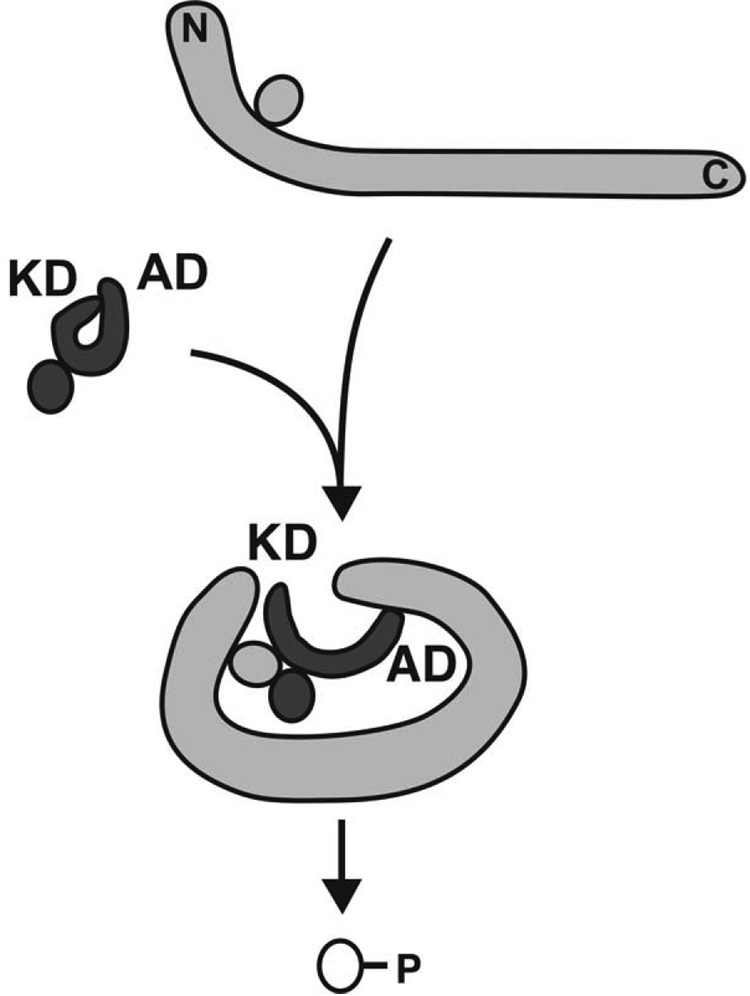
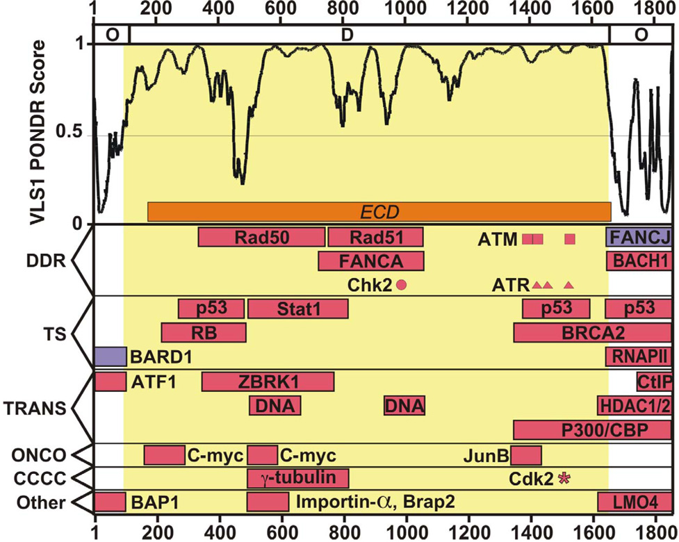
Regulation, recognition and cell signaling involve the coordinated actions of many players. Signaling scaffolds, with their ability to bring together proteins belonging to common and/or interlinked pathways, play crucial roles in orchestrating numerous events by coordinating specific interactions among signaling proteins. This review examines the roles of intrinsic disorder (ID) in signaling scaffold protein function. Several well-characterized scaffold proteins with structurally and functionally characterized ID regions are used here to illustrate the importance of ID for scaffolding function. These examples include scaffolds that are mostly disordered, only partially disordered or those in which the ID resides in a scaffold partner. Specific scaffolds discussed include RNase, voltage-activated potassium channels, axin, BRCA1, GSK-3beta, p53, Ste5, titin, Fus3, BRCA1, MAP2, D-AKAP2 and AKAP250. Among the mechanisms discussed are: molecular recognition features, fly-casting, ease of encounter complex formation, structural isolation of partners, modulation of interactions between bound partners, masking of intramolecular interaction sites, maximized interaction surface per residue, toleration of high evolutionary rates, binding site overlap, allosteric modification, palindromic binding, reduced constraints for alternative splicing, efficient regulation via posttranslational modification, efficient regulation via rapid degradation, protection of normally solvent-exposed sites, enhancing the plasticity of interaction and molecular crowding. We conclude that ID can enhance scaffold function by a diverse array of mechanisms. In other words, scaffold proteins utilize several ID-facilitated mechanisms to enhance function, and by doing so, get more functionality from less structure.


Figures

Fig. 1
Fig. 1
PONDR® VL-XT prediction of RNase E showing that the C-terminal half of the protein (residues 483–1061) is predicted to largely IDed (PONDR® scores > 0.5) except for four short regions (A–D) that contain order-promoting residues. These regions correlated to experimentally determined minimal motifs required for self-association (region A) and for binding structured RNAs (region B), enolase (region C) and PNPase (region D).
Fig. 2
Fig. 2. ID-mediated association of PSD-95 and Kv channels
A. The Kv channels (black) are embedded in the postsynaptic membrane (light grey) and attached to the PSD-95 scaffold\(dark grey) by C-terminal PDZ domains (half circles) that are attached to the main body of the channel by IDed linkers (dotted lines). Open grey circles represent a much reduced subset of additional PSD-95-associtated proteins. Portions of the figure were based on Kim et al. (Kim and Sheng, 2004) and Magidovich et al. (Magidovich et al., 2006). The N-terminal ball and chain domains are depicted in the channel blocking position with the IDed linker also depicted as dotted lines. B. VLS1 PONDR® prediction of Shaker potassium voltage-gated channel protein (SwissProt accession number P08510) illustrating the substantial length of predicted ID (PONDR® scores > 0.5) located at both termini. The IDed regions depicted as dotted lines in panel A are highlighted with grey shading.
Fig. 3
Fig. 3. ID and interaction regions for axin
All data are plotted to scale by residue number (x-axis). Predictions of ID are shown in the upper half of each panel. Combined output from charge/hydropathy and CDF two-state prediction analysis (see text) is depicted in the topmost bar with predicted order and predicted ID labeled O and D, respectively. Regions that have been Experimentally Characterized as Disordered (see text) are indicated with orange bars labeled ECD. The large central IDR predicted by VLS1 is highlighted in yellow. Experimentally determined binding regions derived from structures (blue) and molecular biology methods (red) are shown in the bottom half of each panel. Binding regions are grouped by associated pathway as labeled on the y-axis. Pathway abbreviations are explained in the text. Note that regions are shown only once but may participate in more that one pathway. The following regions are from experiments using from mouse isoform 2 (the shorter, more prevalent form, Swiss-Prot accession number O35625-2) or mapped onto that sequence using bl2seq (human and mouse axin share 86% identity and 90% similarity)(Tatusova and Madden, 1999): β-catenin (Xing et al., 2003); GSK-3 β (Dajani et al., 2003); APC (Spink et al., 2000); Dix (homodimerization)(RCSB Protein Data Bank ID 1WSP, Shibata et al. 2006); Smad7 (Liu et al., 2006); PP2A, CKI, Axam, Smad3, dishevelled (Dvl), protein inhibitor of activated STAT (PIAS), Diversin, I-mfa, low density lipoprotein receptor-related protein 5 (LRP5) and LRP 6 from the excellent review by Luo (Luo and Lin, 2004); Arakadia and Smad7 (Liu et al., 2006); homeodomain-interacting protein kinase-2 (HIPK2) and p53 (Rui et al., 2004); Ccd1 (Wong et al., 2004); D and I (homodimerization) (Luo et al., 2005); mitogen-activated protein kinase kinase kinase 1 (MEKK1) and MEKK4 (Luo et al., 2003); small ubiquitin-related modifier conjugating enzymes (E6) (Rui et al., 2002).
Fig. 4
Fig. 4. Mechanism of axin-dependent activation of HIPK2 kinase activity
HIPK2 is held in an inactive state by interactions between its kinase and axin-binding domains (KD and AD, respectively). Upon binding axin, inhibition is relieved and the KD is able to phosphorylate both the HIPK2-bound p53 molecule and the one bound to axin. Binding sites of HIPK2 and p53 on axin are depicted approximately to scale (see Fig. 3). Based on Fig. 8 of Rui et al. (Rui et al., 2004).
Fig. 5
Fig. 5. ID and interaction regions for BCRA1
Layout and annotations are the same as for Fig. 3. Two domains of BRCA1, BRCT (which binds FNACJ) (Clapperton et al., 2004; Gaiser et al., 2004; Litman et al., 2005) and RING (which bind BARD1) (Brzovic et al., 2003), have known 3-D structures. The remaining evidence for protein-protein interactions is based on molecular biology evidence. These regions are: BRCA2 (Chen et al., 1998); HDAC1 & 2 (Yarden and Brody, 1999); BAP1 (Jensen et al., 1998); CtIP (Li et al., 1999a); p53 (Chen et al., 2006); ZBRK1 (Zheng et al., 2000); c-Myc, retinoblastoma protein (RB), DNA, Rad 50 & 51, FANCA, and JunB (Mark et al., 2005); Importin-α, BRAP2, STAT1, BACH1, ATF1, LMO4, p300/CBP RNA polemerase II (RNAPII) and γ-tubulin (Thompson and Schild, 2002). Sites phosphorylated by Chk2, ATM, ATR and CDK2 are shown as is indicated by a circle, boxes triangles, and an asterisk, respectively (Mark et al., 2005; Ruffner et al., 1999).
Fig. 6
Fig. 6. Differential and competitive binding modes of MAPKK Ste7 and the Ste5 scaffold for sites on Fus3
A. Ste5 binds Fus3 in a bipartite manner. The two Ste5 interacting regions are separated by a IDed linker. By binding to both the N- and C-lobes of Fus3 and being restricted by the length of the linker, Ste5 alters the positioning of the lobes and consequently the active site configuration which results in altered Fus3 activity. The IDed linker connecting the bipartite binding peptides is depicted as a dotted line. B. Canonical binding of Ste7 to the C-lobe of Fus3 does not alter the relationship between the lobes or alter the conformation of the active site and thus does not alter Fus3 activity. Dotted line represents the position of the upper (N-lobe) of Fus3 as depicted in panel A. C. Sequence comparison of the structurally characterized binding modes of two peptides that bind the C-lobe of Fus3. Far1 binds in a canonical orientation as does the Ste7 peptide. However, the Ste5 peptide that binds to the C-lobe does so in an opposite orientation. D. Both peptides display arginine, proline and leucine side chains as recognition features on one face of the polyproline II helix. This figure is based on Figure 2 of Bhattacharyya et al. (Bhattacharyya et al., 2006).
Fig. 7
Fig. 7
The SH3 domains (ribbon structures) of three different hypothetical interacting proteins (green circle, blue square and red pentagon) binding in either class I or class II modes to SH3 elements in the PVEK region of a titin molecule. The PVEK regions of titin isoforms can include both bipolar SH3 elements, which encode overlapping antiparallel ligand sequences (BBBBBBBBB), or unipolar binding sites (UUUUUUUU). Nebulin has a demonstrated ability to bind bipolar ligand sequences in either orientation and is represented here by the green circle. In translation from the class I binding mode (A) to the class II binding mode (B), the SH3 domain flips and rotates. Note that, due to the PPII conformation of the SH3 element, the per-residue offset between the antiparallel class I and class II domains would entail a 60° rotation about the centerline of the ligand sequence. In this simple cartoon, inter-protein interactions (black arrows) can take place between adjacent SH3-containing proteins (green and red in A and blue and green in B). Note that, in living cells, it is likely that such relationships would be dynamic with fractional stoichiometric distributions among alternate binding sites (Ma et al., 2006). C. PONDR® VLS1 prediction for titin (SwissProt accession number Q8WZ42) reflecting homogeneity of residue composition favoring ID (PONDR® scores > 0.5) along most of its length.
Fig. 8
Fig. 8. PONDR® VLS1 predictions for three AKAP scaffolds
A. AKAP250, B. MAP2 and C. D-AKAP2 (Q02952, P15146 and O43572 SwissProt accession numbers respectively).

Similar articles

Cited by

References

    1. Albert R, Jeong H, Barabasi AL. Error and attack tolerance of complex networks. Nature. 2000;406:378–382. - PubMed
    1. Andrade MA, Bork P. HEAT repeats in the Huntington's disease protein. Nat Genet. 1995;11:115–116. - PubMed
    1. Andrade MA, Perez-Iratxeta C, Ponting CP. Protein repeats: structures, functions, and evolution. J Struct Biol. 2001;134:117–131. - PubMed
    1. Aoki C, Siekevitz P. Ontogenetic changes in the cyclic denosine 3',5'-monophosphate-stimulatable phosphorylation of cat visual cortex proteins, particularly of microtubule-associated protein 2 (MAP 2): effects of normal and dark rearing and of the exposure to light. J Neurosci. 1985;5:2465–2483. - PMC - PubMed
    1. Avalos JL, Celic I, Muhammad S, Cosgrove MS, Boeke JD, Wolberger C. Structure of a Sir2 enzyme bound to an acetylated p53 peptide. Mol Cell. 2002;10:523–535. - PubMed

Publication types