Review
. 2008 Aug;58(2):88-111.
doi: 10.1016/j.phrs.2008.06.007. Epub 2008 Jun 24.

The immune system and cardiac repair

Affiliations
Review

The immune system and cardiac repair

Nikolaos G Frangogiannis. Pharmacol Res. 2008 Aug.

Abstract

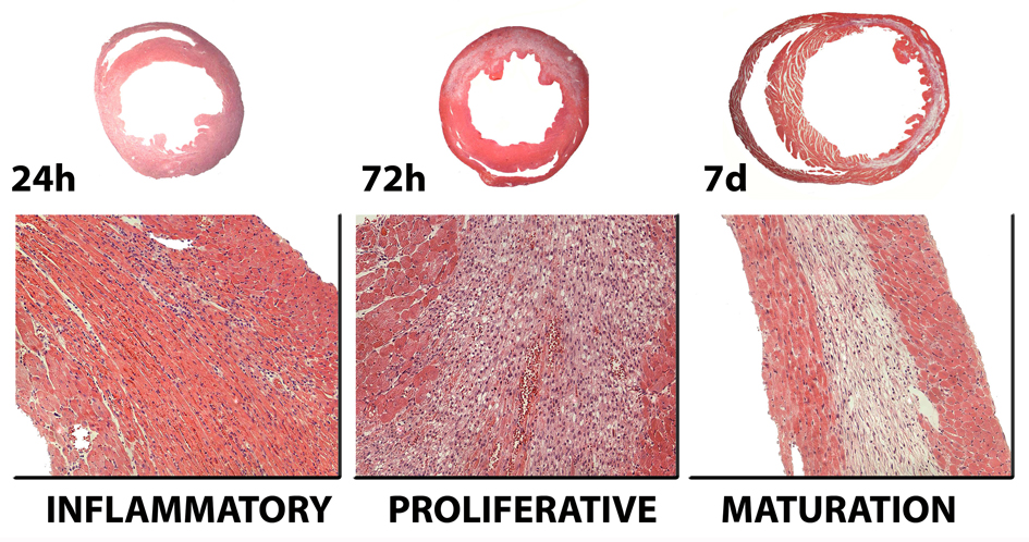
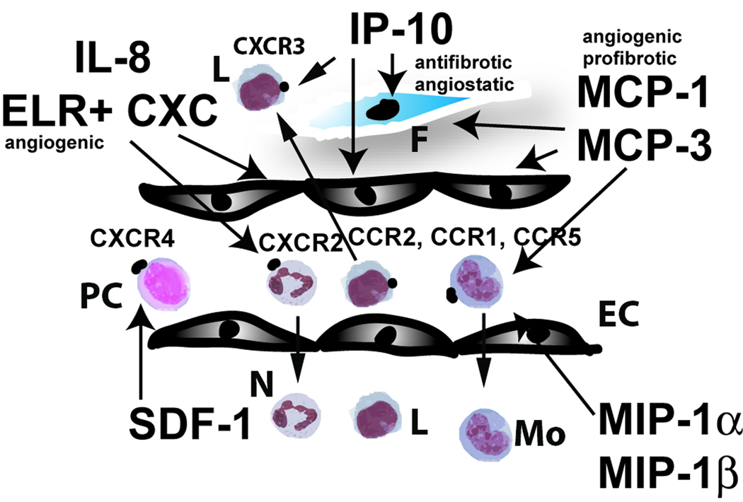
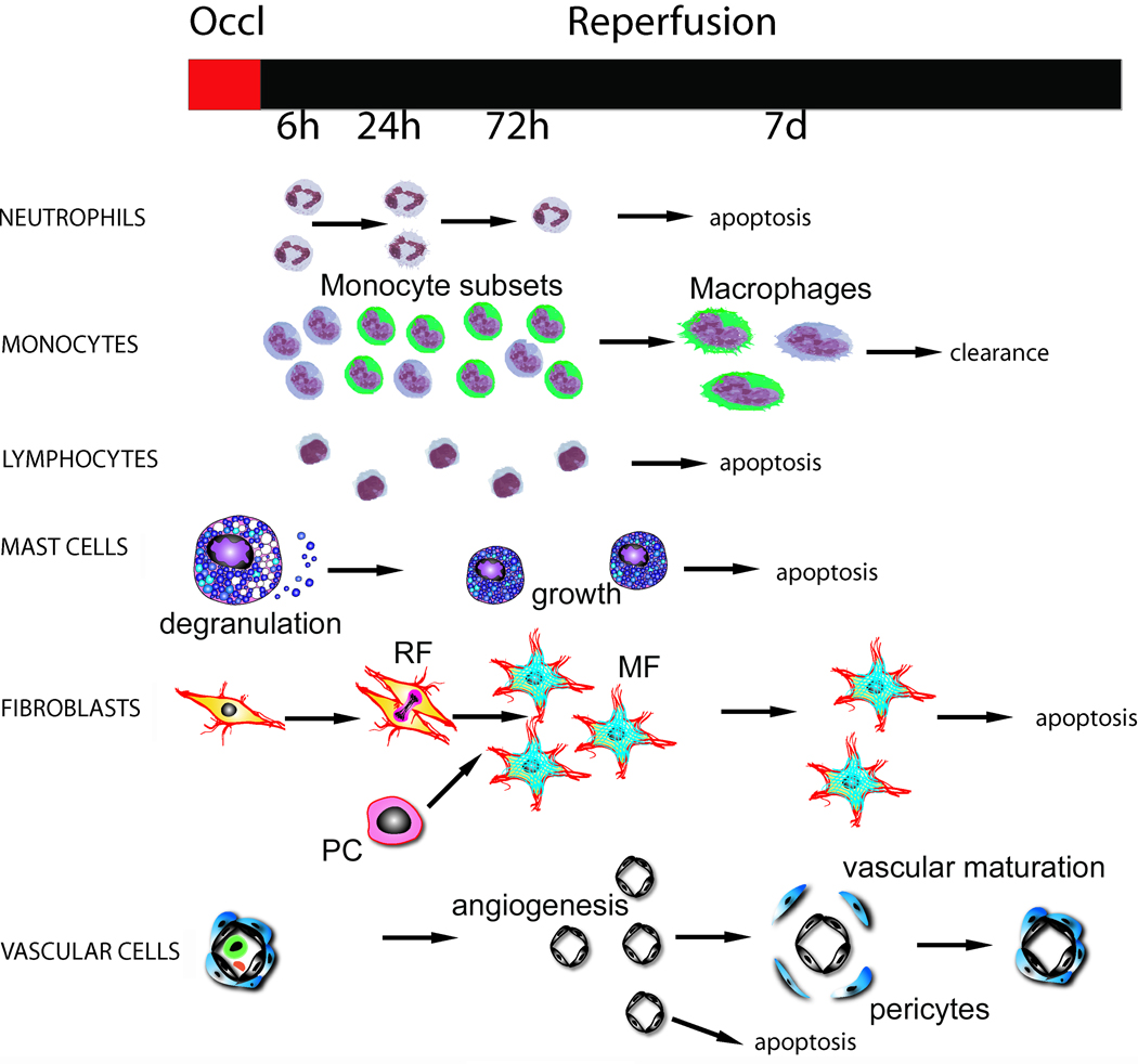
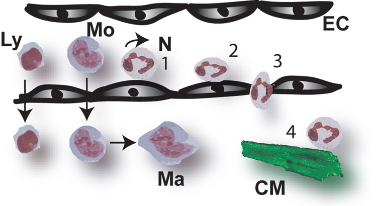
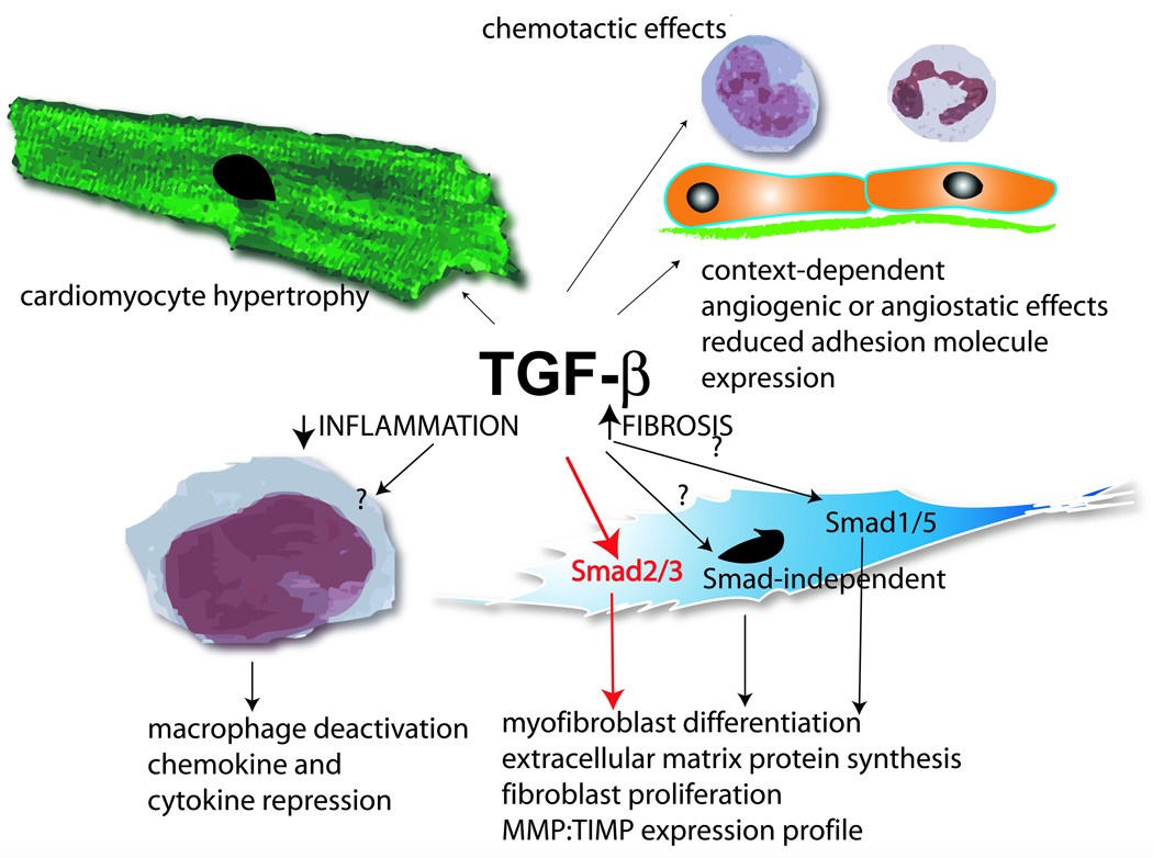
Myocardial infarction is the most common cause of cardiac injury and results in acute loss of a large number of myocardial cells. Because the heart has negligible regenerative capacity, cardiomyocyte death triggers a reparative response that ultimately results in formation of a scar and is associated with dilative remodeling of the ventricle. Cardiac injury activates innate immune mechanisms initiating an inflammatory reaction. Toll-like receptor-mediated pathways, the complement cascade and reactive oxygen generation induce nuclear factor (NF)-kappaB activation and upregulate chemokine and cytokine synthesis in the infarcted heart. Chemokines stimulate the chemotactic recruitment of inflammatory leukocytes into the infarct, while cytokines promote adhesive interactions between leukocytes and endothelial cells, resulting in transmigration of inflammatory cells into the site of injury. Monocyte subsets play distinct roles in phagocytosis of dead cardiomyocytes and in granulation tissue formation through the release of growth factors. Clearance of dead cells and matrix debris may be essential for resolution of inflammation and transition into the reparative phase. Transforming growth factor (TGF)-beta plays a crucial role in cardiac repair by suppressing inflammation while promoting myofibroblast phenotypic modulation and extracellular matrix deposition. Myofibroblast proliferation and angiogenesis result in formation of highly vascularized granulation tissue. As the healing infarct matures, fibroblasts become apoptotic and a collagen-based matrix is formed, while many infarct neovessels acquire a muscular coat and uncoated vessels regress. Timely resolution of the inflammatory infiltrate and spatial containment of the inflammatory and reparative response into the infarcted area are essential for optimal infarct healing. Targeting inflammatory pathways following infarction may reduce cardiomyocyte injury and attenuate adverse remodeling. In addition, understanding the role of the immune system in cardiac repair is necessary in order to design optimal strategies for cardiac regeneration.


Figures

Figure 1
Figure 1. Infarct healing is closely intertwined with ventricular remodeling
The healing response can be divided in three overlapping phases. During the inflammatory phase, chemokines and cytokines are induced in the infarct and marked leukocyte infiltration is noted. Neutrophils and macrophages clear the wound from dead cells and matrix debris. During the proliferative phase of healing, activated macrophages release cytokines and growth factors leading to formation of highly-vascularized granulation tissue. At this stage expression of pro-inflammatory mediators is suppressed, while fibroblasts and endothelial cells proliferate. Activated myofibroblasts produce extracellular matrix proteins and an extensive microvascular network is formed. Maturation of the scar follows: fibroblasts and vascular cells undergo apoptosis and a collagen-based scar is formed. As the infarct heals, dilative remodeling of the infarcted ventricle is noted (top panel). The time course of the cellular events presented in this figure is based on a reperfused model of infarction in the mouse. Large animals and humans exhibit delayed healing in comparison with rodents.
Figure 2
Figure 2. The role of the chemokines in cardiac injury
Both CC and CXC chemokines are induced in the infarcted myocardium. CXC chemokines that contain the ELR motif (such as IL-8) induce neutrophil infiltration and may exert angiogenic effects. In contrast, ELR-negative CXC chemokines (such as IP-10) have angiostatic and antifibrotic properties. Early IP-10 upregulation in the infarcted heart may inhibit granulation tissue formation until the wound is debrided from dead cells and a provisional matrix necessary to support fibroblast and endothelial cell migration is formed. In addition, IP-10 may play a role in recruitment of effector T cells. On the other hand, the CXC chemokine SDF-1 may induce infiltration of CXCR4+ progenitor cells enhancing tissue regeneration and angiogenesis. The best studied CC chemokine is MCP-1, a potent mononuclear cell chemoattractant with angiogenic and pro-fibrotic properties. MCP-1 deficiency is associated with attenuated adverse remodeling at the expense of delayed clearance of the infarct from dead cardiomyocytes. MCP-3, MIP-1α and MIP-1β are also induced in the infarct; however, their role in cardiac injury and repair remains unknown (Symbols: PC, immature progenitor cells; N, neutrophils; Mo, monocytes; L, lymphocytes; EC, endothelial cells; F, fibroblasts).
Figure 3
Figure 3. The cell biology of healing myocardial infarction
Infarct healing is dependent on the sequential infiltration of the injured myocardium with neutrophils, mononuclear cells, mast cells, fibroblasts and vascular cells (see text for more details). This is a dynamic and superbly orchestrated process: recruitment or proliferation of each cell type is followed by activation. Various cell populations have distinct but overlapping functions. Orchestration of the sequence of cellular events in the healing infarct is dependent on timely apoptosis of specific cell types. The time course presented here is based on reperfused mouse myocardial infarction; large mammals and humans exhibited a delayed inflammatory and reparative response (Symbols: RF, resident fibroblasts; PC, progenitor cells; MF, myofibroblasts).
Figure 4
Figure 4. Infiltration of the infarcted myocardium with leukocytes is dependent on a sequence of adhesive interactions
During the inflammatory phase of infarct healing, chemoattractants for neutrophils, monocytes and lymphocytes are upregulated in the injured myocardium. Local release of inflammatory mediators results in endothelial cell activation and surface expression of adhesion molecules. The selectin family of adhesion molecules mediates the initial capture of leukocytes from the rapidly flowing bloodstream to the blood vessel. Selectins promote leukocyte attachment and rolling (1) at shear stresses characteristic of post-capillary venules. Although rolling is a prerequisite for eventual firm adherence to blood vessels under conditions of flow, selectin-dependent adhesion of leukocytes does not lead to firm adhesion (2) and transmigration (3) unless another set of adhesion molecules, the integrins, is engaged. The mechanisms responsible for leukocyte transmigration into the tissues are poorly understood. In vitro studies have demonstrated that infiltrating neutrophils are capable of inducing cytotoxic injury to cardiomyocytes (4); this process is dependent on adhesive interactions between activated neutrophils and cytokine-stimulated cardiomyocytes. Whether neutrophil-mediated injury significantly contributes to cardiomyocyte death following infarction remains controversial (Symbols: Ly, lymphocyte; Mo, monocyte; Ma, macrophage; N, neutrophil; EC, endothelial cell; CM, cardiomyocyte).
Figure 5
Figure 5. Vascular maturation through acquisition of a pericyte coat regulates infarct angiogenesis
Panels A and B show dual immunohistochemical staining of reperfused canine infarcts for the endothelial cell marker CD31 (black) and α-smooth muscle actin (red). During the proliferative phase of healing (A) the infarct contains a large number of capillaries and several dilated pericyte-poor vessels (arrow - “mother vessels”). As the scar matures (B), some vessels acquire a mural cell coat (B- arrowheads), while uncoated vessels regress. PDGF-BB/PDGFR-β interactions are critically involved in vascular maturation following myocardial infarction.
Figure 6
Figure 6. The complex role of TGF-β signaling in myocardial infarction
Evidence suggests that TGF-β plays a dual role in cardiac injury, contributing to resolution of inflammation through deactivation of macrophages, while exerting fibrogenic actions. Thus TGF-β may be the “master switch” responsible for transition from inflammation to fibrosis. TGF-β is a highly pleiotropic mediator capable of modulating the phenotype of all cells involved in cardiac repair. Evidence suggests that TGF-β induces cardiomyocyte hypertrophy, has complex, context-dependent angiogenic and angiostatic effects and stimulates leukocyte chemotaxis, while suppressing endothelial cell adhesion molecule synthesis. Because of the complex and multifunctional role of TGF-β, dissection of the signaling pathways responsible for specific actions is critical in order to identify therapeutic targets. It appears that the fibrogenic effects of TGF-β in the healing infarct are, at least in part, dependent on Smad3 signaling. The role of the Smad1/5 and Smad-independent pathways remains unknown.
Figure 7
Figure 7. Through its unique composition, the infarct border zone may protect the non-infarcted myocardium from expansion of inflammatory injury
Immunohistochemical staining of canine myocardial infarction demonstrates a strikingly selective deposition of the matricellular protein Thrombospondin (TSP)-1 (arrows) in the infarct border zone (BZ). TSP-1 is a key TGF-β activator, has direct anti-inflammatory effects mediated through CD47 and exerts potent angiostatic effects. Thus, TSP-1 induction in the border zone may serve as a “barrier” preventing expansion of the inflammatory reaction into the non-infarcted myocardium (C). (Symbol: I, infarct)

References

    1. Tavazzi L. Clinical epidemiology of acute myocardial infarction. Am Heart J. 1999;138:S48–S54. - PubMed
    1. Jennings RB, Murry CE, Steenbergen C, Jr, Reimer KA. Development of cell injury in sustained acute ischemia. Circulation. 1990;82:II2–II12. - PubMed
    1. Pfeffer MA, Braunwald E. Ventricular remodeling after myocardial infarction. Experimental observations and clinical implications. Circulation. 1990;81:1161–1172. - PubMed
    1. Opie LH, Commerford PJ, Gersh BJ, Pfeffer MA. Controversies in ventricular remodelling. Lancet. 2006;367:356–367. - PubMed
    1. Cohn JN, Ferrari R, Sharpe N. Cardiac remodeling--concepts and clinical implications: a consensus paper from an international forum on cardiac remodeling. Behalf of an International Forum on Cardiac Remodeling. J Am Coll Cardiol. 2000;35:569–582. - PubMed

Publication types