Review
. 2009 Feb 6;158(3):983-94.
doi: 10.1016/j.neuroscience.2008.06.025. Epub 2008 Jun 19.

Diverse roles of matrix metalloproteinases and tissue inhibitors of metalloproteinases in neuroinflammation and cerebral ischemia

Affiliations
Review

Diverse roles of matrix metalloproteinases and tissue inhibitors of metalloproteinases in neuroinflammation and cerebral ischemia

E Candelario-Jalil et al. Neuroscience. .

Abstract

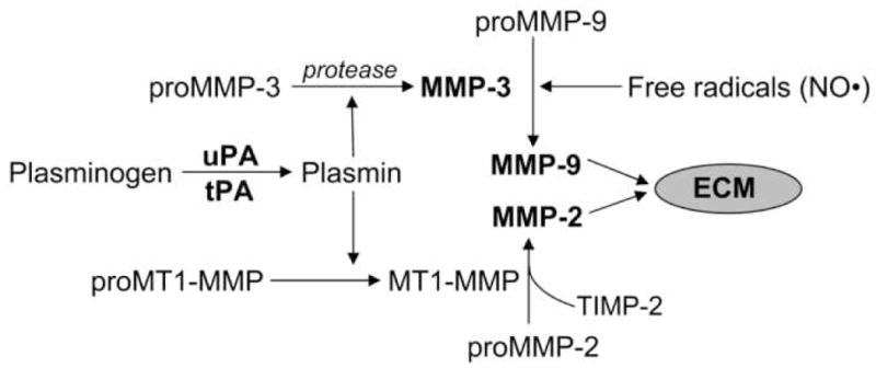
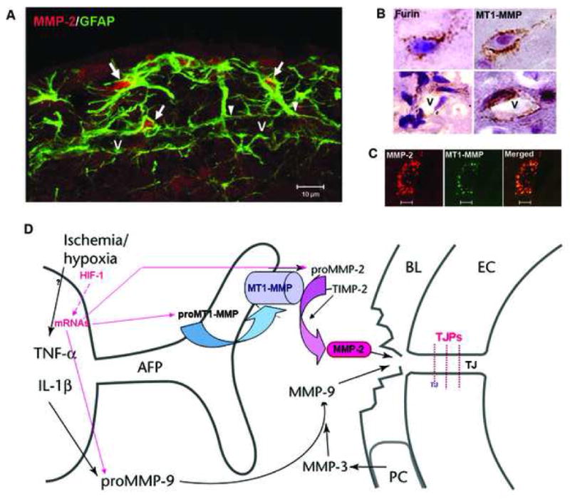
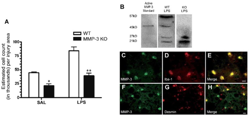
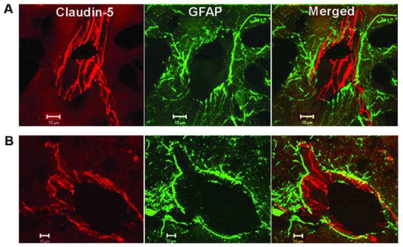
Regulation of the extracellular matrix by proteases and protease inhibitors is a fundamental biological process for normal growth, development and repair in the CNS. Matrix metalloproteinases (MMPs) and the tissue inhibitors of metalloproteinases (TIMPs) are the major extracellular-degrading enzymes. Two other enzyme families, a disintegrin and metalloproteinase (ADAM), and the serine proteases, plasminogen/plasminogen activator (P/PA) system, are also involved in extracellular matrix degradation. Normally, the highly integrated action of these enzyme families remodels all of the components of the matrix and performs essential functions at the cell surface involved in signaling, cell survival, and cell death. During the inflammatory response induced in infection, autoimmune reactions and hypoxia/ischemia, abnormal expression and activation of these proteases lead to breakdown of the extracellular matrix, resulting in the opening of the blood-brain barrier (BBB), preventing normal cell signaling, and eventually leading to cell death. There are several key MMPs and ADAMs that have been implicated in neuroinflammation: gelatinases A and B (MMP-2 and -9), stromelysin-1 (MMP-3), membrane-type MMP (MT1-MMP or MMP-14), and tumor necrosis factor-alpha converting enzyme (TACE). In addition, TIMP-3, which is bound to the cell surface, promotes cell death and impedes angiogenesis. Inhibitors of metalloproteinases are available, but balancing the beneficial and detrimental effects of these agents remains a challenge.


Figures

Fig. 1
Fig. 1
Mechanisms of activation of MMPs. Plasmin plays an active role in the activation of MMPs. MT1-MMP (MMP-14) binds to TIMP-2 and proMMP-2 leading to the formation of catalytically active MMP-2. ProMMP-9 is activated by MMP-3 and free radicals (nitric oxide, NO•). Plasmin has been shown to activate proMMP-3. Recently, it was shown that there is an intracellular mechanism of activation of MMP-3 which involves a serine protease other than furin (Choi et al., 2008). Although proMMP-3 possesses the furin recognition sequence, the cleavage would leave 9 amino acids belonging to the prodomain resulting in a form which is not catalytically active (Choi et al., 2008).
Fig. 2
Fig. 2
(A) Confocal immunohistochemistry shows GFAP-positive astrocytes around a vessel (V) that express MMP-2 (arrows) in intact rat brain tissue. The arrowheads indicate the astrocyte endfeet around the vessel. (B) Expression of furin and MT1-MMP in brain cells and around vessels (V). (C) Confocal images show the co-localization of MMP-2 and MT1-MMP immunohistochemistry in brain cells. (D) Schematic drawing to show that the activation of MMP-2 occurs through the action of the trimolecular complex during the early opening of the BBB in 3h-reperfusion after 90 min MCAO. In the astrocytic foot processes (AFP), the membrane-type 1 matrix metalloproteinase (MT1-MMP) joins with tissue inhibitor of metalloproteinases-2 (TIMP-2) to activate proMMP-2 in a spatially constrained manner close to the basal lamina (BL). In the BL are the pericytes (PC). The endothelial cells (EC) have tight junctions (TJ). The activated MMP-2 has direct access to the portion of the BL beneath the AFP and components of the BL are degraded. The manner in which this disruption of the BL leads to increased permeability is unclear since the role of the BL in maintaining the integrity of the blood vessel is uncertain.
Fig. 3
Fig. 3
(A) Stereology for neutrophil counts 24 h after LPS or saline injection. Counts of MPO-immunoreactive neutrophils were greater in the LPS-injected caudate for both WT and MMP-3 KO. However, in the MMP-3 KO, there were significantly fewer neutrophils in the caudate than in WT in both saline and LPS-injected hemispheres (*P < 0.01, **P < 0.001; n = 4 for both WT and KO). Panels B–H: Western blotting and immunohistochemical staining for MMP-3, pericytes, and microglia. (B) Western blotting for MMP-3 showed the proform at 57 kDa and the active form at 45 kDa along with two lower, unidentified bands. The MMP-3 KO mouse did not show the 57 or 45-kDa bands of MMP-3. (C–E) MMP-3 colocalized with Iba-1-immunoreactive microglia/macrophages. (C) MMP-3 staining in LPS-injected WT; (D) Iba-1 immunostaining; (E) merged image of C and D. (F–H) MMP-3 was also seen in pericytes stained for desmin in LPS-injected WT. (F) MMP-3 staining; (G) Pericyte staining; (H) merged composite of panels F and G. Scale bars are 10 μm. Adapted from Gurney et al., 2006.
Fig. 4
Fig. 4
Confocal micrographs showing claudin-5 immunoreactivity after 3 h of reperfusion following 90 min of MCAO in the rat. (A) The nonischemic side show that the claudin-5 (Cy-3) in blood vessels is separated from the astrocytes (GFAP-FITC) surrounding them. The merged images show that the claudin-5 and astrocytes are separate. (B) In the ischemic hemisphere, there is fragmentation and degeneration of the claudin-5 immunoreactivity. Co-localization of claudin-5 and GFAP was seen in the ischemic hemisphere. Adapted from Yang et al., 2007.
Fig. 5
Fig. 5
(A) T2 weighted image (B) diffusion-weighted (DW) contrast image, and (C) color coded permeability coefficient map for BB1101 treated (top row) and control (bottom row) rats. The ischemia can be seen as a hyperintense lesion (arrow) on T2 weighted images. A 53.1 and 48.4% reduction in the ADC value on the ischemic side (region of interest on lesion) compared with that in the matching contralateral side was observed in BB1101-treated and control rats, respectively, after 3 h of reperfusion. The arrows on diffusion-weighted contrast image point at ischemic regions with diffusion changes. Color-coded permeability maps show clearly the regions of high (arrow) and low permeability in treated and control rats. Taken from Sood et al., 2008.
Fig. 6
Fig. 6
(A) Effect of rtPA on mortality in animals with different intervals of ischemia and reperfusion: Mortality is shown in percent with the numbers of animals dying and the number of animals studied shown in parentheses above the bars. When reperfusion was delayed to 6 hours, mortality was increased markedly in rtPA-treated animals (**P<0.01). Treatment with BB-94 reduced rtPA-associated mortality significantly (*P<0.05). Control animals had MCAO without rtPA treatment. (B) Opening of the BBB as measured by sucrose space in rats with 6 hours of ischemia and 1 hour of reperfusion (I6R1). Compared with sham-operated animals, BBB permeability was markedly increased in untreated rats (**P<0.01). BB-94 given 2 and 5 hours after MCAO markedly decreased BBB opening (*P<0.01). Adapted from Pfefferkorn and Rosenberg, 2003.
Fig. 7
Fig. 7
Lesion size in timp-3 +/+ versus timp-3 −/− at 3 days of reperfusion following 30 min transient MCAO. Timp-3 +/+ and timp-3 −/− mice were subjected to 30 min transient MCAO followed by 3 days of reperfusion. Coronal sections were stained for degenerating neurons using Fluoro-Jade (a, b) or stained for apoptotic nuclei using TUNEL (c, d). Infarct volume in timp-3 +/+ versus timp-3 −/− mice as assessed by morphometric analysis of Fluoro-Jade-stained histological sections, (e) n=5 mice per strain, *P<0.001. (f) Average lesion area per histological section in rostrocaudal direction in timp-3 +/+ versus timp-3 −/− mice. Taken from Wetzel et al., 2008.

References

    1. Amantea D, Russo R, Gliozzi M, Fratto V, Berliocchi L, Bagetta G, Bernardi G, Corasaniti MT. Early upregulation of matrix metalloproteinases following reperfusion triggers neuroinflammatory mediators in brain ischemia in rat. Int Rev Neurobiol. 2007;82:149–169. - PubMed
    1. Asahi M, Asahi K, Jung JC, del Zoppo GJ, Fini ME, Lo EH. Role for matrix metalloproteinase 9 after focal cerebral ischemia: effects of gene knockout and enzyme inhibition with BB-94. J Cereb Blood Flow Metab. 2000;20:1681–1689. - PubMed
    1. Asahi M, Sumii T, Fini ME, Itohara S, Lo EH. Matrix metalloproteinase 2 gene knockout has no effect on acute brain injury after focal ischemia. Neuroreport. 2001a;12:3003–3007. - PubMed
    1. Asahi M, Wang X, Mori T, Sumii T, Jung JC, Moskowitz MA, Fini ME, Lo EH. Effects of matrix metalloproteinase-9 gene knock-out on the proteolysis of blood-brain barrier and white matter components after cerebral ischemia. J Neurosci. 2001b;21:7724–7732. - PMC - PubMed
    1. Ballabh P, Braun A, Nedergaard M. The blood-brain barrier: an overview: structure, regulation, and clinical implications. Neurobiol Dis. 2004;16:1–13. - PubMed

Publication types

MeSH terms

Substances