. 2008 Sep;111(3-5):240-6.
doi: 10.1016/j.jsbmb.2008.06.008. Epub 2008 Jun 22.

Human prostate stromal cells stimulate increased PSA production in DHEA-treated prostate cancer epithelial cells

Affiliations

Human prostate stromal cells stimulate increased PSA production in DHEA-treated prostate cancer epithelial cells

Julia T Arnold et al. J Steroid Biochem Mol Biol. 2008 Sep.

Abstract

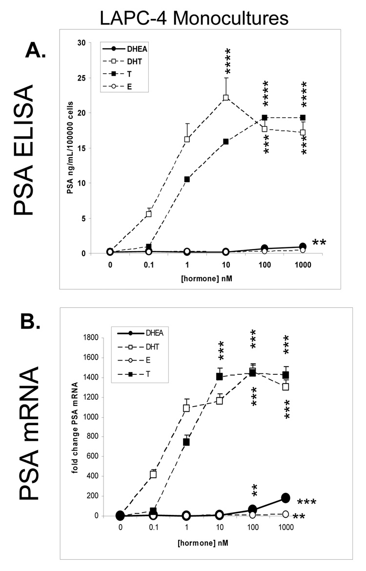
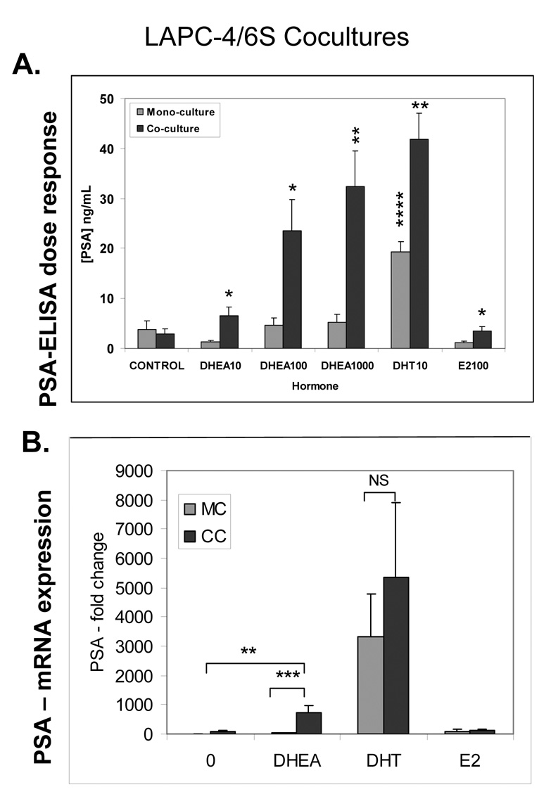
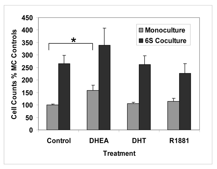
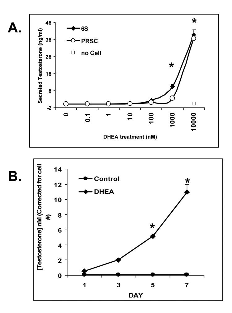
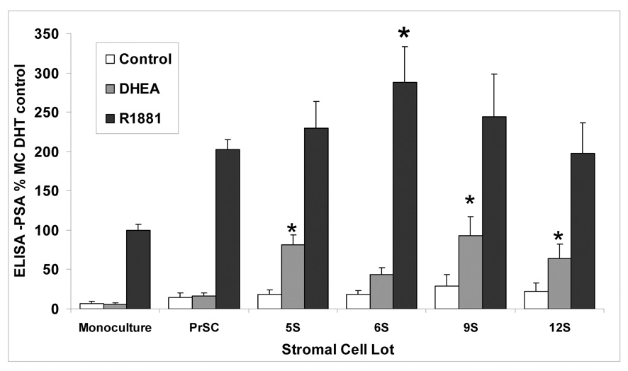
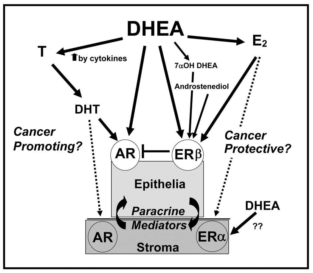
Dehydroepiandrosterone (DHEA) is commonly used as a dietary supplement and may affect prostate pathophysiology when metabolized to androgens and/or estrogens. Human prostate LAPC-4 cancer cells with a wild type androgen receptor (AR) were treated with DHEA, androgens dihydrotestosterone (DHT), T, or R1881), and E2 and assayed for prostate specific antigen (PSA) protein and gene expression. In LAPC-4 monocultures, DHEA and E2 induced little or no increase in PSA protein or mRNA expression compared to androgen-treated cells. When prostate cancer-associated (6S) stromal cells were added in coculture, DHEA stimulated LAPC-4 cell PSA protein secretion to levels approaching induction by DHT. Also, DHEA induced 15-fold more PSA mRNA in LAPC-4 cocultures than in monocultures. LAPC-4 proliferation was increased 2-3-fold when cocultured with 6S stromal cells regardless of hormone treatment. DHEA-treated 6S stromal cells exhibited a dose- and time-dependent increase in T secretion, demonstrating stromal cell metabolism of DHEA to T. Coculture with non-cancerous stroma did not induce LAPC-4 PSA production, suggesting a differential modulation of DHEA effect in a cancer-associated prostate stromal environment. This coculture model provides a research approach to reveal detailed endocrine, intracrine, and paracrine signaling between stromal and epithelial cells that regulate tissue homeostasis within the prostate, and the role of the tumor microenvironment in cancer progression.


Figures

Figure 1
Figure 1. PSA gene and protein expression in LAPC-4 cells in monoculture
A. PSA protein determination by ELISA Cells were plated in triplicate at a density of 5 × 10⁵ cells/well in 24 well dishes, in 1mL of Treatment Media. After 3 days, cells were treated with control DHEA, DHT, T, and E2, at 0.1nM, 1nM, 10nM, 100nM, and 1000nM in Treatment Media with a final concentration of CDS at 1%. After 3 days, media with hormone was replaced and allowed to condition for 48 hours. Conditioned media was collected and assayed by ELISA for PSA concentrations. Cell numbers were estimated using the MTT assay scaled to a 24 well format. Graphs illustrate mean ± SD values of PSA, averaged from three experiments. B. PSA gene expression determination by Real Time RT-PCR LAPC-4 cells were plated in triplicate in 6 well plates at a density of 2 × 10⁶ cells/well, in Treatment Media for 3 days prior to treatment with control, DHEA, DHT, T, or E2 at same doses as above. RNA samples were extracted, reverse transcribed and assayed for PSA gene expression by Real Time PCR and expressed as fold change PSA mRNA. Graphs illustrate mean ± SD values averaged from three experiments.
Figure 2
Figure 2. Prostate stromal effects on LAPC-4 PSA protein and gene expression
A. Prostate stromal induction of LAPC-4 PSA protein production Cocultures of LAPC-4 cells and 6S stromal cells were prepared. LAPC-4 cells were seeded in triplicate onto Millipore PICM 12mm inserts coated with a film of Matrigel at a density of 5×10⁵ cells/well. Stromal cells (6S) cells were seeded in triplicate at 1×10⁵ / well of 24 well plates. Cells were grown separately in media containing 2% CDS for 2 or 3 days prior to combining in coculture and addition of DHEA (10, 100 or 1000 nM) DHT (10 nM) and E2 (100 nM) and were incubated for 3 days. Media containing hormones was replaced and allowed to condition for 48 hours. Conditioned media were collected assayed by ELISA for free PSA. Cell numbers were estimated using the MTT assay scaled to a 24 well format. Assay was performed three times and mean and SE are represented.. P values are represented as follows: **** P<0.0001,, ** P<0.01, *P<0.05 B. Prostate stromal induction of LAPC-4 PSA gene expression LAPC-4 cells were seeded in triplicate 30mm inserts pre-coated with Matrigel film at a density of 2×10⁶ cells/insert. 6S cells were plated on 60 mm dish at 5×10⁵ / dish. After 2–3 days, cells were combined and treated with 100nM DHEA, 100nM E2, or 10 nM DHT for 2 days, and then harvested to extract RNA and perform reverse transcription and realtime PCR. Assay was performed three times and mean and SE are represented. Graphs illustrate mean ± SD values averaged from three experiments performed in triplicate. P values are represented as follows: *** P<0.001, ** P<0.01.
Figure 3
Figure 3. Hormone effects on LAPC-4 Cell Proliferation in monoculture and coculture: LAPC-4 proliferation is increased in coculture with 6S primary prostate stromal cells
LAPC-4 cells were seeded at 106 cells per Matrigel-film-coated 30mm insert. Primary prostate (6S) stromal cells were seeded in triplicate at 5×10⁵ / 60 mm tissue culture plastic dish. After 3 days, inserts containing the LAPC-4 cells were added to the 60 mm dish of stromal cells and hormones were added at 0, 100nM DHEA, 10nM DHT or 10 nM R1881 and allowed to coculture for 3 days. LAPC-4 cells were harvested by trypsinization and counted using a Beckman Coulter counter. Cell counts are expressed as percentage of the monoculture control. Assay was performed three times and means and SE are represented, *P<0.01.
Figure 4
Figure 4. Stromal Cell Metabolism of DHEA to Testosterone:DHEA-treated primary 6S and PrSC stromal cells increase T secretion in a dose and time dependent manner
Stromal cells (6S or PrSC) were plated in triplicate in 24 well plates and pretreated with Treatment Media for 2 days. (A). Dose response; DHEA was added at doses 0.1 to 10,000 nM, for 5 days. B. Time response; Cells treated with control of no hormone or DHEA at 100 nM for 1, 3, 5, and 7 days. Conditioned Media was collected and assayed for testosterone by ELISA. T values were corrected for cell number by running a parallel MTT assay. Assay was performed three times and mean and SE are represented.
Figure 5
Figure 5. Effects of various stromal cell lots on LAPC-4 PSA Level induced by DHEA and R1881 compared with effects in monocultured LAPC-4 cells
Coculture assays for ELISA determination of LAPC-4 PSA were prepared as before using stromal lots 5S, 6S, 9S, 12S, or PrSC. Hormones were added at 100nM DHEA treatment or (10nM) R1881. Values are represented as percent change in LAPC-4 PSA level induced by various stromal cells. Assay was performed three times and mean and SE are represented (*P< 0.05 compared to MC same hormone treatment).
Figure 6
Figure 6. Endocrine- Intracrine- Paracrine- model of interactions between prostate stromal and epithelial cells affecting the pathways of DHEA metabolism in prostate tissues
The complexity of the effects of DHEA in prostate tissues includes metabolism of DHEA to androgenic or estrogenic ligands, the potential cytokine contribution to increased DHEA metabolism, both stromal and epithelial components, the expression of AR and ER β or (ERα in stromal cells) and their downstream effectors, the hormone-induced paracrine signaling and shows the importance of the stromal component as a contributor to DHEA metabolism. Androgenic ligands may promote progression to cancer by increasing growth, IGF axis and PSA production. Conversely, the estrogenic ligands, mediated by ER beta may protect from cancer by antagonizing androgenic pathways. Each of these factors plays an important role in defining the effects of DHEA on the prostate. These factors can be manipulated in the co-culture model provided to determine DHEA mechanisms as well as elucidate paracrine -intracrine - endocrine prostate stromal-epithelial signals important in responses to hormones.

References

    1. Belanger A, Candas B, Dupont A, Cusan L, Diamond P, Gomez JL, Labrie F. Changes in serum concentrations of conjugated and unconjugated steroids in 40-to 80-year-old men. J Clin Endocrinol Metab. 1994;79:1086–1090. - PubMed
    1. Alesci S, Manoli I, Blackman MR. Dehydrodepiandrosterone (DHEA) In: Coates P, Blackman MR, Cragg G, Levine M, Moss J, White J, editors. Encyclopedia of Dietary Supplements. 1st ed. New York, NY: Marcel Dekker, Inc.; 2005. pp. 167–176.
    1. Klein H, Bressel M, Kastendieck H, Voigt KD. Androgens, adrenal androgen precursors, and their metabolism in untreated primary tumors and lymph node metastases of human prostatic cancer. Am J Clin Oncol. 1988;11 Suppl 2:S30–S36. - PubMed
    1. Voigt KD, Bartsch W. Intratissular androgens in benign prostatic hyperplasia and prostatic cancer. J Steroid Biochem. 1986;25:749–757. - PubMed
    1. Labrie F, Belanger A, Luu-The V, Labrie C, Simard J, Cusan L, Gomez JL, Candas B. DHEA and the intracrine formation of androgens and estrogens in peripheral target tissues: its role during aging. Steroids. 1998;63:322–328. - PubMed

Publication types