. 2008 Jul 15;105(28):9627-32.
doi: 10.1073/pnas.0801963105. Epub 2008 Jul 9.

Functional architecture of inositol 1,4,5-trisphosphate signaling in restricted spaces of myoendothelial projections

Affiliations

Functional architecture of inositol 1,4,5-trisphosphate signaling in restricted spaces of myoendothelial projections

Jonathan Ledoux et al. Proc Natl Acad Sci U S A. .

Abstract

Calcium (Ca(2+)) release through inositol 1,4,5-trisphosphate receptors (IP(3)Rs) regulates the function of virtually every mammalian cell. Unlike ryanodine receptors, which generate local Ca(2+) events ("sparks") that transmit signals to the juxtaposed cell membrane, a similar functional architecture has not been reported for IP(3)Rs. Here, we have identified spatially fixed, local Ca(2+) release events ("pulsars") in vascular endothelial membrane domains that project through the internal elastic lamina to adjacent smooth muscle membranes. Ca(2+) pulsars are mediated by IP(3)Rs in the endothelial endoplasmic reticulum of these membrane projections. Elevation of IP(3) by the endothelium-dependent vasodilator, acetylcholine, increased the frequency of Ca(2+) pulsars, whereas blunting IP(3) production, blocking IP(3)Rs, or depleting endoplasmic reticulum Ca(2+) inhibited these events. The elementary properties of Ca(2+) pulsars were distinct from ryanodine-receptor-mediated Ca(2+) sparks in smooth muscle and from IP(3)-mediated Ca(2+) puffs in Xenopus oocytes. The intermediate conductance, Ca(2+)-sensitive potassium (K(Ca)3.1) channel also colocalized to the endothelial projections, and blockage of this channel caused an 8-mV depolarization. Inhibition of Ca(2+) pulsars also depolarized to a similar extent, and blocking K(Ca)3.1 channels was without effect in the absence of pulsars. Our results support a mechanism of IP(3) signaling in which Ca(2+) release is spatially restricted to transmit intercellular signals.


Conflict of interest statement

The authors declare no conflict of interest.

Figures

Fig. 1.
Fig. 1.
Ca2+ pulsars colocalized with IEL holes. (A) (a) IEL autofluorescence shows the presence of “holes” in the IEL. (b) Initiation sites of Ca2+ pulsars from the composite image correspond to holes in the IEL (red arrows). The yellow arrows indicate pulsar sites not associated with detectable IEL holes. (Scale bar, 10 μm.) (c) Histogram illustrating the distance between Ca2+ pulsar initiation sites and IEL holes in endothelium (n = 357 pulsar sites). (B) Time course of a three-dimensional Ca2+ pulsar originating from within an IEL hole (white circle) shown in the leftmost image. (Scale bar, 5 μm.) (C) Ca2+ pulsars from a pressurized artery (80 mmHg) expressing GCaMP2. (Ca) An endothelial cell and its nucleus are outlined (dotted lines), with the initiation sites (Cb and Cc) indicated by red arrows. (Scale bar, 10 μm.) See also Movie S1 and Movie S2
Fig. 2.
Fig. 2.
Kinetics and repetitive occurrences of Ca2+ pulsars. (A) Average of 10 images of a field of endothelial cells from mesenteric arteries of a GCaMP2-expressing mouse. The red arrow indicates the initiation site of Ca2+ pulsars shown in B and C. (Scale bar, 10 μm.) (B) Life span of a Ca2+ pulsar is shown. The field of view corresponds to the green square in A (Scale bar, 10 μm.) (C) Repetitive occurrence of Ca2+ pulsars at one site expressed as a line-scan analysis along the yellow line in A. (Scale bar, 5 s.) (D) Representative traces illustrating Ca2+ pulsar kinetics originating from two different sites (red and blue lines). See also Movie S1
Fig. 3.
Fig. 3.
Ca2+ pulsars originating from IP3-sensitive stores. Removal of extracellular Ca2+ (A) or ryanodine (C) did not affect Ca2+ pulsars. Inhibition of SERCA with CPA (B), of IP3Rs with xestospongin C (D), or of PLC with U73122 (E) decreased Ca2+ pulsars. (F) A data summary of pharmacological experiments targeting the source of Ca2+ pulsars is given. (n = 6, 5, 4, 6, and 3 arteries for 0 Ca2+, CPA, ryanodine, xestospongin C, and U73122, respectively; *, P < 0.05). For A–E, different colors represent F/Fo in ROIs over different pulsar sites in the endothelium. Calcium pulsars were recorded for 2 min, followed by variable incubation times (0 Ca, 5 min; Ry, 35 min; CPA, 15 min; xestospongin C, 40 min; U73122, 15 min) and then 2 min of recording in the drug treatment (see Methods Summary).
Fig. 4.
Fig. 4.
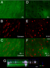
Localization of ER and IP3Rs within IEL holes. Images show immunostaining for ER protein calnexin (red) (A–C) and for IP3Rs (red) (D–F) at the level of the IEL (green). (C and F) Superimposed images reveal that ER and IP3R are highly concentrated inside distinct IEL holes. (Scale bar, 5 μm.) (G) A three-dimensional view along the z axis (2.9 μm) shows densities of IP3R-positive fluorescence (white) projecting through the depth of the IEL (green). (Scale bar, 1 μm.)
Fig. 5.
Fig. 5.
Ca2+ pulsars hyperpolarizing the endothelium membrane through activation of KCa3.1 channels. (A) (a) CPA (10 μM; black bar) depolarizes the endothelial membrane potential, and subsequent addition of ChTX (300 nM; white bar) had no effect. (b) Summary of membrane potential experiments with CPA using microelectrode and perforated patch techniques. Additionally, in microelectrode experiments, CPA exposure was followed by ChTX. Subsequent exposure to 60 mM KCl depolarized the endothelial membrane potential to −20 ± 2 mV. n = 6 and 4 for microelectrode and perforated patch recordings, respectively. (B) (a) ChTX (300 nM; black bar) depolarizes the endothelial membrane potential, and subsequent addition of CPA (10 μM; white bar) had no effect. (b) A summary of microelectrode experiments with ChTX followed by CPA is given. Subsequent exposure to 60 mM KCl depolarized the endothelial membrane potential to −22 ± 2 mV (n = 5; *, P < 0.05). All microelectrode experiments were carried out in the presence of paxilline (500 nM) and nitrendipine (100 nM). (C) (a and b) Immunostaining for KCa3.1 (red) at the level of the IEL (green) is shown. (c) The superimposed image reveals distinct densities of KCa3.1 within and around IEL holes. (Scale bar, 5 μm.) (d) A three-dimensional view along the z axis (3.1 μm) shows densities of KCa3.1-positive fluorescence (white) projecting through the depth of the IEL (green). (Scale bar, 1 μm.)
Fig. 6.
Fig. 6.
Model of endothelial Ca2+ pulsars. IP3R-dense ER stores follow portions of the endothelial cell membrane that evaginate through holes in the IEL and interface with underlying SM cell membranes. Repetitive localized Ca2+ events (pulsars) originate from these deep Ca2+ stores that are regionally delimited to the myoendothelial junction and the base of the endothelial cell. These ongoing dynamic Ca2+ signals are driven by constitutive IP3 production and are inherently dependent on the level of endothelial stimulation. The left detail depicts a single endothelial projection through the IEL. KCa3.1 channels in the plasma membranes of these endothelial projections are in very close proximity to Ca2+ pulsars, eliciting persistent Ca2+-dependent hyperpolarization of the membrane potential at the myoendothelial junctions. The right detail illustrates the endothelial influence on the SM membrane potential at the myoendothelial interaction site where Ca2+ pulsars would activate KCa3.1 channels and hyperpolarize the endothelial membrane. This hyperpolarization can be transmitted to the SM through gap junction channels or by activation of SM Kir channels by K+ ions released by endothelial KCa3.1 channels. Membrane hyperpolarization promotes relaxation of SM through a decrease in voltage-dependent calcium channel open probability.

References

    1. Nelson MT, et al. Relaxation of arterial smooth muscle by calcium sparks. Science. 1995;270:633–637. - PubMed
    1. Feletou M, Vanhoutte PM. Endothelium-derived hyperpolarizing factor:Where are we now? Arterioscler Thromb Vasc Biol. 2006;26:1215–1225. - PubMed
    1. Kohler R, Hoyer J. The endothelium-derived hyperpolarizing factor: Insights from genetic animal models. Kidney Int. 2007;72:145–150. - PubMed
    1. Kansui Y, Garland CJ, Dora KA. Enhanced spontaneous Ca2+ events in endothelial cells reflect signalling through myoendothelial gap junctions in pressurized mesenteric arteries. Cell Calcium. in press. - PMC - PubMed
    1. McSherry IN, et al. A role for heterocellular coupling and EETs in dilation of rat cremaster arteries. Microcirculation. 2006;13:119–130. - PubMed

Publication types

MeSH terms

Substances

LinkOut - more resources