. 2008 Jul 22;105(29):10209-14.
doi: 10.1073/pnas.0712144105. Epub 2008 Jul 9.

Respiratory syncytial virus uses a Vps4-independent budding mechanism controlled by Rab11-FIP2

Affiliations

Respiratory syncytial virus uses a Vps4-independent budding mechanism controlled by Rab11-FIP2

Thomas J Utley et al. Proc Natl Acad Sci U S A. .

Abstract

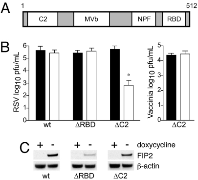
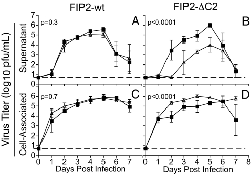
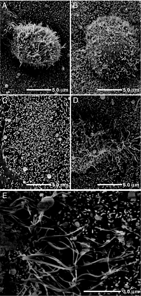
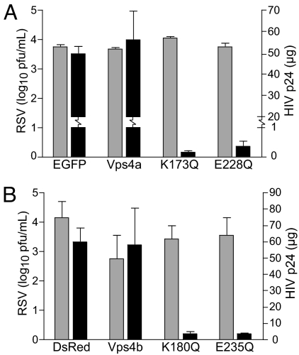
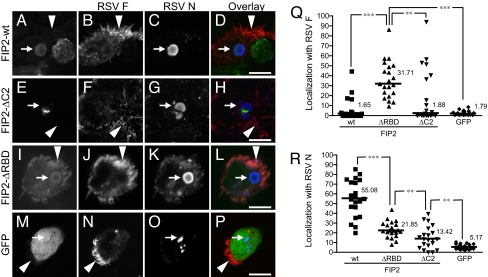
Respiratory syncytial virus (RSV) infects polarized epithelia, which have tightly regulated trafficking because of the separation and maintenance of the apical and basolateral membranes. Previously we established a link between the apical recycling endosome (ARE) and the assembly of RSV. The current studies tested the role of a major ARE-associated protein, Rab11 family interacting protein 2 (FIP2) in the virus life cycle. A dominant-negative form of FIP2 lacking its N-terminal C2 domain reduced the supernatant-associated RSV titer 1,000-fold and also caused the cell-associated virus titer to increase. These data suggested that the FIP2 C2 mutant caused a failure at the final budding step in the virus life cycle. Additionally, truncation of the Rab-binding domain from FIP2 caused its accumulation into mature filamentous virions. RSV budding was independent of the ESCRT machinery, the only well-defined budding mechanism for enveloped RNA viruses. Therefore, RSV uses a virus budding mechanism that is controlled by FIP2.


Conflict of interest statement

The authors declare no conflict of interest.

Figures

Fig. 1.
Fig. 1.
FIP2 facilitates production of supernatant-associated virus. (A) FIP2 contains an N-terminal C2 domain, a MVb-binding domain, and three NPF motifs, followed by a C-terminal Rab11-binding domain. MDCK cells stably transfected with DNA encoding FIP2-GFP variants (FIP2-WT; C2 domain deletion, FIP2-ΔC2; or RBD deletion, FIP2-ΔRBD) under doxycycline repression, expressing (open square) or not expressing (filled square), were infected with RSV or Vaccinia virus. (B) Five days after infection, supernatants were collected, and the virus titer was measured by plaque assay. Representative data from three independent experiments are shown. (C) Relative expression levels for each FIP2 variant in either the presence or absence of doxycycline along with β-actin loading control. *, P = 0.002; Wilcoxon rank sum test. Data shown are mean ±SD.
Fig. 2.
Fig. 2.
FIP2-ΔC2 delays and reduces RSV budding. The MDCK FIP2-WT cell line (A and C) or FIP2-ΔC2 cell line (B and D) was infected with RSV. Viral titer was measured in both expressing (open triangle) and not expressing (filled square) cells over a 7-day period for both supernatant-associated virus (A and B) and cell-associated virus (C and D). Dotted lines indicate the limit of detection. Least-squares regression was used to estimate the best-fitting quadratic curves for viral titer over time, and P values were computed by using likelihood ratio tests comparing these curves between expressing and not expressing cells. Triplicate values were determined; error bars indicate ±SD.
Fig. 3.
Fig. 3.
FIP2 facilitates termination of RSV filament growth and virus budding. (A, B, D, and E) Scanning EM was performed to view the length of RSV filaments at the apical surface of MDCK cells expressing FIP2-WT, a dominant negative (A), an MVb-tail construct (B), or FIP2-ΔC2 (D or E) (images from separate cells at two resolutions). (C) Uninfected cells expressing FIP2-ΔC2.
Fig. 4.
Fig. 4.
RSV uses a Vps4-independent budding mechanism. Cells (293T) transfected with Vps4a constructs: pEGFP vector (EGFP) or Vps4a-EGFP (Vps4a), or dominant-negative mutants Vps4a-K173Q-EGFP (K173Q) or Vps4a-E228Q-EGFP (E228Q) (A), or Vps4b constructs: pDsRed vector (DsRed) or Vps4b-DsRed (Vps4b), or dominant-negative mutants Vps4b-K180Q-DsRed (K180Q) or Vps4a-E235Q-DsRed (E235Q) (B). Cells were infected 24 h after transfection at a moi of 1.0 with either WT RSV or HIVNL4−3. Supernatant virus was collected 48 h after infection and assayed for either RSV by viral titer (gray bars) or HIV p24 by ELISA (black bars).
Fig. 5.
Fig. 5.
FIP2 associates with both viral inclusions and viral filaments. (A–P) MDCK cells expressing FIP2-GFP variants [FIP2-WT (A–D), FIP2-ΔC2 (E–H), or FIP2-ΔRBD (I–L)] infected with RSV or MDCK cells infected with recombinant RSV expressing GFP (M–P) were stained by indirect immunofluorescence for both RSV F (B, F, J, and N) and RSV N (C, G, K, and O). In the overlays (D, H, L, and P), FIP2-GFP variants and GFP are pseudocolored green, RSV F is pseudocolored red, and RSV N is pseudocolored blue. The arrows with tails indicate viral inclusion body location, whereas the arrows without tails indicate an area of dense viral filament formation. (Q and R) Percentage of the total integrated pixel intensity indicating colocalization of FIP2-GFP variants or GFP with RSV F (Q) or RSV N (R) were calculated. The median value is shown. ***, P < 0.0001; **, P ≤ 0.009; Wilcoxon rank sum test. No significant difference was measured among WT, ΔC2, and GFP groups in graph Q. Additional comparisons on graph R demonstrate that the WT differs significantly from ΔC2 and GFP (both P < 0.0001); ΔRBD differed significantly from GFP (P < 0.0001).

References

    1. Wright PF, et al. Growth of respiratory syncytial virus in primary epithelial cells from the human respiratory tract. J Virol. 2005;79:8651–8654. - PMC - PubMed
    1. Zhang L, Peeples ME, Boucher RC, Collins PL, Pickles RJ. Respiratory syncytial virus infection of human airway epithelial cells is polarized, specific to ciliated cells, and without obvious cytopathology. J Virol. 2002;76:5654–5666. - PMC - PubMed
    1. Roberts SR, Compans RW, Wertz GW. Respiratory syncytial virus matures at the apical surfaces of polarized epithelial cells. J Virol. 1995;69:2667–2673. - PMC - PubMed
    1. Bachi T, Howe C. Morphogenesis and ultrastructure of respiratory syncytial virus. J Virol. 1973;12:1173–1180. - PMC - PubMed
    1. Bachi T. Direct observation of the budding and fusion of an enveloped virus by video microscopy of viable cells. J Cell Biol. 1988;107:1689–1695. - PMC - PubMed

Publication types

MeSH terms

Substances

LinkOut - more resources