. 2008 Jul 15;105(28):9588-93.
doi: 10.1073/pnas.0712099105. Epub 2008 Jul 9.

Alpha-Helix folding in the presence of structural constraints

Affiliations

Alpha-Helix folding in the presence of structural constraints

Janne A Ihalainen et al. Proc Natl Acad Sci U S A. .

Abstract

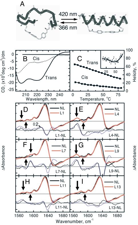
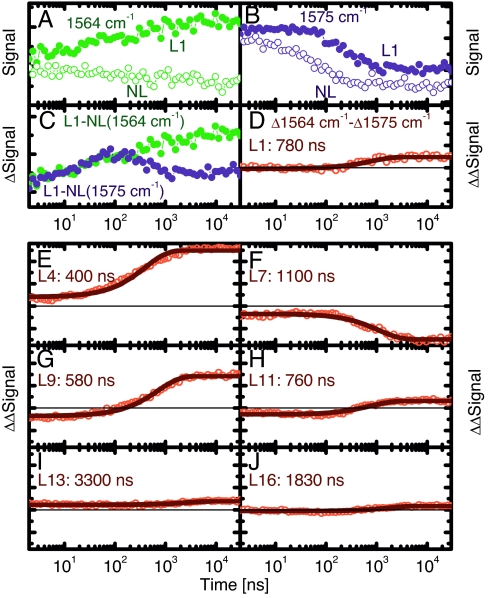
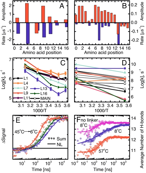
We have investigated the site-specific folding kinetics of a photoswitchable cross-linked alpha-helical peptide by using single (13)C = (18)O isotope labeling together with time-resolved IR spectroscopy. We observe that the folding times differ from site to site by a factor of eight at low temperatures (6 degrees C), whereas at high temperatures (45 degrees C), the spread is considerably smaller. The trivial sum of the site signals coincides with the overall folding signal of the unlabeled peptide, and different sites fold in a noncooperative manner. Moreover, one of the sites exhibits a decrease of hydrogen bonding upon folding, implying that the unfolded state at low temperature is not unstructured. Molecular dynamics simulations at low temperature reveal a stretched-exponential behavior which originates from parallel folding routes that start from a kinetically partitioned unfolded ensemble. Different metastable structures (i.e., traps) in the unfolded ensemble have a different ratio of loop and helical content. Control simulations of the peptide at high temperature, as well as without the cross-linker at low temperature, show faster and simpler (i.e., single-exponential) folding kinetics. The experimental and simulation results together provide strong evidence that the rate-limiting step in formation of a structurally constrained alpha-helix is the escape from heterogeneous traps rather than the nucleation rate. This conclusion has important implications for an alpha-helical segment within a protein, rather than an isolated alpha-helix, because the cross-linker is a structural constraint similar to those present during the folding of a globular protein.


Conflict of interest statement

The authors declare no conflict of interest.

Figures

Fig. 1.
Fig. 1.
Photoswitchable peptide and its steady-state spectra at room temperature. (A) Schematic drawing of the photoswitchable peptide in its cis (Left) and trans (Right) conformations. (B) CD-spectra of the peptide in its complete trans and cis states at room temperature. (C) The helicities in the cis and trans conformations at various temperatures. The trans spectrum was obtained in darkness, and the cis-spectrum was obtained under 365-nm illumination together with an estimate that 75% of the molecules are in the cis conformation (based on the UV-vis absorbance difference). (Inset) The first derivative of the trans CD data reveals the inflection point. (D–I) FTIR-difference spectra between the trans (under 436-nm illumination) and cis state (under 365-nm illumination) of each peptide at room temperature. Shown are the difference between not-labeled (NL) and labeled (LX, labeled at residue X) samples taken from the FTIR-spectra (thin black line) and from the late delay-time pump-probe spectra 30 μs after photoswitching (thin blue line). The arrows point to the frequency position from which the site-specific kinetics has been extracted.
Fig. 2.
Fig. 2.
Site-specific folding signals at 19°C. (A and B) Kinetic traces of the L1 and NL at 1,564 cm−1 (A) and at 1575 cm−1 (B). (C) The difference of the L1 and NL signals at these wavelengths. (D) The difference between the resulting signals together with its single exponential fit (solid line). (E–J) Site-selective folding signals of the other sites. The solid lines are single-exponential fits of the curves with the resulting time constant indicated.
Fig. 3.
Fig. 3.
Summary of the experimental (A, C, and E) and MD (B, D, and F) results. (A and B) Amplitudes (upward, red) and corresponding rates (downward, blue) at 19°C (A) and 8°C (B). Note that site 7 in the experiment, and sites 13–16 in the MD simulations, have inverted amplitudes. (C and D) Site-selective folding rates as a function of inverse temperature. In D, black circles are used for residues not measured experimentally. (E) Sum of all site signals (symbols) and amide I′ signal of the nonlabeled peptide (solid lines) at 6°C, 19°C, and 45°C. (F) Average number of hydrogen bonds along the MD simulations: 100 MD runs at 8°C with stretched exponential fit h(t) = 13.6 − 1.6exp(−t/29 ns)0.39 (blue), 50 MD runs at 57°C with single-exponential fit h(t) = 12.2 − 1.4exp(−t/6 ns) (red), and 50 MD runs of peptide without cross-linker at 8°C with single-exponential fit h(t) = 13.6 − 0.9exp(−t/2 ns) (magenta). The standard deviations are almost all <0.3 units. The experimental data in E are normalized, and the background has been removed, whereas the corresponding MD data in F are on their original scale.
Fig. 4.
Fig. 4.
The network analysis (22, 29) of the 100 MD runs at 281 K shows parallel folding channels. Each node (i.e., conformation) of the network represents a secondary structure string (39), and a link is a direct transition (within 20 ps) observed in the MD runs. The surface of each node is proportional to its statistical weight, and only the 1,387 nodes with at least 200 snapshots (≈96.7% of total sampling) are shown to avoid overcrowding. (A) The free-energy basins, i.e., native (green nodes) and metastable states [identified by kinetic grouping analysis (29) using a commitment time of 10 ns to group conformations that interconvert rapidly], are shown with different colors, and their characteristics are listed in Table S1. Within each basin, nodes and intrabasin links are shown with the same color, and interbasin links are colored in gray. White diamonds indicate the starting points of 82 of the 100 folding runs, whereas the remaining 18 runs reached directly the most populated node (i.e., fully formed α-helix, large green circle) and are not shown. An enlarged version of the network is shown in Fig. S2. (B–D) Nodes are colored according to the values of the mean first passage time (29) from the most populated node (white squares) of individual metastable states to all other nodes. The time scale goes from 0 (yellow) to >2 μs (blue). The coloring shows that the unfolded state is kinetically partitioned, and the folded (i.e., fully α-helical) state acts as a hub. Visits to unfolded metastable states different from the starting one require a much longer time than reaching the folded state. Representative structures of the folded state and each metastable state are shown by flexible tubes of variable diameter reflecting conformational disorder, with α-helical turns in green, loop segments in gray, N terminus in blue, and cysteine side chains in yellow for emphasizing the position of the linker.

References

    1. Eaton WA, Henry ER, Hofrichter J, Mozzarelli A. Is cooperative oxygen binding by hemoglobin really understood? Nat Struct Biol. 1999;6:351–358. - PubMed
    1. Creighton TE. Proteins. New York: Freeman; 1993.
    1. Zimm BH, Bragg JK. Theory of the phase transition between helix and random coil in polypeptide chains. J Chem Phys. 1959;31:526–535.
    1. Lifson S, Roig A. On the theory of helix–coil transition in polypeptides. J Chem Phys. 1961;34:1963–1974.
    1. Scholtz JM, Qian H, York EJ, Stewart JM, Baldwin RL. Parameters of helix–coil transition theory for alanine-based peptides of varying chain lengths in water. Biopolymers. 1991;31:1463–1470. - PubMed

Publication types

LinkOut - more resources