. 2008 Jul 15;105(28):9715-20.
doi: 10.1073/pnas.0803697105. Epub 2008 Jul 9.

Tools for neuroanatomy and neurogenetics in Drosophila

Affiliations

Tools for neuroanatomy and neurogenetics in Drosophila

Barret D Pfeiffer et al. Proc Natl Acad Sci U S A. .

Abstract

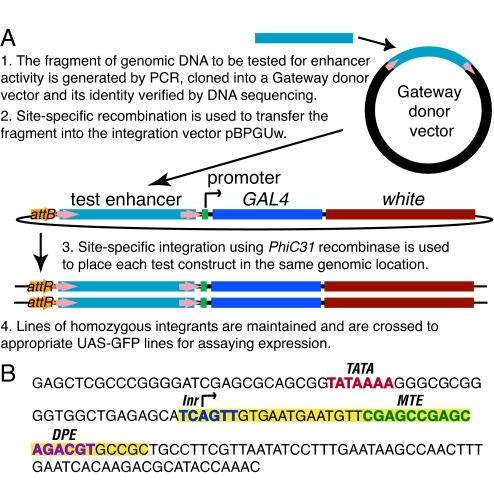
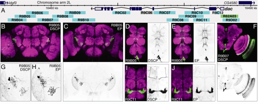
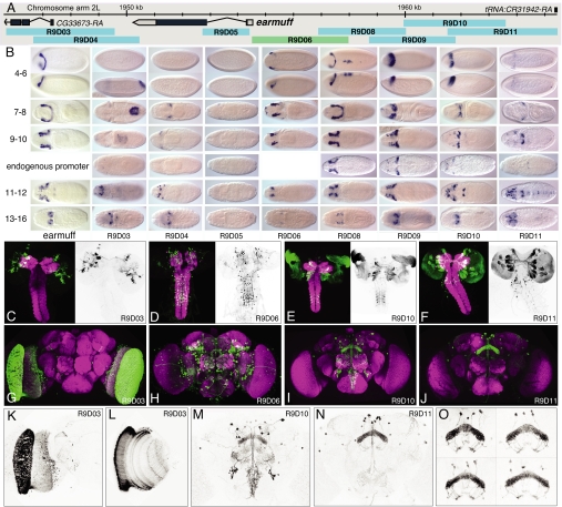
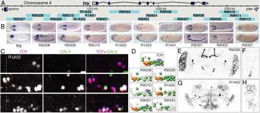
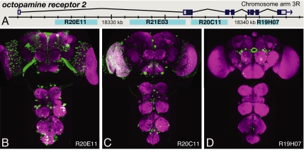
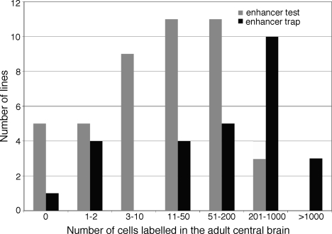
We demonstrate the feasibility of generating thousands of transgenic Drosophila melanogaster lines in which the expression of an exogenous gene is reproducibly directed to distinct small subsets of cells in the adult brain. We expect the expression patterns produced by the collection of 5,000 lines that we are currently generating to encompass all neurons in the brain in a variety of intersecting patterns. Overlapping 3-kb DNA fragments from the flanking noncoding and intronic regions of genes thought to have patterned expression in the adult brain were inserted into a defined genomic location by site-specific recombination. These fragments were then assayed for their ability to function as transcriptional enhancers in conjunction with a synthetic core promoter designed to work with a wide variety of enhancer types. An analysis of 44 fragments from four genes found that >80% drive expression patterns in the brain; the observed patterns were, on average, comprised of <100 cells. Our results suggest that the D. melanogaster genome contains >50,000 enhancers and that multiple enhancers drive distinct subsets of expression of a gene in each tissue and developmental stage. We expect that these lines will be valuable tools for neuroanatomy as well as for the elucidation of neuronal circuits and information flow in the fly brain.


Conflict of interest statement

The authors declare no conflict of interest.

Figures

Fig. 1.
Fig. 1.
Strategy for constructing transgenic lines to test DNA segments for enhancer activity. (A) Diagram of the vectors and sequential cloning steps. (B) Sequence of the Drosophila synthetic core promoter (DSCP). Sequences highlighted in yellow were added to the promoter of the eve gene. The positions of known promoter motifs are indicated.
Fig. 2.
Fig. 2.
Testing fragments from the dac genetic region for enhancer activity with both the DSCP and the endogenous dac promoter. (A) Diagram of the chromosomal region surrounding the gene showing the structures of the transcription unit and those of adjacent genes (data taken from D. melanogaster genome sequence Release 4.1). The extent and position of the DNA segments that were tested for enhancer activity are shown as light blue bars below the map; R62A03 (green) is a promoter fusion, and we did not obtain data for R9C06 (gray). Total (B–E and G–J) or partial projections (F) of confocal images of the brain (B, C, F–J) or ventral nerve cord (D and E) of 2- to 5-day old adults of the indicated transgenic line. Fragments were tested with either the DSCP (B, D, F, G, and I) or the dac endogenous promoter (EP) (C, E, H, and J). In the color images, gene expression driven by the enhancer fragment is shown in green and the neuropil is counterstained in magenta (see SI Methods for details). Embryonic expression patterns are shown in Fig. S3. The gray scale images show only the enhancer-driven expression.
Fig. 3.
Fig. 3.
Patterns generated by fragments of the earmuff gene in embryos, larvae, and adults. (A) The genomic map of the earmuff locus. (B) Expression of the earmuff gene in embryonic stages 4–6, 7–8, 9–10, 11–12, and 13–16 visualized by whole mount in situ hybridization with a probe to earmuff mRNA shown adjacent to the expression produced by the fragments R9D03, R9D04, R9D05, R9D08, R9D09, R9D10, and R9D11 when placed in the enhancer test vector and fragment R9D06 as a promoter fusion. Transgene expression is visualized by whole mount in situ hybridization with a probe to GAL4 mRNA. The enhancer constructs shown use the DSCP; for stages 9–10, we also show data obtained with the endogenous earmuff promoter. Dorsal views are shown except for stages 4–6, where a lateral view is also shown below the dorsal view; anterior is at Left. (C, D, E, and F) Expression driven by the indicated fragment in late third instar larvae. The clusters of labeled cells seen in F represent distinct lineages of secondary neurons; this labeling is not maintained in the adult (J). (G–O) Expression in the adult brain of the indicated lines. A total projection (K) and single optical section (L) of the optic lobes of the brain shown in G. (O) Expression in the fan-shaped body of line R9D11 in four different brains.
Fig. 4.
Fig. 4.
Patterns generated by fragments of the toy gene and a comparison with the expression pattern of the endogenous toy gene in the embryo. (A) Genomic map of the toy locus and the positions of the tested fragments. (B) Expression of the endogenous toy mRNA and the expression of GAL4 mRNA driven by the indicated nine fragments shown in stage 13–16 embryos; the other nine fragments shown in A did not drive detectable expression at this stage. Dorsal (Upper) and lateral (Lower) views are shown; anterior is at Left. (C) Endogenous toy protein (magenta in the merged image) and nuclear localized GFP (green in the merged image) expression driven by the R1A02 fragment in an abdominal CNS hemisegment of a stage 16 embryo (anterior, up; midline, dashed line). Three focal planes are shown: deep (Top), intermediate (Middle), and superficial (Bottom); see Fig. S2 for similar data on other fragments. (D) Diagrams of endogenous toy-positive neurons and the subset of toy-positive neurons in which each indicated fragment drives expression (deep neurons, blue; intermediate neurons, green; superficial neurons, orange); each fragment also drives expression in a reproducible set of toy-negative neurons that are not shown in these diagrams. The TM neurons are indicated by the red arrowheads. (E–H) Total projections of confocal images of the adult brain showing enhancer fragment driven expression in the brain (E and G) or ventral nerve cord (F and H) of lines R9G08 (E and F) and R1A02 (G and H).
Fig. 5.
Fig. 5.
Distinct expression patterns generated by fragments of the octopamine receptor 2 gene. (A) Diagram of the genomic locus. (B–D) Expression driven by the indicated fragments in the adult brain and ventral nerve cord.
Fig. 6.
Fig. 6.
The patterns driven by individual fragments generally contain fewer cells than those found in enhancer trap lines. The gray bars in the histogram show the number of cells found in the patterns within the central brain of the adult generated by the 44 enhancer fragments shown in blue in Figs. 2A (dac), 3A (earmuff), 4A (toy), and 5A (octopamine receptor 2) genes. The black bars show the number of cells found in 27 enhancer trap lines that were chosen randomly from the unpublished collection of J. Simpson and B. Ganetsky and are typical of the type of patterns seen in other enhancer trap collections (8).

References

    1. Baker BS, Taylor BJ, Hall JC. Are complex behaviors specified by dedicated regulatory genes? Reasoning from Drosophila. Cell. 2001;105:13–24. - PubMed
    1. Hiromi Y, Kuroiwa A, Gehring WJ. Control elements of the Drosophila segmentation gene fushi tarazu. Cell. 1985;43:603–613. - PubMed
    1. Harding K, Hoey T, Warrior R, Levine M. Autoregulatory and gap gene response elements of the even-skipped promoter of Drosophila. EMBO J. 1989;8:1205–1212. - PMC - PubMed
    1. Goto T, Macdonald P, Maniatis T. Early and late periodic patterns of even skipped expression are controlled by distinct regulatory elements that respond to different spatial cues. Cell. 1989;57:413–422. - PubMed
    1. Levine M, Tjian R. Transcription regulation and animal diversity. Nature. 2003;424:147–151. - PubMed

Publication types