. 2008 Jul 22;105(29):10221-6.
doi: 10.1073/pnas.0803453105. Epub 2008 Jul 11.

In silico identified CCR4 antagonists target regulatory T cells and exert adjuvant activity in vaccination

Affiliations

In silico identified CCR4 antagonists target regulatory T cells and exert adjuvant activity in vaccination

Jagadeesh Bayry et al. Proc Natl Acad Sci U S A. .

Abstract

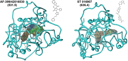
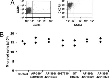
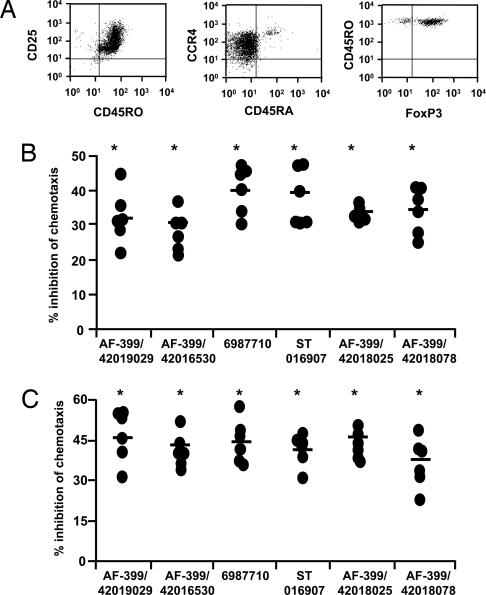
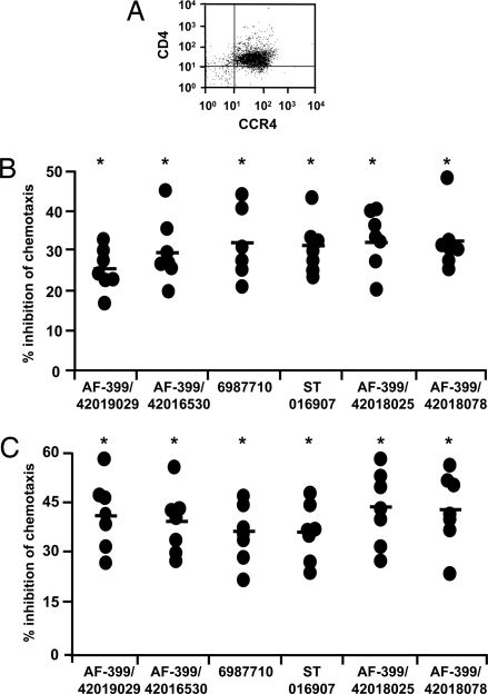
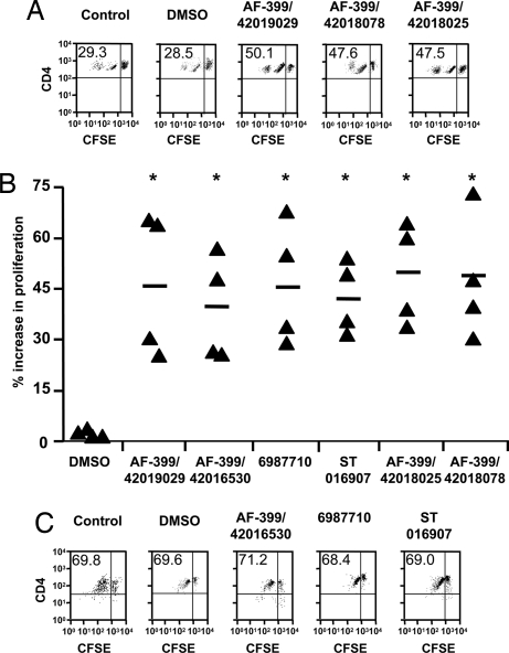
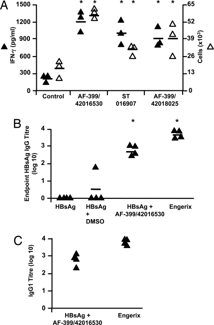
Adjuvants are substances that enhance immune responses and thus improve the efficacy of vaccination. Few adjuvants are available for use in humans, and the one that is most commonly used (alum) often induces suboptimal immunity for protection against many pathogens. There is thus an obvious need to develop new and improved adjuvants. We have therefore taken an approach to adjuvant discovery that uses in silico modeling and structure-based drug-design. As proof-of-principle we chose to target the interaction of the chemokines CCL22 and CCL17 with their receptor CCR4. CCR4 was posited as an adjuvant target based on its expression on CD4(+)CD25(+) regulatory T cells (Tregs), which negatively regulate immune responses induced by dendritic cells (DC), whereas CCL17 and CCL22 are chemotactic agents produced by DC, which are crucial in promoting contact between DC and CCR4(+) T cells. Molecules identified by virtual screening and molecular docking as CCR4 antagonists were able to block CCL22- and CCL17-mediated recruitment of human Tregs and Th2 cells. Furthermore, CCR4 antagonists enhanced DC-mediated human CD4(+) T cell proliferation in an in vitro immune response model and amplified cellular and humoral immune responses in vivo in experimental models when injected in combination with either Modified Vaccinia Ankara expressing Ag85A from Mycobacterium tuberculosis (MVA85A) or recombinant hepatitis B virus surface antigen (rHBsAg) vaccines. The significant adjuvant activity observed provides good evidence supporting our hypothesis that CCR4 is a viable target for rational adjuvant design.


Conflict of interest statement

The authors declare no conflict of interest.

Figures

Fig. 1.
Fig. 1.
In silico modeling of CCR4 antagonists. Representative illustrations of two small molecule CCR4 antagonists AF-399/42016530 and ST 016907 docked by GOLD into the homology model of CCR4. The diagram depicts a view looking down on the protein in the membrane from outside the cell. Residues making principal van der Waals contacts are shown in full; the remainder of CCR4 is shown as a single ribbon after the amino acid backbone. Antagonists are visualized with a surrounding hydrophobic Connolly surface. Pictures generated by using Sybyl7.3. Values in parentheses denote molecular weight.
Fig. 2.
Fig. 2.
Assessment of the specificity of CCR4 antagonists. (A) Expression of chemokine receptors CCR4 and CXCR4 by CCRF-CEM cells. (B) CCR4 antagonists do not inhibit CXCL12-mediated chemotaxis of CCRF-CEM cells. Data (n = 2) show the number of migrated cells in response to 3 nM CXCL12 in the absence (Control) or presence of 2 μM CCR4 antagonists AF-399/42019029, AF-399/42016530, 6987710, ST 016907, AF-399/42018025, and AF-399/420018078.
Fig. 3.
Fig. 3.
CCR4 antagonists block CCL22- and CCL17-mediated migration of human peripheral blood CD4+CD25+ regulatory T cells. (A) Expression of CCR4, CD25, CD45RO, CD45RA, and FoxP3 on Tregs. (B and C) Inhibition by CCR4 antagonists of CCL22 (1.2 nM)-mediated (B) and CCL17 (1.2 nM)-mediated (C) chemotaxis of Tregs. Data show the percent inhibition of chemotaxis by the indicated CCR4 antagonists (10 nM) for six donors. Percent inhibition of chemotaxis by CCR4 antagonists was calculated as follows: [(no. cells migrated in the presence of DMSO − no. cells migrated in the presence of antagonist)/no. cells migrated in the presence of DMSO] × 100. Mean values are indicated with a horizontal bar. *, P < 0.05 compared with DMSO controls.
Fig. 4.
Fig. 4.
CCR4 antagonists inhibit CCL22- and CCL17-mediated migration of human Th2 cells. (A) The expression of CD4 and CCR4 on in vitro-generated Th2 cells. (B and C) Percent inhibition by CCR4 antagonists (10 nM) of CCL22 (1.2 nM)-mediated (B) and CCL17 (1.2 nM)- mediated (C) migration of Th2 cells (n = 7 donors). Mean values are indicated with a horizontal bar. *, P < 0.05 compared with DMSO controls.
Fig. 5.
Fig. 5.
CCR4 antagonists boost DC-mediated human CD4+ T cell proliferation by blocking Treg recruitment. (A) CFSE profiles of DC-stimulated T cells treated with medium alone (Control), or with solvent (DMSO) or representative CCR4 antagonists (10 nM). The upper right quadrant represents undivided cells, whereas upper left quadrant represents cells that have divided and therefore diluted CFSE fluorescence. The values denote percent of cells that have undergone division. (B) The percent increase in DC-mediated T cell division upon exposure to CCR4 antagonists compared with controls. Percentage enhancement of T cell proliferation by CCR4 antagonists was calculated as follows: [(% divided cells in the presence of antagonist − % divided cells in control)/% divided cells in control] × 100. Statistical significance as analyzed by Mann–Whitney test is denoted by * (P < 0.05 compared with DMSO). (C) CCR4 antagonists do not modify mature DC-mediated proliferation of CD4+CD45RA+ naïve T cells lacking both Tregs in the population and expression of CCR4. The values denote percent of cells that have undergone division.
Fig. 6.
Fig. 6.
CCR4 antagonists amplify immunogenicity of vaccines in vivo. (A) Assessment of T cell response in mice 6 days after MVA85A vaccination in the presence of ≈2.5 μM CCR4 antagonists (AF-399/42016530, ST 016907, and AF-399/42018025) or DMSO control. IFN-γ production by splenocytes in response to PPD was analyzed by measuring IFN-γ in the supernatants (filled triangles, pg/ml) and ELISPOT assay (open triangles, ×103 cells per spleen). Similar results were obtained in two or three independent experiments. (B) IgG responses against rHBsAg as measured by ELISA 14 days after the second vaccination of mice with rHBsAg, rHBsAg plus DMSO, rHBsAg plus AF-399/42016530 (≈2.5 μM), or Engerix-B. Four mice per group were tested individually. *, P < 0.05 compared with DMSO controls. (C) Anti-HBsAg IgG responses elicited by Engerix-B or rHBsAg plus AF-399/42016530 are of IgG1 subclass.

References

    1. Guy B. The perfect mix: Recent progress in adjuvant research. Nat Rev Microbiol. 2007;5:505–517. - PubMed
    1. Schijns VE, Degen WG. Vaccine immunopotentiators of the future. Clin Pharmacol Ther. 2007;82:750–755. - PubMed
    1. Davies MN, Flower DR. Harnessing bioinformatics to discover new vaccines. Drug Discov Today. 2007;12:389–395. - PubMed
    1. Iellem A, et al. Unique chemotactic response profile and specific expression of chemokine receptors CCR4 and CCR8 by CD4(+)CD25(+) regulatory T cells. J Exp Med. 2001;194:847–853. - PMC - PubMed
    1. Miyara M, Sakaguchi S. Natural regulatory T cells: Mechanisms of suppression. Trends Mol Med. 2007;13:108–116. - PubMed

Publication types

MeSH terms

LinkOut - more resources