. 2008 Jul 15;105(28):9657-62.
doi: 10.1073/pnas.0801527105. Epub 2008 Jul 10.

Par3/Par6 polarity complex coordinates apical ectoplasmic specialization and blood-testis barrier restructuring during spermatogenesis

Affiliations

Par3/Par6 polarity complex coordinates apical ectoplasmic specialization and blood-testis barrier restructuring during spermatogenesis

Elissa W P Wong et al. Proc Natl Acad Sci U S A. .

Abstract

The Par3/Par6/aPKC and the CRB3/Pals1/PATJ polarity complexes are involved in regulating apical ectoplasmic specialization (ES) and blood-testis barrier (BTB) restructuring in the testis. Par6 was a component of the apical ES and the BTB. However, its level was considerably diminished at both sites at stage VIII of the cycle. Par6 also formed a stable complex with Pals1 and JAM-C (a component of the apical ES) in normal testes. When rats were treated with adjudin to induce apical ES restructuring without compromising the BTB, Par6 staining virtually disappeared at the apical ES in misaligned spermatids before their depletion. Additionally, the Par6/Pals1 complex became tightly associated with Src kinase, rendering a loss of association of the Par6/Pals1 complex with JAM-C, thereby destabilizing apical ES to facilitate spermatid loss. Primary Sertoli cell cultures with established functional BTB, but without apical ES, were next used to assess the Par6-based complex on BTB dynamics. When either Par6 or Par3 was knocked down by RNAi in Sertoli cell epithelium, a significant loss of the corresponding protein by approximately 60% in cells vs. controls was detected, alongside with a decline in aPKC after Par6, but not Par3, knockdown. This Par3 or Par6 knockdown also led to a transient loss of selected BTB proteins at the cell-cell interface, thereby compromising the BTB integrity. These findings illustrate that the Par6/Par3-based polarity complex likely coordinates the events of apical ES and BTB restructuring that take place concurrently at the opposing ends of adjacent Sertoli cells in the seminiferous epithelium during spermatogenesis.


Conflict of interest statement

The authors declare no conflict of interest.

Figures
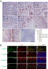
Fig. 1.
Fig. 1.
Par6 is localized at the apical ES and BTB in adult rat testes. (A) (a–h) Frozen testis-sections were stained by using an anti-Par6 antibody (see Table S1) vs. control (i) stained with normal rabbit serum. (a) A low-magnification section of an adult rat testis is shown. (b–e) Magnified views of representative tubules at different stages of the epithelial cycle as shown in the Upper Insets at lower magnification. Par6 was localized at the apical ES (see c and the magnified area in c Inset) in almost all stages except that at late-stage VIII just before spermiation, it was greatly diminished (see d vs. c). Lower Insets in b–d are magnified views of the boxed regions. (f–h) Par6 was also localized prominently at the BTB sites (see black arrowheads in g and h) except that the Par6 staining at the BTB was also diminished at stage VIII (see a and d and f vs. g and h). (j) An immunoblot wherein 80 μg of protein from lysates of testis (T), Sertoli cell (SC), and germ cell (GC) was probed with an anti-Par6 antibody. [Scale bars: a (also applies to Upper Insets in b–e), 100 μm; b (also applies to c–h), 20 μm; b Lower Inset (also applies to Lower Insets in c and d), 10 μm; and i, 40 μm.] (B) Colocalization of Par6 (red) with apical ES protein nectin-3 (a–d, green) and BTB proteins [occludin (e–h), N-cadherin (i–l), and γ-catenin (m–p) (green)] was performed. Nuclei were stained with DAPI (blue). [Scale bars: a (also applies to b–d), 10 μm; e (also applies to f–h), 5 μm; and i (also applies to j–p), 10 μm.]
Fig. 2.
Fig. 2.
Loss of Par6 at the apical ES in misoriented spermatids during adjudin-induced anchoring junction restructuring. (A) (Left) Normal testes were immunostained with an anti-Par6 antibody. Par6 was associated with elongating/elongated spermatids at the apical ES with spermatid heads pointing uniformly toward the basement membrane as illustrated schematically (Right). The black arrows depict the properly oriented spermatid heads maintained by the apical ES. (B) Twelve hours after adjudin treatment, groups of elongating/elongated spermatids were noted to become misoriented with their heads pointing toward the tubule lumen (red arrows in the schematic drawing, Right). Par6 staining associated with the misoriented spermatids was considerably diminished as seen in four randomly selected areas (denoted by black arrowheads) which were enlarged and shown in i–iv for comparison between correctly (i) or incorrectly (ii–iv) oriented spermatids. (C) Nearly all elongating/elongated spermatids were depleted from the epithelium by 2 days after adjudin treatment and found in the lumen with Par6 still associated with spermatids. (D) Control section using normal rabbit serum instead of anti-Par6 antibody. (E–G) Semithin testis sections illustrating a representative stage VI tubule from normal testes (E) and 12 h after adjudin treatment (F and G). Spermatids were found in the tubule lumen (see asterisk in F and G) but absent in normal testes (E vs. F and G), and misaligned spermatids were found in the epithelium (see black arrowheads in F and G). [Scale bars: A (also applies to B–D), 80 μm; i (also applies to ii–iv), 10 μm; and E (also applies to F–G), 8 μm.]
Fig. 3.
Fig. 3.
Adjudin-induced misorientation of elongating spermatids and ultrastructural changes at the apical ES. (A) An electron micrograph from a normal rat testis showing an elongating spermatid (Nu, nucleus; Ac, acrosome) anchored onto a Sertoli cell (SC) in which the actin filament bundles (black arrowheads) were sandwiched between cisternae of endoplasmic reticulum (ER) and two apposing plasma membranes of Sertoli and germ cells (see apposing white arrowheads). (B) The Sertoli cell nucleus (n) located near the basement membrane is clearly visible in this testis 12 h after adjudin treatment, with the heads of two misoriented elongating spermatids pointing toward the tubule lumen, one of these is boxed and magnified in C. (C) The actin filament bundles at the apical ES were either defragmenting (see black arrowheads) or already disintegrated (see asterisks). (Scale bars: A, 0.1 μm; B, 2 μm; and C, 0.1 μm.)
Fig. 4.
Fig. 4.
Changes in the association of Pals1 with polarity proteins during adjudin-induced spermatid misorientation. (A) Testes lysates (700 μg) from rats treated with adjudin at specified time points were used for Co-IP with an anti-JAM-C, anti-Src kinase or anti-Par6 antibody and immunoblotted with an anti-Pals1 antibody. A decrease in protein–protein association between JAM-C and Pals1 was observed by 6 h after adjudin treatment when elongating/elongated spermatids began to undergo misorientation. An increase in protein–protein association for Par6–Pals1 and Src kinase–Pals1 was detected at 6 and 12 h, respectively. (B) This histogram illustrates the composite results of three independent sets of experiments. Protein–protein association at 0 h was arbitrarily set at 1. Each bar is mean ± SD. *, P < 0.05 by one-way ANOVA.
Fig. 5.
Fig. 5.
Knockdown of Par6 in Sertoli cells results in changes in the steady-state proteins levels of aPKC and BTB-associated proteins. (A and B) Sertoli cells, such as those used for RNAi experiments (see below), were cultured for 3 days with functional BTB [arrowheads; and the finger-like cytoplasmic processes (asterisks), which are typical features of Sertoli cells cultured in vitro (A)], which was confirmed by quantifying TER across the cell epithelium on Matrigel-coated bicameral units (B). Data are mean ± SD of n = 3. SC, Sertoli cell; LD, lipid droplet. (C) Par6 or Par3 was knocked down by transfecting cells with specific siRNA duplex against Par6 or Par3 vs. control siRNA duplex and analyzed by immunoblotting 4 days thereafter, illustrating Par6 or Par3 was knocked down by ≈60% vs. control. Note that the Par6 siRNA duplex was directed against the 37-kDa isoform only, whereas the Par3 siRNA duplex was against all three Par3 isoforms (100, 150, and 180 kDa). (D and E) These histograms are composite results of data such as those shown in C normalized against actin. Relative protein levels of control siRNA were arbitrarily set at 1 against which statistical analysis was performed. Each bar is mean ± SD of n = 3. *, P < 0.05; **, P < 0.01.
Fig. 6.
Fig. 6.
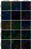
Normal protein distribution at the Sertoli–Sertoli cell interface is disrupted after knockdown of Par3 or Par6. Primary Sertoli cells were transfected with Cy3-labeled (red) nontargeting control (Left), Par3 siRNA duplex (Center), or Par6 siRNA duplex (Right). Two days after transfection, cells were subjected to calcium switch by first starving cells in DMEM without growth factors for 1 h followed by incubation in medium with 4 mM EGTA for 3 h and returned to normal DMEM with growth factors for 7 h. Cells were stained with the indicated TJ and basal ES markers (green) at the BTB. Loss of Par3 and Par6 resulted in a disruption of JAM-A and α-catenin at the cell–cell contact sites. Silencing of Par6 and Par3 also caused a loss of N-cadherin and ZO-1 at the cell–cell contact sites, respectively. (Scale bar: 5 μm.)
Fig. 7.
Fig. 7.
A hypothesis depicting the regulation of apical ES and BTB restructuring by Par3/Par6 at stage VIII of the epithelial cycle. (A) At the apical ES before spermiation, Par6/Pals1 forms an adhesion complex with JAM-C to facilitate spermatid attachment to Sertoli cells. The presence of the Par3/Par6/aPKC complex also facilitates BTB closure by stabilizing the N-cadherin- and JAM-C-based adhesion complexes. (B) Par3 and Par6 possibly regulate the disassembly of apical ES and BTB at stage VIII of the epithelial cycle as follows: (1) At the apical ES, the tighter association among Pals1, Src kinase, and Par6 (may also be coupled with a transient loss and/or redistribution of Par6) plausibly sequesters the polarity protein complexes from JAM-C, destabilizing the JAM-C adhesion complex, leading to spermiation. (2) When the level of Par6 at the BTB is down-regulated, such as by cytokines, this leads to a decrease in aPKC level. Subsequently, N-cadherin, α-catenin, and JAM-A are degraded or endocytosed, leading to a transient BTB opening to facilitate the transit of preleptotene spermatocytes (PS) across the BTB. (3) Similarly, down-regulation of Par3 results in redistribution of junction proteins (α-catenin, ZO-1, and JAM-A) via endocytosis or degradation, destabilizing the BTB, contributing to its opening. Unshaded target proteins represent molecules that are either degraded or endocytosed. Sp, elongated spermatid.

References

    1. Macara IG. Parsing the polarity code. Nat Rev Mol Cell Biol. 2004;5:220–231. - PubMed
    1. Shin K, Fogg VC, Margolis B. Tight junctions and cell polarity. Annu Rev Cell Dev Biol. 2006;22:207–235. - PubMed
    1. Suzuki A, Ohno S. The Par-aPKC system: Lessons in polarity. J Cell Sci. 2006;119:979–987. - PubMed
    1. Shin K, Wang Q, Margolis B. PATJ regulates directional migration of mammalian epithelial cells. EMBO J. 2007;8:158–164. - PMC - PubMed
    1. Mruk DD, Cheng CY. Sertoli–Sertoli and Sertoli–germ cell interactions and their significance in germ cell movement in the seminiferous epithelium during spermatogenesis. Endocr Rev. 2004;25:747–806. - PubMed

Publication types