. 2008 Oct;95(7):3340-8.
doi: 10.1529/biophysj.108.138479. Epub 2008 Jul 11.

Metallic nanoparticles used to estimate the structural integrity of DNA motifs

Affiliations

Metallic nanoparticles used to estimate the structural integrity of DNA motifs

Jiwen Zheng et al. Biophys J. 2008 Oct.

Abstract

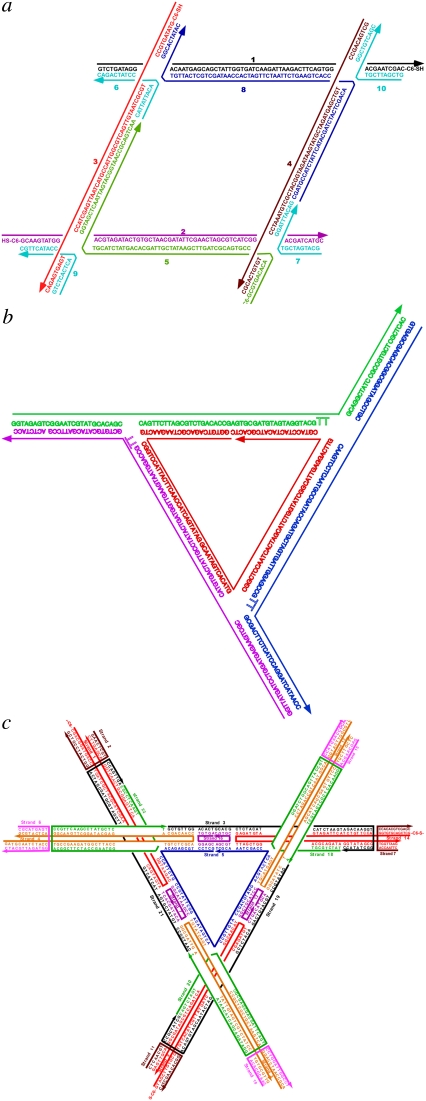
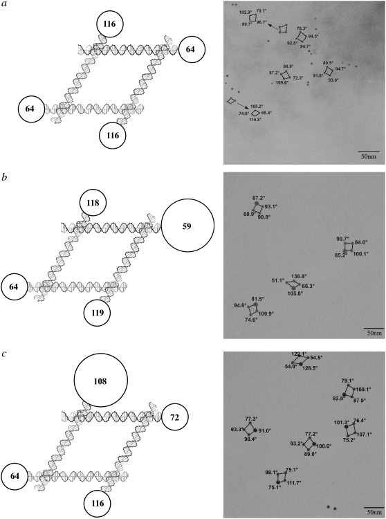
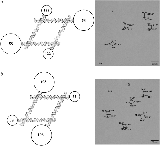
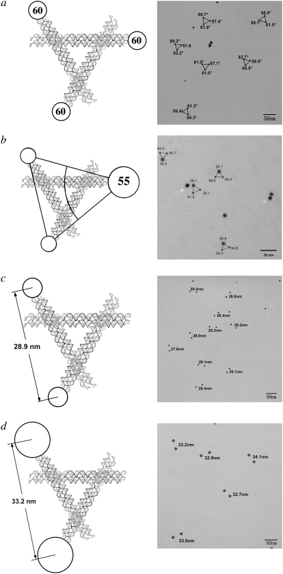
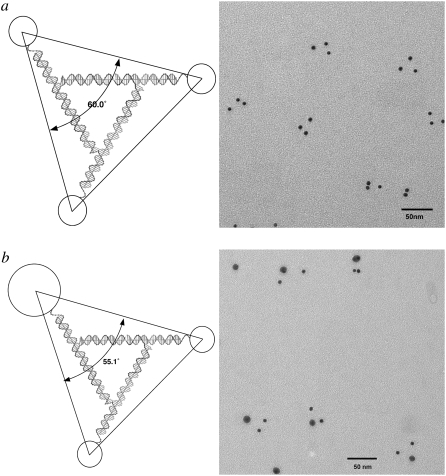
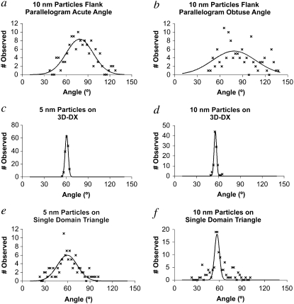
Branched DNA motifs can be designed to assume a variety of shapes and structures. These structures can be characterized by numerous solution techniques; the structures also can be inferred from atomic force microscopy of two-dimensional periodic arrays that the motifs form via cohesive interactions. Examples of these motifs are the DNA parallelogram, the bulged-junction DNA triangle, and the three-dimensional-double crossover (3D-DX) DNA triangle. The ability of these motifs to withstand stresses without changing geometrical structure is clearly of interest if the motif is to be used in nanomechanical devices or to organize other large chemical species. Metallic nanoparticles can be attached to DNA motifs, and the arrangement of these particles can be established by transmission electron microscopy. We have attached 5 nm or 10 nm gold nanoparticles to every vertex of DNA parallelograms, to two or three vertices of 3D-DX DNA triangle motifs, and to every vertex of bulged-junction DNA triangles. We demonstrate by transmission electron microscopy that the DNA parallelogram motif and the bulged-junction DNA triangle are deformed by the presence of the gold nanoparticles, whereas the structure of the 3D-DX DNA triangle motif appears to be minimally distorted. This method provides a way to estimate the robustness and potential utility of the many new DNA motifs that are becoming available.


Figures

FIGURE 1
FIGURE 1
The sequences of the molecules used here. (a) The parallelogram. (b) The bulged-junction triangle. (c) The DX triangle. “C6-S-” indicates the particle site of attachment.
FIGURE 2
FIGURE 2
DNA parallelograms with gold nanoparticles. The expected angles between the edges of the parallelograms are shown in the circles in the schematics at left and are indicated on the TEM pictures at right. The circles are scaled to indicate nanoparticle size. (a) Four 5 nm gold nanoparticles. For clarity, some angles are displaced in the TEM picture. (b) Three 5 nm nanoparticles and one 10 nm nanoparticle near an acute angle. Note the second parallelogram from the bottom, where the 10 nm particle flanks an obtuse angle. (c) Three 5 nm nanoparticles and one 10 nm nanoparticle near an obtuse angle. Note the bottom image, where the 10 nm particle flanks an acute angle.
FIGURE 3
FIGURE 3
DNA parallelograms with two 5 nm particles and two 10 nm particles. The same conventions apply as in Fig. 2. (a) The 10 nm particles flank acute angles. The bottom image is nearly ideal, but the other images are distorted significantly. (b) The 10 nm particles flank obtuse angles. Distortions are seen in several of the parallelograms, most prominently in the bottom image, where the expected distribution is reversed.
FIGURE 4
FIGURE 4
3D-DX triangles with two or three particles attached. (a) Three 5 nm particles attached. Note that the triangles are not significantly distorted from ideality. (b) Two 5 nm particles and one 10 nm particle attached. Only minor distortions are seen. (c) Two 5 nm particles attached. This asymmetric distribution of particles produces separations close to the expected values. (d) Two 10 nm particles attached. Even 10 nm particles attached asymmetrically do not produce separations indicative of motif distortion.
FIGURE 5
FIGURE 5
Bulged-junction triangles with three particles attached. (a) Three 5 nm particles. (b) Two 5 nm particles and one 10 nm particle. In each case, a triangle can be seen that is near the expected shape, but most triangles are highly distorted.
FIGURE 6
FIGURE 6
Angular distributions observed and fit by a Gaussian function. The parallelogram distributions (a) and (b) are very broad, as is the distribution of the single domain triangle with three 5 nm particles (e). The distribution of the triangle with a 10 nm particle is narrower but is badly fit by a single Gaussian (f). The 3D-DX distributions are very narrow (c) and (d). The parameters describing the distributions are listed in Table 1.

Similar articles

Cited by

References

    1. Chen, J., and N. C. Seeman. 1991. The synthesis from DNA of a molecule with the connectivity of a cube. Nature. 350:631–633. - PubMed
    1. Winfree, E., F. Liu, L. A. Wenzler, and N. C. Seeman. 1998. Design and self-assembly of two-dimensional DNA crystals. Nature. 394:539–544. - PubMed
    1. Mao, C., W. Sun, Z. Shen, and N. C. Seeman. 1999. A DNA nanomechanical device based on the B-Z transition. Nature. 397:144–146. - PubMed
    1. Yan, H., X. Zhang, Z. Shen, and N. C. Seeman. 2002. A robust DNA mechanical device controlled by hybridization topology. Nature. 415:62–65. - PubMed
    1. Zheng, J., P. E. Constantinou, C. Micheel, A. P. Alivisatos, R. A. Kiehl, and N. C. Seeman. 2006. 2D nanoparticle arrays show the organizational power of robust DNA motif. Nano Lett. 6:1502–1504. - PMC - PubMed

Publication types

LinkOut - more resources