. 2008 Sep;7(9):1582-90.
doi: 10.1128/EC.00150-08. Epub 2008 Jul 11.

Polo-like kinase is expressed in S/G2/M phase and associated with the flagellum attachment zone in both procyclic and bloodstream forms of Trypanosoma brucei

Affiliations

Polo-like kinase is expressed in S/G2/M phase and associated with the flagellum attachment zone in both procyclic and bloodstream forms of Trypanosoma brucei

Takashi Umeyama et al. Eukaryot Cell. 2008 Sep.

Abstract

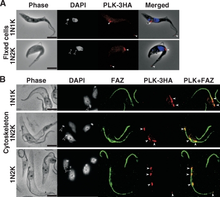
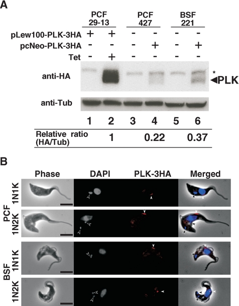
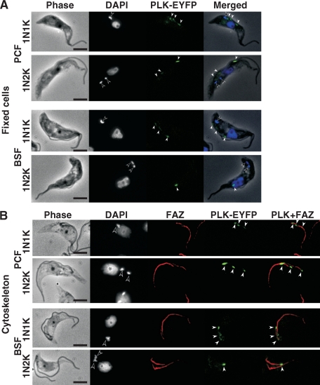
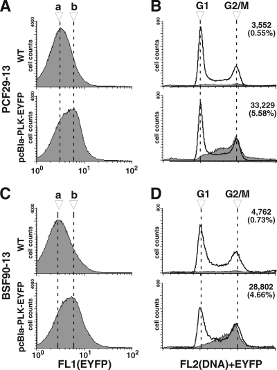
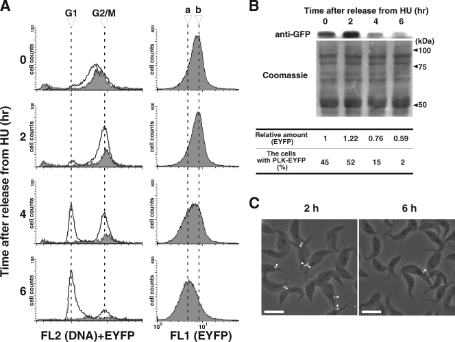
Trypanosoma brucei, the etiologic agent of African sleeping sickness, divides into insect (procyclic) and bloodstream forms. These two forms are subject to distinct cell cycle regulations, with cytokinesis controlled primarily by basal body/kinetoplast segregation in the procyclic form but by mitosis in the bloodstream form. Polo-like kinases (PLKs), known to play essential roles in regulating both mitosis and cytokinesis among eukaryotes, have a homologue in T. brucei, TbPLK, which regulates only cytokinesis. In our previous study, overexpressed triply hemagglutinin-tagged TbPLK (TbPLK-3HA) in the procyclic form localized to a mid-dorsal point and the anterior tip of the cell along the flagellum attachment zone (FAZ). In our current study, TbPLK-3HA expressed at the endogenous level was identified at the same dorsal location of both procyclic and bloodstream forms, albeit it was no longer detectable at the anterior tip of the cell. Endogenously expressed TbPLK fused with an enhanced yellow fluorescent protein (EYFP) localized to the same dorsal location along the FAZs in living procyclic and bloodstream cells. Fluorescence-activated cell sorter analysis of hydroxyurea-synchronized procyclic cells revealed that TbPLK-EYFP emerges during S phase, persists through G(2)/M phase, and vanishes in G(1) phase. An indicated TbPLK-EYFP association with the FAZs of G(2)/M cells may thus represent a timely localization to a potential initiation site of cytokinesis, which agrees with the recognized role of TbPLK in cytokinetic initiation.


Figures

FIG. 1.
FIG. 1.
Overexpressed TbPLK-3HA localized to the ends of the FAZ. (A) Procyclic-form 29-13 cells harboring pLew100-PLK-3HA were cultured for 5 days in the presence of 0.1 μg/ml tetracycline, fixed with 4% paraformaldehyde, and stained with anti-HA antibody (red) and DAPI (blue). (B) The cytoskeleton from the same cell culture was stained with anti-HA antibody for TbPLK-3HA (red), with L3B2 antibody for the FAZ (green), and with DAPI for DNA (blue). Open and closed arrowheads indicate the kinetoplast and TbPLK-3HA signals, respectively. Bars, 5 μm.
FIG. 2.
FIG. 2.
Endogenous TbPLK-3HA localization in the procyclic and bloodstream forms. (A) Western blotting showing TbPLK-3HA expression levels. For the overexpression of TbPLK-3HA, procyclic-form (PCF) 29-13 cells (lanes 1 and 2) harboring pLew100-PLK-3HA were cultured for 1 day in the absence (lane 1) or presence (lane 2) of 0.1 μg/ml tetracycline. PCF 427 cells (lane 4) and bloodstream-form (BSF) 221 cells (lane 6) harboring pcNeo-PLK-3HA in one of the two alleles of the TbPLK genomic locus were used to examine the endogenous TbPLK-3HA. Wild-type PCF 427 and BSF 221 cells were used as negative controls (lanes 3 and 5). Total proteins from 2.5 × 10⁶ cells were applied to Western analysis with anti-HA antibody (top) or anti-α-tubulin antibody as the loading control (bottom). The asterisk indicates a nonspecific band. The ratio of TbPLK-3HA to α-tubulin (Tub), determined using ImageJ software, is indicated under each lane. (B) Immunostaining of endogenous TbPLK-3HA using anti-HA antibody. The 29-13 cells with the endogenous TbPLK-3HA harboring pcNeo-PLK-3HA were fixed with 4% paraformaldehyde and stained with anti-HA antibody (red) and DAPI (blue). Open and closed arrowheads indicate kinetoplast and TbPLK-3HA signals, respectively. Bars, 5 μm.
FIG. 3.
FIG. 3.
Endogenous TbPLK-EYFP localization in both procyclic and bloodstream forms. (A) Procyclic-form (PCF) 29-13 cells or bloodstream-form (BSF) 90-13 cells harboring pcBla-PLK-EYFP were fixed with 4% paraformaldehyde and stained with DAPI for DNA (blue). TbPLK-EYFP was visualized in green. (B) The cytoskeleton from the same cell culture was stained with L3B2 antibody for the FAZ (red), and with DAPI for DNA (blue). TbPLK-EYFP was visualized in green. Open and closed arrowheads indicate kinetoplast and TbPLK-EYFP signals, respectively. Bars, 5 μm.
FIG. 4.
FIG. 4.
Endogenous TbPLK-EYFP was expressed in S- and G2/M-phase cells. Procyclic-form (PCF) 29-13 cells or bloodstream-form (BSF) 90-13 cells without (wild type [WT]) or with pcBla-PLK-EYFP were stained with PI and analyzed by flow cytometry (one representative experiment out of three). (A and C) Histograms of the cells without or with TbPLK-EYFP monitored by green fluorescence (FL1 channel). Dashed lines a and b indicate the peak locations in the histograms of cells without and with pcBla-PLK-EYFP, respectively. (B and D) Histograms for cells sorted by the DNA contents (FL2 channel) from total cells are indicated by solid lines. To determine the cells showing fluorescence from TbPLK-EYFP, a gate was defined to exclude most of the autofluorescent cells from the group of cells having no TbPLK-EYFP, and the same gate was applied to the cells with TbPLK-EYFP. Histograms of the DNA contents of cells that fell within the gate are indicated by gray color. Of 500,000 events that were analyzed, the number (along with the percentage) of events that fell within the gate is indicated at the top right of each histogram.
FIG. 5.
FIG. 5.
Hydroxyurea (HU) synchronization of procyclic-form cells expressing endogenous TbPLK-EYFP. Procyclic-form 29-13 cells with pcBla-PLK-EYFP were treated with 0.2 mM HU overnight, washed twice with growth medium, and collected at the times indicated (0, 2, 4, and 6 h). The collected cells were fixed with ethanol, stained with propidium iodide, and analyzed by flow cytometry. (A) Histograms on the left indicate results from cell sorting for DNA contents (FL2 channel [solid line]). Cells showing fluorescence from TbPLK-EYFP (gray area) were processed in a way similar to that described for Fig. 4. Histograms on the right show results from cell sorting by TbPLK-EYFP fluorescence (FL1 channel). Dashed lines a and b indicate the peak locations in the histograms from the cells collected at 6 h and 2 h after release from HU treatment, respectively. (B) Western blot showing the changing TbPLK-EYFP levels after release from HU treatment. Total proteins from 106 cells stained with anti-GFP antibody (top) or Coomassie brilliant blue G-250 as a loading control (bottom) were applied to the Western analysis. The relative amount of TbPLK-EYFP, determined using ImageJ software, is indicated under each lane. The population of cells expressing PLK-EYFP on the dorsal side (n > 120) is also indicated under each lane. (C) Merged phase-contrast (gray) and PLK-EYFP fluorescence (white, arrowheads) images from the cells at 2 h (predominantly G2/M cells) and 6 h (predominantly G1 cells) after their release from HU treatment. Bars, 10 μm.

References

    1. Abrieu, A., T. Brassac, S. Galas, D. Fisher, J. C. Labbe, and M. Doree. 1998. The Polo-like kinase Plx1 is a component of the MPF amplification loop at the G2/M-phase transition of the cell cycle in Xenopus eggs. J. Cell Sci. 1111751-1757. - PubMed
    1. Alexandru, G., F. Uhlmann, K. Mechtler, M. A. Poupart, and K. Nasmyth. 2001. Phosphorylation of the cohesin subunit Scc1 by Polo/Cdc5 kinase regulates sister chromatid separation in yeast. Cell 105459-472. - PubMed
    1. Barr, F. A., H. H. Sillje, and E. A. Nigg. 2004. Polo-like kinases and the orchestration of cell division. Nat. Rev. Mol. Cell Biol. 5429-440. - PubMed
    1. Berriman, M., E. Ghedin, C. Hertz-Fowler, G. Blandin, H. Renauld, D. C. Bartholomeu, N. J. Lennard, E. Caler, N. E. Hamlin, B. Haas, U. Bohme, L. Hannick, M. A. Aslett, J. Shallom, L. Marcello, L. Hou, B. Wickstead, U. C. Alsmark, C. Arrowsmith, R. J. Atkin, A. J. Barron, F. Bringaud, K. Brooks, M. Carrington, I. Cherevach, T. J. Chillingworth, C. Churcher, L. N. Clark, C. H. Corton, A. Cronin, R. M. Davies, J. Doggett, A. Djikeng, T. Feldblyum, M. C. Field, A. Fraser, I. Goodhead, Z. Hance, D. Harper, B. R. Harris, H. Hauser, J. Hostetler, A. Ivens, K. Jagels, D. Johnson, J. Johnson, K. Jones, A. X. Kerhornou, H. Koo, N. Larke, S. Landfear, C. Larkin, V. Leech, A. Line, A. Lord, A. Macleod, P. J. Mooney, S. Moule, D. M. Martin, G. W. Morgan, K. Mungall, H. Norbertczak, D. Ormond, G. Pai, C. S. Peacock, J. Peterson, M. A. Quail, E. Rabbinowitsch, M. A. Rajandream, C. Reitter, S. L. Salzberg, M. Sanders, S. Schobel, S. Sharp, M. Simmonds, A. J. Simpson, L. Tallon, C. M. Turner, A. Tait, A. R. Tivey, S. Van Aken, D. Walker, D. Wanless, S. Wang, B. White, O. White, S. Whitehead, J. Woodward, J. Wortman, M. D. Adams, T. M. Embley, K. Gull, E. Ullu, J. D. Barry, A. H. Fairlamb, F. Opperdoes, B. G. Barrell, J. E. Donelson, N. Hall, C. M. Fraser, S. E. Melville, and N. M. El-Sayed. 2005. The genome of the African trypanosome Trypanosoma brucei. Science 309416-422. - PubMed
    1. Briggs, L. J., P. G. McKean, A. Baines, F. Moreira-Leite, J. Davidge, S. Vaughan, and K. Gull. 2004. The flagella connector of Trypanosoma brucei: an unusual mobile transmembrane junction. J. Cell Sci. 1171641-1651. - PubMed

Publication types

MeSH terms