. 2008 Sep;148(1):235-45.
doi: 10.1104/pp.108.122754. Epub 2008 Jul 11.

The Omp85-related chloroplast outer envelope protein OEP80 is essential for viability in Arabidopsis

Affiliations

The Omp85-related chloroplast outer envelope protein OEP80 is essential for viability in Arabidopsis

Ramesh Patel et al. Plant Physiol. 2008 Sep.

Abstract

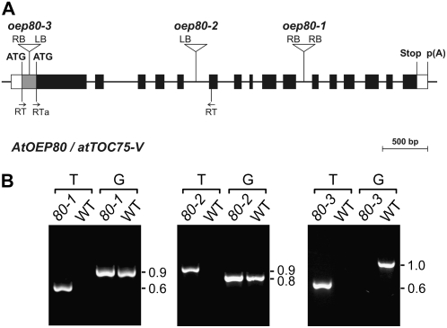
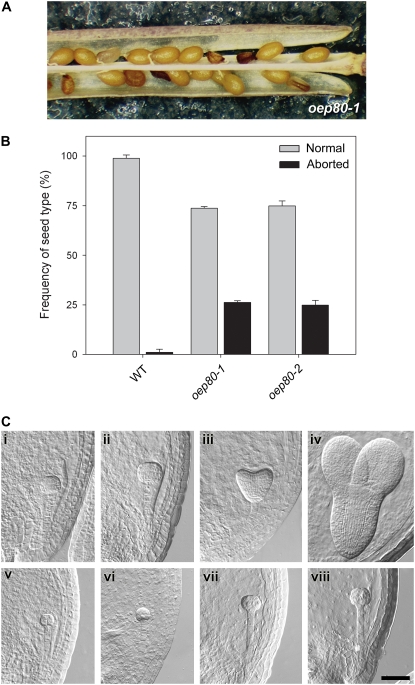
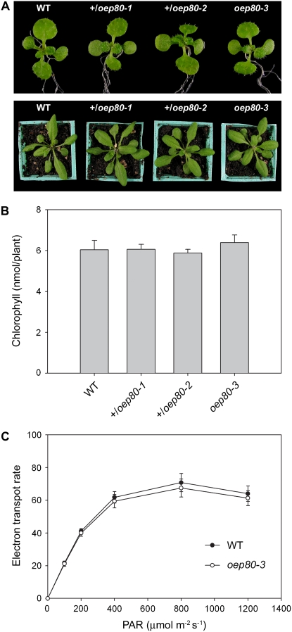
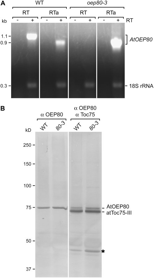
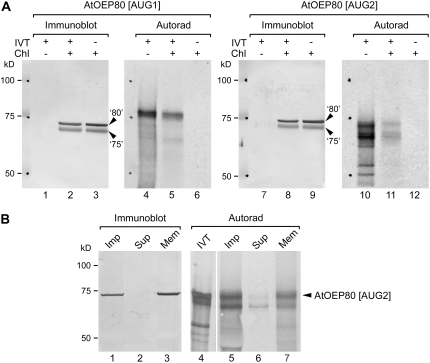
beta-Barrel proteins of the Omp85 (Outer membrane protein, 85 kD) superfamily exist in the outer membranes of Gram-negative bacteria, mitochondria, and chloroplasts. Prominent Omp85 proteins in bacteria and mitochondria mediate biogenesis of other beta-barrel proteins and are indispensable for viability. In Arabidopsis (Arabidopsis thaliana) chloroplasts, there are two distinct types of Omp85-related protein: Toc75 (Translocon at the outer envelope membrane of chloroplasts, 75 kD) and OEP80 (Outer Envelope Protein, 80 kD). Toc75 functions as a preprotein translocation channel during chloroplast import, but the role of OEP80 remains elusive. We characterized three T-DNA mutants of the Arabidopsis OEP80 (AtOEP80) gene. Selectable markers associated with the oep80-1 and oep80-2 insertions segregated abnormally, suggesting embryo lethality of the homozygous genotypes. Indeed, no homozygotes were identified among >100 individuals, and heterozygotes of both mutants produced approximately 25% aborted seeds upon self-pollination. Embryo arrest occurred at a relatively late stage (globular embryo proper) as revealed by analysis using Nomarski optics microscopy. This is substantially later than arrest caused by loss of the principal Toc75 isoform, atToc75-III (two-cell stage), suggesting a more specialized role for AtOEP80. Surprisingly, the oep80-3 T-DNA (located in exon 1 between the first and second ATG codons of the open reading frame) did not cause any detectable developmental defects or affect the size of the AtOEP80 protein in chloroplasts. This indicates that the N-terminal region of AtOEP80 is not essential for the targeting, biogenesis, or functionality of the protein, in contrast with atToc75-III, which requires a bipartite targeting sequence.


Figures

Figure 1.
Figure 1.
Basic characterization of the AtOEP80 T-DNA insertion mutants. A, Schematic showing the structure of the AtOEP80 gene and the location of each T-DNA insertion. Protein-coding exons are represented by black boxes and untranslated regions by white boxes; introns are represented by thin lines between the boxes. The gray area at the 5′ end of exon 1 represents a putative untranslated region, or alternatively encodes a nonessential cleavable peptide. Locations of primers used for RT-PCR analysis (RT; Fig. 4A) are shown. T-DNA insertion sites are indicated precisely, but the insertion sizes are not to scale. ATG, Potential translation initiation codons; Stop, translation termination codon; p(A), polyadenylation site. B, Analysis of mutant genotypes by PCR. Genomic DNA extracted from wild-type and mutant plants (oep80-1, 80-1; oep80-2, 80-2; and oep80-3, 80-3) was analyzed by PCR. Appropriate T-DNA- and AtOEP80-gene-specific primers were employed. Two different primer combinations were used: The first (T) comprised one T-DNA primer (RB for oep80-1; LB for oep80-2 and oep80-3) and one gene-specific primer (reverse in the case of oep80-1 and oep80-3; forward in the case of oep80-2); the second (G) comprised two gene-specific primers flanking the T-DNA insertion site. The results shown for oep80-1 and oep80-2 are representative of those obtained for all antibiotic-resistant plants tested; amplification using both T and G indicated the presence of both mutant and wild-type alleles, respectively, and demonstrated that the plants were heterozygous. Results shown for oep80-3 are representative of those obtained for all homozygotes tested; absence of amplification using the G primers indicated that the wild-type allele was not present. Sizes of the amplicons are indicated at right (in kb).
Figure 2.
Figure 2.
Embryo lethality of the oep80-1 and oep80-2 mutations. A, Appearance of aborted seeds within mature siliques of oep80-1 heterozygous plants. The aborted seeds are smaller in size than the normal seeds and have a darker, shriveled appearance. B, Frequency of aborted seeds within mature siliques of wild-type, oep80-1, and oep80-2 plants. The data shown are means (±sd) derived from analyses of three or four siliques from each of three to six independent plants per genotype. C, Analysis of embryo development in oep80-1 using Nomarski optics. Equivalent developmental series for normal (i–iv) and mutant (v–viii) embryos within immature oep80-1 heterozygous siliques. Normal embryos: i, early globular stage; ii, late globular stage; iii, heart stage; iv, torpedo stage. Corresponding mutant embryos from the same siliques: v, proembryo stage; vi, early globular stage; vii and viii, raspberry-like globular stage. Embryo cell stage names refer to the morphology of the embryo proper. All images are at the same magnification. Bar = 50 μm.
Figure 3.
Figure 3.
Phenotypic analysis of the oep80 mutants. A, Plants of the indicated genotypes were grown on selective medium (except for the wild type) in vitro for 8 d, rescued to nonselective medium, and then photographed on day 14 (top). Additional similar plants were transferred to soil on day 14 and then allowed to grow for a further 10-d period prior to photography (bottom). Representative plants are shown in both cases. B, Chlorophyll concentrations in 14-d-old plants grown as described in A were determined photometrically. Values shown are means (±se) derived from 16 independent samples per genotype, each one containing six plants. Units are nmol chlorophyll a + b per plant. C, Analysis of photosynthesis in the oep80-3 mutant. Light response curves of photosynthetic electron transport rates in wild-type and mutant plants were determined by measuring chlorophyll fluorescence. Values were recorded at different irradiances of photosynthetically active radiation (PAR), ranging from 0 to 1,200 μmol photons m−2 s−1. Units for the data shown are μmol electrons m−2 s−1, assuming that 84% of the incident light is absorbed and that the transport of each electron utilizes two photons (Meyer et al., 1997; Aronsson et al., 2007). Measurements were done on fully grown leaves from 10 different 29-d-old plants per genotype grown under identical conditions. Values shown are means (±sd).
Figure 4.
Figure 4.
Analyses of mRNA and protein expression in the oep80-3 mutant. A, Analysis of AtOEP80 mRNA expression. Total RNA extracted from wild-type and homozygous oep80-3 mutant plants was analyzed by RT-PCR. Each reaction contained two primer pairs: The first specifically amplified a 1.1-kb fragment from the wild-type AtOEP80 transcript (locations of the RT primers used are indicated in Fig. 1A) or a 0.9-kb fragment from the wild-type and oep80-3 mutant transcripts (the forward RT primer was replaced with RTa in this case; see Fig. 1A); the second amplified a 315-bp fragment derived from 18S rRNA and served as a positive control. Reactions lacking reverse transcriptase (−RT) were included as negative controls. Images from different portions of the same gel are separated by vertical lines. Sizes of the amplicons are indicated at left (in kb). B, Analysis of AtOEP80 protein expression. Isolated chloroplast samples (equivalent to 20 μg [left] or 10 μg [right] chlorophyll) were separated by SDS-PAGE and then analyzed by immunoblotting using antiserum against AtOEP80 only (left), or a mixture of antisera against AtOEP80 and psToc75 (right). Protein bands corresponding to AtOEP80 and atToc75-III are indicated at right. Positions of molecular mass standards are indicated at left (sizes in kD); note that the 75-kD standard migrates more slowly than atToc75-III and at approximately the same speed as endogenous AtOEP80. A 40-kD protein band that was nonspecifically recognized by the psToc75 antiserum is indicated with an asterisk. Images from different portions of the same gel are separated by a vertical line.
Figure 5.
Figure 5.
Electrophoretic mobility comparisons between proteins imported into Arabidopsis chloroplasts in vitro and endogenous AtOEP80. A, Radiolabeled long (AtOEP80 [AUG1]; 732 residues) and short (AtOEP80 [AUG2]; 680 residues) forms of the AtOEP80 protein were generated by in vitro translation using different cDNA templates. These were incubated with Arabidopsis chloroplasts under import conditions and then the chloroplasts were recovered. In vitro translation products equivalent to 10% of the amount added to each import assay (IVT+/Chl−), Arabidopsis chloroplasts containing imported, radiolabeled proteins (IVT+/Chl+), and equivalent chloroplast samples lacking imported, radiolabeled protein (IVT−/Chl+) were resolved side-by-side using SDS-PAGE, blotted onto the same membrane, and then analyzed either by probing with AtOEP80 and psToc75 antisera (Immunoblot) or by autoradiography (Autorad). The positions of endogenous AtOEP80 and atToc75-III proteins are indicated at right (‘80’ and ‘75’, respectively). Positions of molecular mass standards are indicated at left (sizes in kD). Under the conditions used, the endogenous AtOEP80 protein migrated slower than atToc75-III, whereas both proteins ran faster than the 75-kD marker protein. B, In vitro translated, radiolabeled AtOEP80 (AtOEP80 [AUG2]; 680 residues) was incubated with Arabidopsis chloroplasts under import conditions. One-half of the recovered chloroplast sample was subjected to alkaline extraction using 0.1 m Na2CO3 and separated into soluble and membrane fractions as described (Inoue and Potter, 2004). In vitro translation products equivalent to 5% of the amount used for the import assay (IVT), unfractionated Arabidopsis chloroplasts containing imported, radiolabeled protein (Imp), and the supernatant (Sup) and membrane (Mem) fractions obtained after alkaline extraction were resolved side-by-side using SDS-PAGE, blotted onto the same membrane, and then analyzed either by probing with AtOEP80 antiserum (Immunoblot) or by autoradiography (Autorad). The position of imported AtOEP80 is indicated at right. Positions of molecular mass standards are indicated at left (sizes in kD); note that the 75-kD marker runs significantly slower than atToc75-III on a 7.5% SDS-PAGE gel.

References

    1. Apuya NR, Yadegari R, Fischer RL, Harada JJ, Goldberg RB (2002) RASPBERRY3 gene encodes a novel protein important for embryo development. Plant Physiol 129 691–705 - PMC - PubMed
    1. Apuya NR, Yadegari R, Fischer RL, Harada JJ, Zimmerman JL, Goldberg RB (2001) The Arabidopsis embryo mutant schlepperless has a defect in the chaperonin-60α gene. Plant Physiol 126 717–730 - PMC - PubMed
    1. Aronsson H, Boij P, Patel R, Wardle A, Töpel M, Jarvis P (2007) Toc64/OEP64 is not essential for the efficient import of proteins into chloroplasts in Arabidopsis thaliana. Plant J 52 53–68 - PubMed
    1. Aronsson H, Combe J, Jarvis P (2003) Unusual nucleotide-binding properties of the chloroplast protein import receptor, atToc33. FEBS Lett 544 79–85 - PubMed
    1. Aronsson H, Jarvis P (2002) A simple method for isolating import-competent Arabidopsis chloroplasts. FEBS Lett 529 215–220 - PubMed

Publication types