. 2008 Sep;148(1):223-34.
doi: 10.1104/pp.108.120527. Epub 2008 Jul 11.

A novel RNA-binding protein associated with cell plate formation

Affiliations

A novel RNA-binding protein associated with cell plate formation

Lian Ma et al. Plant Physiol. 2008 Sep.

Abstract

Building a cell plate during cytokinesis in plant cells requires the participation of a number of proteins in a multistep process. We previously identified phragmoplastin as a cell plate-specific protein involved in creating a tubulovesicular network at the cell plate. We report here the identification and characterization of a phragmoplastin-interacting protein, PHIP1, in Arabidopsis (Arabidopsis thaliana). It contains multiple functional motifs, including a lysine-rich domain, two RNA recognition motifs, and three CCHC-type zinc fingers. Polypeptides with similar motif structures were found only in plant protein databases, but not in the sequenced prokaryotic, fungal, and animal genomes, suggesting that PHIP1 represents a plant-specific RNA-binding protein. In addition to phragmoplastin, two Arabidopsis small GTP-binding proteins, Rop1 and Ran2, are also found to interact with PHIP1. The zinc fingers of PHIP1 were not required for its interaction with Rop1 and phragmoplastin, but they may participate in its binding with the Ran2 mRNA. Immunofluorescence, in situ RNA hybridization, and green fluorescent protein tagging experiments showed the association of PHIP1 with the forming cell plate during cytokinesis. Taken together, our data suggest that PHIP1 is a novel RNA-binding protein and may play a unique role in the polarized mRNA transport to the vicinity of the cell plate.


Figures
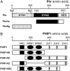
Figure 1.
Figure 1.
Functional domains and deletion constructs of phragmoplastin and PHIP1. A, Phragmoplastin contains three functional domains: the dynamin homology 1 (DYN1), DYN2, and a GTPase effector domain (GED). The PhrHc fragment containing DYN2 and part of GED is sufficient for interaction with PHIP1. Relative positions of restriction enzyme sites on the phragmoplastin cDNA are indicated: Bg2, BglII; Hc2, HincII; BH1, BamHI. B, PHIP1 contains one KRD, two RRMs, and three CCHC-type ZnFs. Constructs containing different motifs were assayed for interaction with phragmoplastin and Rop1. PHIP-KRZ lacking the N terminus and the third ZnF was isolated from the library screen using phragmoplastin as bait. Deletion fragments containing KRD and RRM retained the ability to interact with both phragmoplastin and Rop1. Phr, Full-length phragmoplastin; PhrHc, the HincII-BamHI fragment of phragmoplastin; PhrBg, the BglII fragment of phragmoplastin. ZnF1, ZnF2, and ZnF3 indicate the three ZnFs present in the C terminus of PHIP1.
Figure 2.
Figure 2.
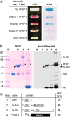
PHIP1 interacts with Rop1 in the yeast two-hybrid system and in vitro. A, Rop1 (RopWT), its dominant positive mutant (RopGTP), or its dominant negative mutant (RopGDP) was expressed in yeast cells using the pAS2 bait vector (see “Materials and Methods”). Plasmids were transformed to Y190 cells harboring pACT-PHIP1. Transformants were streaked on a propagation medium (+His), and the replica was stained for X-Gal activity (X-Gal). Plasmids pAS-Phr and pAS2 (vector alone) were used as positive and negative controls, respectively. B, Rop1 and phragmoplastin were purified through a GST tag (lanes 2 and 3). GST alone served as a negative control (lane 1). PHIP-KRZ, the nearly full-length PHIP1 that was isolated from the original library screen, was synthesized and labeled with [35S]Met using the in vitro transcription/translation system (lane 4). [35S]PHIP1 was incubated with either Rop1 or phragmoplastin bound to agarose beads. Proteins that were retained on the beads were eluted and resolved by SDS-PAGE. The gel stained with Coomassie Brilliant Blue R-250 was photographed (PAGE) and autoradiographed (Autoradiograph). Hb, Hemoglobin that is a by-product of the in vitro labeling system; M, molecular mass standards in kilodaltons. Asterisks indicate the bands of the pull-down 35S-labeled PHIP1 (*PHIP1) on the autoradiograph and their relative positions on the PAGE gel. Arrowheads indicate the position of G-Rop1. C, Schematic representation of the protein components shown in B and their calculated molecular mass in kilodaltons. [See online article for color version of this figure.]
Figure 3.
Figure 3.
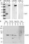
Western-blot analysis of PHIP1 in Arabidopsis plants. A, Specificity of polyclonal antibodies to PHIP1. Five micrograms of purified GST (negative control; lane 1) and GST-PHIP1 (lane 2) was resolved on 10% SDS-PAGE and stained with Coomassie Brilliant Blue R-250 (left). M, Molecular mass standards in kilodaltons. Similar gels containing less sample (100 ng of protein per lane) were blotted onto nitrocellulose membranes and probed with polyclonal antibodies to PHIP1 (middle) or the preimmune serum (preim. s.; right). Protein bands were visualized on x-ray film after incubation with horseradish peroxidase-conjugated second antibody (goat antibody against rabbit IgG). B, Association of PHIP1 with the membranes. Arabidopsis young seedlings (10 d old) were used to prepare soluble fraction (lane 1; Total S) and membrane pellet (lane 2; Total P). The total membrane pellet was further extracted with 1% Triton X-100 (lanes 3 and 4), 100 mm Na2CO3 (pH 11.5; lanes 5 and 6), or 1 m NaI (lanes 7 and 8). Molecular masses of protein markers (in kilodaltons) and the position of PHIP1 are indicated. Note that PHIP1 is present only in the membrane pellet (lane 2), can be partially extracted from the membrane pellet by Triton X-100 (lane 3), and can be removed from the membranes with high-pH solution (lane 5) or chaotropic agent (lane 7).
Figure 4.
Figure 4.
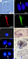
Subcellular localization of PHIP1 and Ran2 mRNA at cytokinesis. A to D, Direct immunofluorescence localization of PHIP1. Onion root tip cells were fixed in paraformaldehyde and reacted to antibodies against Arabidopsis PHIP1 (α-PHIP1) or soybean phragmoplastin (α-Phr). Antibodies to PHIP1 were conjugated with fluorescent dye Alexa 594 and exhibited red fluorescence, whereas phragmoplastin antibodies conjugated with Alexa 488 emitted green fluorescence. Nuclear DNA was stained with DAPI. The bright-field image (Light) was taken using Normarski optics. Arrows indicate the position of the cell plate. E to J, Whole-mount in situ RNA hybridization. Tobacco BY-2 cells were fixed and reacted with digoxigenin-labeled Ran2 RNA probes, which were detected by anti-digoxigenin antibody conjugated with alkaline phosphatase (F, H, and J). Nuclear DNA (N) was stained with DAPI and imaged under a UV filter (E, G, and I). A nondividing cell (E and F) and a cytokinetic cell (G and H) with the cell plate (CP) are shown. The Ran2 sense RNA probe was used as a negative control (I and J). Bar = 10 μm.
Figure 5.
Figure 5.
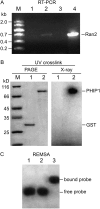
PHIP1 binds Ran2 mRNA. A, RT-PCR products of Ran2 mRNA bound to PHIP1. Primers specific to Ran2 were used for PCR amplification. Lane 1, PCR product using Arabidopsis total RNA without performing RT (negative control). Lane 2, RT-PCR product using Arabidopsis total RNA (positive control). Lane 3, RT-PCR product using RNA bound to the GST beads (negative control). Lane 4, RT-PCR product using RNA extracted from the GST-PHIP1 beads. M, DNA ladder in kilobases. B, UV cross-linking assay of PHIP1 with 32P-labeled Ran2 mRNA. Left panel, Coomassie Brilliant Blue R-250 staining of SDS-PAGE. Lane 1, Cross-linking product of GST with the [32P]Ran2 mRNA probe (negative control). M, Molecular mass standards in kilodaltons. Lane 2, Cross-linking product of GST-PHIP1 with [32P]Ran2 mRNA. Right panel, X-ray autoradiograph of the gel shown at left. C, RNA electrophoretic mobility-shift assay (REMSA) of [32P]Ran2 mRNA with PHIP1. Lane 1, Free [32P]Ran2 mRNA probe. Lane 2, [32P]Ran2 mRNA incubated with GST protein (negative control). Lane 3, [32P]Ran2 mRNA incubated with GST-PHIP1. Note that the mobility of the [32P]Ran2 mRNA probe shifted in the presence of PHIP1.
Figure 6.
Figure 6.
Proposed role of PHIP1 in the polarized RNA transport during cell plate formation. Phragmoplastin (Phr) is associated with the Golgi-derived vesicles that are transported to the cell plate along the phragmoplast microtubules (PMT). PHIP1 contains RRMs and ZnFs and binds specifically with the Ran2 mRNA (RNA) and possibly with a specific group of other cell plate formation-related mRNAs. The interaction between phragmoplastin and PHIP1 allows the delivery of these mRNAs to the vicinity of the cell plate, where they are translated into proteins (such as Ran2). N, Newly separated daughter nuclei; V, Golgi-derived cell plate vesicles.

Similar articles

Cited by

References

    1. Ainger K, Avossa D, Morgan F, Hill SJ, Barry C, Barbarese E, Carson JH (1993) Transport and localization of exogenous myelin basic protein mRNA microinjected into oligodendrocytes. J Cell Biol 123 431–441 - PMC - PubMed
    1. Berg JM (1986) Potential metal-binding domains in nucleic acid binding proteins. Science 232 485–487 - PubMed
    1. Bess JW Jr, Powell PJ, Issaq HJ, Schumack LJ, Grimes MK, Henderson LE, Arthur LO (1992) Tightly bound zinc in human immunodeficiency virus type 1, human T-cell leukemia virus type I, and other retroviruses. J Virol 66 840–847 - PMC - PubMed
    1. Bouget FY, Gerttula S, Quatrano RS (1995) Spatial redistribution of poly(A)+ RNA during polarization of the Fucus zygote is dependent upon microfilaments. Dev Biol 171 258–261 - PubMed
    1. Bouget FY, Gerttula S, Shaw S, Quatrano RS (1996) Localization of actin mRNA during the establishment of cell polarity and early cell divisions in Fucus embryos. Plant Cell 8 189–201 - PMC - PubMed

Publication types

MeSH terms