Review
. 2008 Sep;18(9):664-78.
doi: 10.1093/glycob/cwn061. Epub 2008 Jul 11.

Strategies for carbohydrate recognition by the mannose 6-phosphate receptors

Affiliations
Review

Strategies for carbohydrate recognition by the mannose 6-phosphate receptors

Nancy M Dahms et al. Glycobiology. 2008 Sep.

Abstract

The two members of the P-type lectin family, the 46 kDa cation-dependent mannose 6-phosphate receptor (CD-MPR) and the 300 kDa cation-independent mannose 6-phosphate receptor (CI-MPR), are ubiquitously expressed throughout the animal kingdom and are distinguished from all other lectins by their ability to recognize phosphorylated mannose residues. The best-characterized function of the MPRs is their ability to direct the delivery of approximately 60 different newly synthesized soluble lysosomal enzymes bearing mannose 6-phosphate (Man-6-P) on their N-linked oligosaccharides to the lysosome. In addition to its intracellular role in lysosome biogenesis, the CI-MPR, but not the CD-MPR, participates in a number of other biological processes by interacting with various molecules at the cell surface. The list of extracellular ligands recognized by this multifunctional receptor has grown to include a diverse spectrum of Man-6-P-containing proteins as well as several non-Man-6-P-containing ligands. Recent structural studies have given us a clearer view of how these two receptors use related, but yet distinct, approaches in the recognition of phosphomannosyl residues.


Figures

Fig. 1
Fig. 1
Generation of the Man-6-P tag on N-linked oligosaccharides. Phosphorylation of mannose residues on N-linked oligosaccharides occurs in two steps. First, the GlcNAc-phosphotransferase transfers GlcNAc-1-phosphate from UDP-GlcNAc to the C-6 hydroxyl group of mannose to form the Man-P-GlcNAc phosphodiester intermediate. Second, the uncovering enzyme removes the GlcNAc moiety in the TGN, revealing the Man-6-P phosphomonoester. The five potential sites of phosphorylation are indicated (gray).
Fig. 2
Fig. 2
Schematic diagram of the MRH domain containing proteins. The MPRs, 381-residue Mrl1, and 886-residue LERP are type I transmembrane glycoproteins. The 279-residue CD-MPR exists as a homodimer. The 2499-residue CI-MPR also undergoes oligomerization and most likely exists as a dimer. The Man-6-P binding sites of the CD-MPR (purple) and CI-MPR (green) are indicated. Domains 1 and 2 are outlined in green since the presence of these two domains enhances the affinity of domain 3 for lysosomal enzymes by ∼1000-fold (Hancock, Yammani, et al. 2002). The CD-MPR contains a single high affinity Man-6-P binding site per polypeptide. In contrast, the CI-MPR contains three carbohydrate recognition sites: two high affinity sites are localized to domains 1–3 and domain 9 and one low affinity site is contained within domain 5. The IGF-II (gold) and plasminogen (Plg)/uPAR (blue) binding sites are also indicated. The fibronectin type II repeat present in domain 13 is outlined in yellow since its presence increases the affinity of domain 11 for IGF-II by ∼10-fold. The red arrow indicates the location of a proteolytically sensitive cleavage site between domains 6 and 7 (Westlund et al. 1991). The 528-residue glucosidase II β subunit, 667-residue OS-9, and 483-residue XTP3-B/Erlectin are soluble resident ER proteins. Glucosidase II β subunit and the yeast ortholog of OS-9 contain a C-terminal ER retention signal (HDEL). The 305-residue GlcNAc-phosphotransferase γ subunit, in complex with the catalytic α/β subunits, is enriched in cis-Golgi cisternae.
Fig. 3
Fig. 3
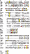
Sequence alignment of the Man-6-P binding sites of the bovine CI-MPR and bovine CD-MPR. (A) Structure-based amino acid sequence alignment of the extracytoplasmic region of the CD-MPR and domains 3, 5, and 9 of the CI-MPR. The secondary structure of domain 3 of the CI-MPR and the CD-MPR are shown, with arrows representing the β-strands and the cylinder representing an α-helix. At the N-terminus, domain 3 of the CI-MPR contains two β-strands (−2β and −1β) whereas the CD-MPR contains a single α-helix. The cysteine residues are boxed in yellow. Residues that are within hydrogen bonding distance of Man-6-P, as determined by the crystal structure of the CD-MPR (PDB 1C39) and domains 1–3 of the CI-MPR (PDB 1SZO) are boxed in red, with the four residues essential for Man-6-P binding (i.e., Gln, Arg, Glu, and Tyr) shaded in red. (B) Sequence alignment of domain 3 of the bovine CI-MPR with the single MRH domain of S. cerevisiae Mrl and the five MRH domains of Drosophila LERP. (C) Sequence alignment of domain 3 of the bovine CI-MPR with the MRH domains of human glucosidase II β subunit, human OS-9, human XTP3-B/Erlectin, and human GlcNAc-phosphotransferase γ subunit. The red triangle indicates the position of the putative conserved Gln. Sequence alignments in panels B and C were performed using M-Coffee (Wallace et al. ; Moretti et al. 2007).
Fig. 4
Fig. 4
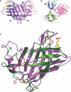
Three-dimensional structure of the CD-MPR and domains 1–3 of the CI-MPR. (A) Crystal structure of the extracytoplasmic region (residues 3–154) of the bovine CD-MPR in the presence of an oligosaccharide, pentamannosyl phosphate (PDB 1C39). Note that only the terminal Man-6-P (gold ball-and-stick model) is shown for clarity. Both monomers (light purple and dark purple) of the CD-MPR dimer are shown in this ribbon diagram. The N-terminus (N) and C-terminus (C) are boxed. (B) Crystal structure of the N-terminal three domains (residues 7–432) of the bovine CI-MPR (PDB 1SZO). The N- and C-terminus of the protein encoding domain 1 (blue), domain 2 (pink), and domain 3 (green) are indicated. The location of Man-6-P (gold ball-and-stick model) is shown. (C) Overlay of the structures of the CD-MPR (purple) and domain 3 (green) of the CI-MPR. The β-strands are sequentially numbered. The location of Man-6-P in the binding pocket (gold ball-and-stick model) is shown. The disulfide bridges are shown in gold, and the N- and C-terminus are boxed.
Fig. 5
Fig. 5
Comparison of the carbohydrate binding pocket of the CD-MPR and domain 3 of the CI-MPR. (A) Ribbon diagram showing the binding site of the CD-MPR (purple) superimposed onto domain 3 (green) of the CI-MPR. The disulfides are shown in gold and Man-6-P (gold ball-and-stick model) is also indicated. The four residues that are essential for Man-6-P binding (i.e., when replaced result in an ∼1,000 reduction in affinity) are circled in red. H105 is located within loop C. R135 is located within the relatively long loop D (dark purple) in the CD-MPR structure. In contrast, loop D (light gray) is short in domain 3. (B) Schematic view of the potential hydrogen bond and ionic interactions between the binding pocket residues of the CD-MPR and Man-6-P (gold ball-and-stick). The binding pocket residues and the single amino acid substitutions that were made are listed for the CD-MPR and domain 3, domain 5, and domain 9 of the CI-MPR. Shaded in light gray are the residues that have not been tested (N104) or when mutated (D103) retained wild-type Man-6-P binding ability. Shaded in blue are the residues that when mutated resulted in receptors with diminished (∼50–150-fold) Man-6-P binding ability as compared to wild-type receptors. Shaded in purple are the residues identified as essential for carbohydrate recognition by the MPRs (i.e., single amino acid substitution of these residues abolished (∼1000-fold) the Man-6-P binding ability of the receptors) (Wendland, Waheed, et al. ; Olson, Hancock, et al. ; Hancock, Haskins, et al. ; Sun et al. ; Chavez et al. 2007).
Fig. 6
Fig. 6
Comparison of the bound and unbound conformations of the CD-MPR. (A) Superimposition of the monomers of the bound (PDB 2RL8, red) and unbound (PDB 2RL7, blue) forms of the CD-MPR. The location of loop D, which exhibits the most dramatic change in position, is indicated. Loops A, B, and C are also labeled. The C- and N-termini are boxed. (B) Superimposition of all Cα atoms of the dimer of the bound (PDB 2RL8, red) and unbound (PDB 2RL7, blue) structures. Between the two conformations, there is an ∼30° scissoring motion between the two subunits of the dimer with respect to the two-fold axis (Z axis) on the XZ plane and an ∼12° twist along the Z-axis pivoting at the center of the dimer molecule. The C- and N-termini are boxed. The location of Man-6-P in the binding pocket (gold ball-and-stick model) is shown.

References

    1. Aeed PA, Elhammer AP. Glycosylation of recombinant prorenin in insect cells: The insect cell line Sf9 does not express the mannose 6-phosphate recognition signal. Biochemistry. 1994;33:8793–8797. - PubMed
    1. Bernasconi R, Pertel T, Luban J, Molinari M. A dual task for the Xbp1-responsive OS-9 variants in the mammalian endoplasmic reticulum: Inhibiting secretion of misfolded protein conformers and enhancing their disposal. J Biol Chem. 2008;283:16446–16454. - PMC - PubMed
    1. Bhamidipati A, Denic V, Quan EM, Weissman JS. Exploration of the topological requirements of ERAD identifies Yos9p as a lectin sensor of misfolded glycoproteins in the ER lumen. Mol Cell. 2005;19:741–751. - PubMed
    1. Blanchard F, Duplomb L, Raher S, Vusio P, Hoflack B, Jacques Y, Godard A. Mannose 6-phosphate/insulin-like growth factor II receptor mediates internalization and degradation of leukemia inhibitory factor but not signal transduction. J Biol Chem. 1999;274:24685–24693. - PubMed
    1. Blanchard F, Raher S, Duplomb L, Vusio P, Pitard V, Taupin JL, Moreau JF, Hoflack B, Minvielle S, Jacques Y, et al. The mannose 6-phosphate/ insulin-like growth factor II receptor is a nanomolar affinity receptor for glycosylated human leukemia inhibitory factor. J Biol Chem. 1998;273:20886–20893. - PubMed

Publication types