. 2008 Sep 5;283(36):24314-25.
doi: 10.1074/jbc.M804800200. Epub 2008 Jul 11.

Analysis of proteinase-activated receptor 2 and TLR4 signal transduction: a novel paradigm for receptor cooperativity

Affiliations

Analysis of proteinase-activated receptor 2 and TLR4 signal transduction: a novel paradigm for receptor cooperativity

Prasad Rallabhandi et al. J Biol Chem. .

Abstract

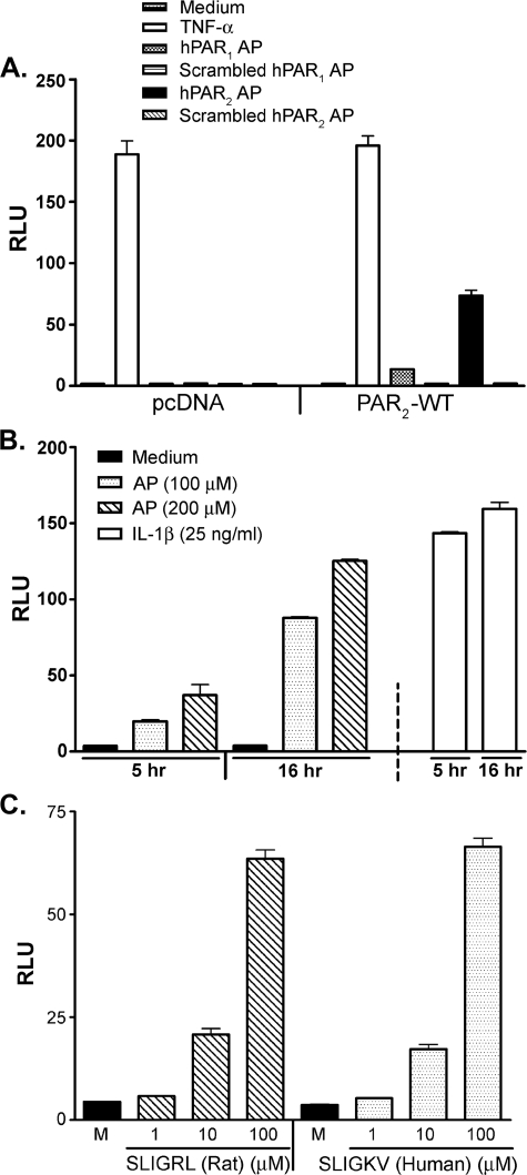
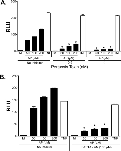
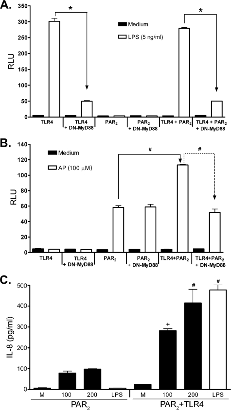
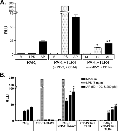
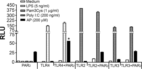
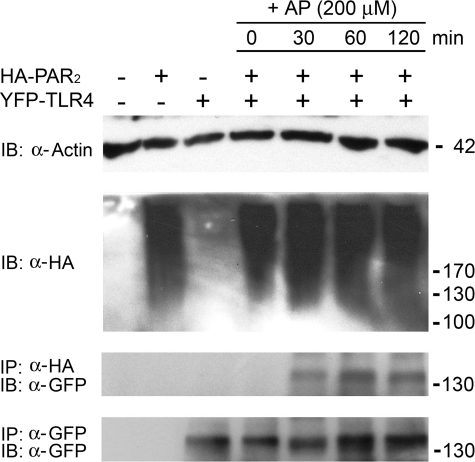
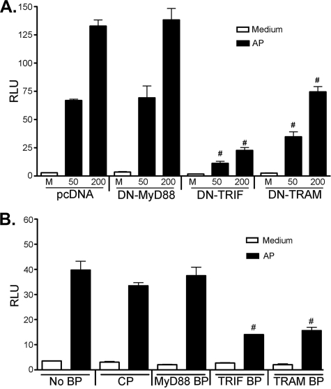
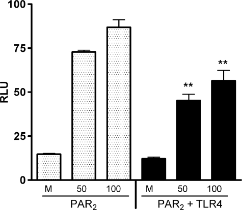
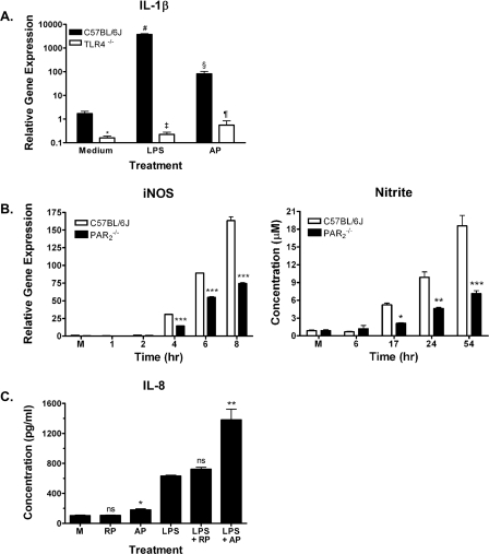
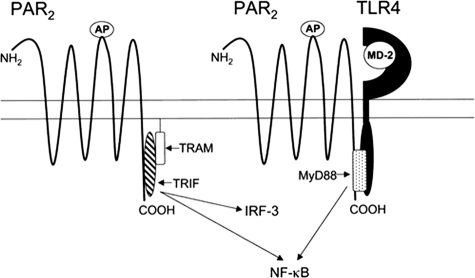
Proteinase-activated receptor 2 (PAR2), a seven-transmembrane G protein-coupled receptor, is activated at inflammatory sites by proteolytic cleavage of its extracellular N terminus by trypsin-like enzymes, exposing a tethered, receptor-activating ligand. Synthetic agonist peptides (AP) that share the tethered ligand sequence also activate PAR2, often measured by Ca2+ release. PAR2 contributes to inflammation through activation of NF-kappaB-regulated genes; however, the mechanism by which this occurs is unknown. Overexpression of human PAR2 in HEK293T cells resulted in concentration-dependent, PAR2 AP-inducible NF-kappaB reporter activation that was protein synthesis-independent, yet blocked by inhibitors that uncouple Gi proteins or sequester intracellular Ca2+. Because previous studies described synergistic PAR2- and TLR4-mediated cytokine production, we hypothesized that PAR2 and TLR4 might interact at the level of signaling. In the absence of TLR4, PAR2-induced NF-kappaB activity was inhibited by dominant negative (DN)-TRIF or DN-TRAM constructs, but not by DN-MyD88, findings confirmed using cell-permeable, adapter-specific BB loop blocking peptides. Co-expression of TLR4/MD-2/CD14 with PAR2 in HEK293T cells led to a synergistic increase in AP-induced NF-kappaB signaling that was MyD88-dependent and required a functional TLR4, despite the fact that AP exhibited no TLR4 agonist activity. Co-immunoprecipitation of PAR2 and TLR4 revealed a physical association that was AP-dependent. The response to AP or lipopolysaccharide was significantly diminished in TLR4(-/-) and PAR2(-/-) macrophages, respectively, and SW620 colonic epithelial cells exhibited synergistic responses to co-stimulation with AP and lipopolysaccharide. Our data suggest a unique interaction between two distinct innate immune response receptors and support a novel paradigm of receptor cooperativity in inflammatory responses.


Figures

FIGURE 1.
FIGURE 1.
HEK293T/PAR2 transfectants respond in a specific manner independent of species of origin of AP. A, HEK293T cells were co-transfected with either the control pcDNA or the untagged human PAR2 construct, together with ELAM (NF-κB)-luciferase and pCMV1-β-galactosidase reporter constructs. Transfected cells were treated with TNF-α (50 ng/ml), 200 μm huPAR1 AP (TFLLR-NH2), or its inactive scrambled peptide or 200 μm huPAR2 AP (SLIGKV-NH2) or its inactive scrambled peptide for 16 h. Reporter activities were measured in cell lysates as described under “Experimental Procedures.” A representative experiment is shown (n = 3). B, HEK293T cells, transfected as described in A, were treated with huPAR2 AP at the indicated concentrations or with human rIL-1β for 5 or 16 h. Cell lysates were assayed for ELAM-luc and β-galactosidase reporter activities. A representative experiment is shown (n = 8). C, HEK293T cells, transfected as described in A, were treated with medium only, rat/mouse PAR2 AP (SLIGRL-NH2), or huPAR2 AP for 16 h. ELAM-luc and β-galactosidase reporter activities were measured in cell lysates. A representative experiment is shown (n = 5). RLU, relative luciferase units; M, medium.
FIGURE 2.
FIGURE 2.
PAR2 activation by AP in HEK293T/huPAR2 transfectants is inhibited by PT and BAPTA-AM. HEK293T cells were co-transfected with huPAR2 construct together with the ELAM-luc and β-galactosidase reporter constructs. Transfectants were pretreated with medium (M) or the indicated concentrations of PT (A) or BAPTA-AM (100 μm)(B) for 1 h. Cells were then further treated with indicated concentrations of huPAR2 AP or TNF-α (TNF, 50 ng/ml) for an additional 16 h. Cell lysates were prepared and reporter activities measured. A representative experiment is shown (n = 5). *, p < 0.001 versus medium-pretreated controls. RLU, relative luciferase units.
FIGURE 3.
FIGURE 3.
Co-expression of TLR4 and PAR2 results in synergistic, MyD88-dependent NF-κB activation in response to PAR2 AP. A and B, HEK293T cells were co-transfected with TLR4 signaling complex (pFLAG-huTLR4, pEF-BOS-HA-huMD-2, pcDNA3-huCD14) and/or huPAR2 constructs, without or with DN-MyD88 construct (200 ng/well). Cells were also co-transfected with the NF-κB-luciferase and β-galactosidase reporter constructs. A, cells were treated with medium only or LPS, although in B the cells were treated with medium only or AP. A representative experiment is shown for A and B (n = 6). C, HEK293T cells were transfected as described above in the absence of reporter constructs. Levels of IL-8 in the supernatants were determined by enzyme-linked immunosorbent assay. A representative experiment is shown (n = 2). RLU, relative luciferase units. *, P < 0.001, #, P < 0.01, and +, P, < 0.05.
FIGURE 4.
FIGURE 4.
Synergy between PAR2 and TLR4 induced by PAR2 AP requires MD-2 and a signaling-competent TLR4. A, HEK293T cells were co-transfected with the huPAR2 construct only or huPAR2, pFLAG-huTLR4, and huCD14, without or with the huMD-2 expression construct. All wells were co-transfected with ELAM-lucandβ-galactosidase reporter constructs. Transfected cells were treated with huPAR2 AP or LPS, and reporter activities were measured. A representative experiment is shown (n = 4). B, HEK293T cells were co-transfected with the huPAR2 construct only, or PAR2 co-transfected with YFP-TLR4-WT or YFP-TLR4-P714H mutant (200 ng/well each) constructs either individually or co-transfected with huPAR2 construct. All wells were also co-transfected with the reporter constructs. Transfected cells were treated with TNF-α, LPS, or huPAR2 AP, and reporter activities were measured. A representative experiment is shown (n = 3). *, p < 0.001, between the PAR2 and PAR2 + YFP-TLR4-WT transfectants; **, p < 0.001, between PAR2 + YFP-TLR4-WT and PAR2 + YFP-P714H-TLR4 transfectants treated with huPAR2 AP. RLU, relative luciferase units.
FIGURE 5.
FIGURE 5.
Co-expression of PAR2 results in synergistic NF-κB activation induced by PAR2 AP with TLR4 but not with TLR2 and TLR3. HEK293T cells were co-transfected with either TLR4 signaling complex, FLAG-tagged TLR2, or FLAG-tagged TLR3 and/or huPAR2 construct. All wells were transfected with the NF-κB-luciferase and pCMV1-β-galactosidase reporter constructs. Transfectants were treated with medium only or respective TLR agonist (LPS for TLR4, S-[2,3-bis(palmitoyloxy)-(2-RS)-propyl]-N-palmitoyl-(R)-Cys-(S)-Ser-(Lys)4-OH, trihydrochloride (Pam3Cys) for TLR2, and poly(I:C) for TLR3), or AP. A representative experiment is shown (n = 3). *, p < 0.001, between the PAR2 and PAR2 + TLR4-WT transfectants. RLU, relative luciferase units.
FIGURE 6.
FIGURE 6.
TLR4 co-immunoprecipitates with PAR2 in a PAR2 AP-dependent manner. HEK293T cells, transfected with either HA-PAR2 vector or YFP-TLR4-WT vector along with MD-2 and CD14 expression vectors, or co-transfected together with HA-PAR2 and YFP-TLR4 complex, were incubated for a further 72 h before washing and lysing as described under “Experimental Procedures.” Immunoprecipitation (IP) and Western blotting (IB) were carried out to analyze the physical interaction between PAR2 and TLR4 proteins. A representative experiment is shown (n = 3). Molecular weight of proteins's represented in kDa (on right).
FIGURE 7.
FIGURE 7.
PAR2-mediated activation of NF-κB is decreased by inhibitors of TRIF and TRAM. HEK293T cells were co-transfected with huPAR2 construct and DN-MyD88, DN-TRIF, or DN-TRAM (50 ng/well) expression vectors (A). Transfected cells were treated for 16 h with the indicated concentrations of huPAR2 AP and ELAM-luc, and β-galactosidase reporter activities were measured. B, HEK293T cells were transfected with huPAR2 construct, ELAM-luc, and β-galactosidase reporter constructs. Transfected cells were pretreated with medium or 40 μm CP or the indicated cell-permeable adapter blocking peptide (BP), and then treated with AP for an additional 16 h, at which time reporter activities were measured. A representative experiment is shown for each experiment (n = 3). #, p < 0.001. RLU, relative luciferase units.
FIGURE 8.
FIGURE 8.
Co-transfection of TLR4 receptor complex with PAR2 inhibits RANTES promoter activity. HEK293T cells were co-transfected with either huPAR2 construct or huPAR2 plus TLR4 signaling complex (pFLAG-huTLR4; pEFBOS-HA-huMD-2; pcDNA3-huCD14). The RANTES-luciferase reporter (500 ng/well) and β-galactosidase vector were also co-transfected. Transfectants were treated with the indicated concentrations of huPAR2 AP for 16 h, and luciferase and β-galactosidase activities were measured. A representative experiment is shown (n = 5). **, p < 0.05. RLU, relative luciferase units.
FIGURE 9.
FIGURE 9.
Macrophages deficient in TLR4 or PAR2 show dysregulated responses to AP or LPS, respectively, and the SW620 intestinal epithelial cell line exhibits a synergistic response to co-stimulation with AP and LPS. A, TLR4–/– macrophages exhibit diminished IL-1β gene induction in response to huPAR2 AP. Macrophages from C57BL/6J or TLR4–/– mice were stimulated with huPAR2 AP (200 μm) or LPS (10 ng/ml) for 2 h, and steady-state IL-1β mRNA was measured. Results represent mean ± S.E. of six separate experiments. *, p < 0.001; Medium (C57BL/6J versus TLR4–/–); #, p < 0.001 Medium versus LPS (C57BL/6J); ‡, p < 0.001 LPS (C57BL/6J versus TLR4–/–); §, p < 0.001 Medium versus AP (C57BL/6J); and ¶, p < 0.001; AP (C57BL/6J versus TLR4–/–). B, PAR –/–2 macrophages exhibit diminished iNOS gene induction and NO• activity in response to LPS. Macrophages from C57BL/6J or PAR –/–2 mice were stimulated for the indicated times with 10 ng/ml LPS for iNOS mRNA or with 100 ng/ml LPS for supernatant analysis for NO. Data are representative of three separate experiments. M, medium control; *, p < 0.05; **, p < 0.01; ***, p < 0.001 (C). SW620 cells respond synergistically to huPAR2 AP (200 μm) and LPS (100 ng/ml) co-stimulation to augment IL-8 secretion (t = 24 h). Data represent the mean ± S.E. of three separate experiments. M, medium control; RP, reverse peptide; ns, not significant; *, p < 0.05 (Medium versus AP); **, p < 0.01 (LPS versus LPS + AP).
FIGURE 10.
FIGURE 10.
Hypothetical model of PAR2 signaling and interaction with TLR4. See “Discussion” for description of model.

Similar articles

Cited by

References

    1. Neurath, H. (1999) Proc. Natl. Acad. Sci. U. S. A. 96 10962–10963 - PMC - PubMed
    1. Turk, B. (2006) Nat. Rev. Drug Discov. 5 785–799 - PubMed
    1. Macfarlane, S. R., Seatter, M. J., Kanke, T., Hunter, G. D., and Plevin, R. (2001) Pharmacol. Rev. 53 245–282 - PubMed
    1. Ramachandran, R., and Hollenberg, M. D. (2008) Br. J. Pharmacol. 153 Suppl. 1, S263–S282 - PMC - PubMed
    1. Hollenberg, M. D., and Compton, S. J. (2002) Pharmacol. Rev. 54 203–217 - PubMed

Publication types

MeSH terms