. 2008 Aug;11(8):932-9.
doi: 10.1038/nn.2153. Epub 2008 Jul 11.

Cell type-specific regulation of DARPP-32 phosphorylation by psychostimulant and antipsychotic drugs

Affiliations

Cell type-specific regulation of DARPP-32 phosphorylation by psychostimulant and antipsychotic drugs

Helen S Bateup et al. Nat Neurosci. 2008 Aug.

Abstract

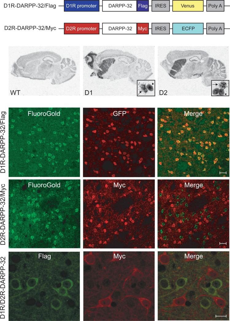
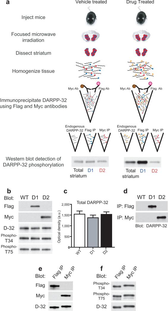
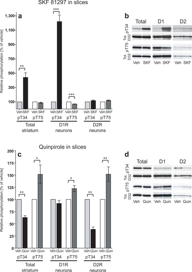
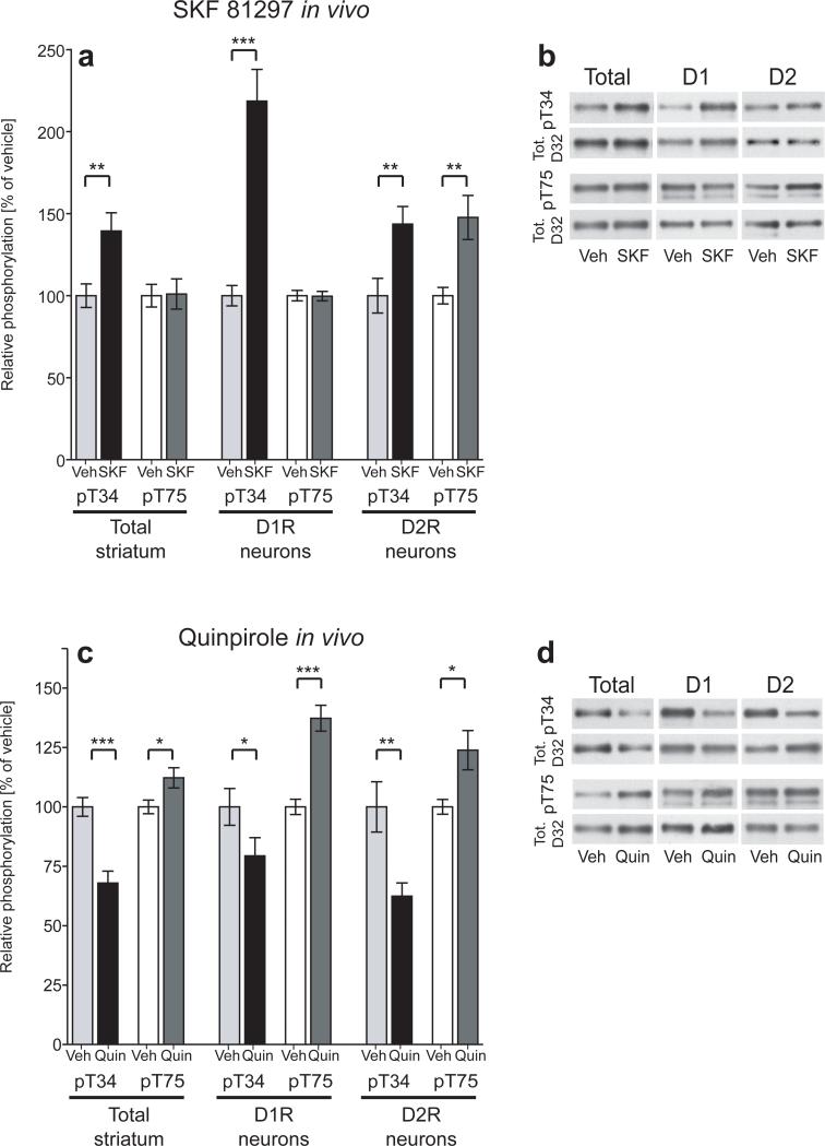
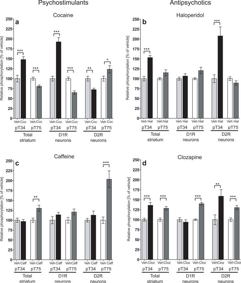
DARPP-32 is a dual-function protein kinase/phosphatase inhibitor that is involved in striatal signaling. The phosphorylation of DARPP-32 at threonine 34 is essential for mediating the effects of both psychostimulant and antipsychotic drugs; however, these drugs are known to have opposing behavioral and clinical effects. We hypothesized that these drugs exert differential effects on striatonigral and striatopallidal neurons, which comprise distinct output pathways of the basal ganglia. To directly test this idea, we developed bacterial artificial chromosome transgenic mice that allowed the analysis of DARPP-32 phosphorylation selectively in striatonigral and striatopallidal neurons. Using this new methodology, we found that cocaine, a psychostimulant, and haloperidol, a sedation-producing antipsychotic, exert differential effects on DARPP-32 phosphorylation in the two neuronal populations that can explain their opposing behavioral effects. Furthermore, we found that a variety of drugs that target the striatum have cell type-specific effects that previous methods were not able to discern.


Figures

Figure 1
Figure 1. Generation of D1R-DARPP-32/Flag and D2R-DARPP-32/Myc mice
(a) Schematic of the modified shuttle vectors used to generate D1R-DARPP-32/Flag and D2R-DARPP-32/Myc mice. IRES, internal ribosome entry site; Poly A, polyadenylation sequence; Venus, venus fluorescent protein; ECFP, enhanced cyan fluorescent protein. (b) In situ hybridization using a 35S labeled antisense probe against GFP which recognizes both Venus and ECFP mRNA shows the expression of the constructs in sagittal brain sections from wild-type (WT), D1R-DARPP-32/Flag (D1), and D2R-DARPP-32/Myc (D2) mice. Insets show magnification of GFP positive striatal cells labeled with silver grains (arrows) and unlabeled cells (arrowheads) after dipping sections in emulsion. (c) FluoroGold retrograde tracer was injected into the substantia nigra pars reticulata. Double immunoflourescence (IF) in striatal sections reveals co-localization of Fluoro-Gold (green) and GFP (red) in the D1R-DARPP-32/Flag mice, scale bar = 20μm. (d) There is no co-localization of Fluoro-Gold (green) and Myc (red) in striatal sections from the D2R-DARPP-32/Myc mice, scale bar = 20μm. (e) IF staining using Flag (green) and Myc (red) antibodies in striatal sections from D1R/D2R-DARPP-32 double transgenic mice showing no co-localization, scale bar = 10μm.
Figure 2
Figure 2. Experimental design and tagged DARPP-32 immunoprecipitation
(a) Schematic of the procedure for in vivo biochemical experiments using D1R/D2R-DARPP-32 double transgenic mice. One feature of this method is the ability to incubate striatal homogenates simultaneously with Myc and Flag antibodies by differential coupling of these antibodies to magnetic beads (brown circles) and affinity gel (pink circles), respectively. Blue circles represent Flag tags, red circles represent Myc tags. “P” indicates phosphorylated protein. (b) Equal amounts of striatal homogenate from wild-type (WT), D1R-DARPP-32/Flag (D1), and D2R-DARPP-32/Myc (D2) single transgenic mice were blotted with Flag, Myc, total DARPP-32, phospho-T34, and phospho-T75 antibodies. (c) Quantification of the average optical density of total DARPP-32 bands from wild-type (WT), D1R-DARPP-32 (D1), and D2R-DARPP-32 (D2) mice (n=3−4 mice per group). Data were normalized to GAPDH loading control (not shown). (d) Striatal homogenates from wild-type (WT), D1R-DARPP-32/Flag (D1), and D2R-DARPP-32/Myc (D2) mice were incubated with Flag (upper panel) and Myc (lower panel) IP antibodies. Equal amounts of IP'd sample was loaded in each lane and blotted with an antibody against total DARPP-32. (e) Striatal homogenate prepared from D1R/D2R-DARPP-32 double transgenic mice was incubated with Flag or Myc antibodies and IP eluates were blotted with either Flag or Myc antibody. Membranes were striped and re-probed with total DARPP-32 antibody (bottom panel) showing equal loading of IP's. (f) Representative blot of the basal level of T34 and T75 phosphorylation in Flag and Myc IP'd samples from D1R/D2R-DARPP-32 mice detected by phospho-specific antibodies. The phospho-T34 blot was stripped and re-probed with total DARPP-32 antibody (bottom panel).
Figure 3
Figure 3. Effects of D1R and D2R agonists on DARPP-32 phosphorylation in striatal slices
(a,c) Bar graphs represent group averages of DARPP-32 phosphorylation from striatal slices incubated with vehicle, SKF 81297 (10 μM, 5 min.), or quinpirole (1 μM, 10 min.). Phospho-T34 and phospho-T75 data were normalized to total DARPP-32 levels and expressed as percent of vehicle control. The left panels represent data from endogenous non-tagged DARPP-32 (Total striatum), the center panels represent data from Flag-tagged DARPP-32 (D1R neurons), and the right panels represent data from Myc-tagged DARPP-32 (D2R neurons). In each experiment 6 striatal slices from 1 mouse were divided into vehicle, SKF 81297, and quinpirole treatment groups and slices from 3 mice were pooled for the analysis of DARPP-32 phosphorylation. Data represent means from 4−8 independent experiments. *, p<.05, **, p<.01, ***, p<.001. (b,d) Representative western blots from treated slices. T34 and T75 phospho-specific blots (top panels) were stripped and re-probed with total DARPP-32 antibody (bottom panels).
Figure 4
Figure 4. Effects of D1R and D2R agonists on DARPP-32 phosphorylation in vivo
(a,c) Bar graphs represent group averages of DARPP-32 phosphorylation from D1R/D2R-DARPP-32 mice injected i.p. with vehicle, SKF 81297 (5mg/kg), or quinpirole (0.2 mg/kg) and sacrificed 15 minutes later. Phospho-T34 and T75 data were normalized to total DARPP-32 levels expressed as percent of vehicle control. The left panels represent data from endogenous non-tagged DARPP-32 (Total striatum), the center panels represent data from Flag-tagged DARPP-32 (D1R neurons), and the right panels represent data from Myc-tagged DARPP-32 (D2R neurons). Each bar represents the mean of 7−12 mice from 2−3 independent experiments. *, p<.05, **, p<.01, ***, p<.001. (b,d) Representative western blots from drug treated D1R/D2R-DARPP-32 mice. T34 and T75 phospho-specific blots (top panels) were stripped and re-probed with total DARPP-32 antibody (bottom panels).
Figure 5
Figure 5. Differential regulation of DARPP-32 phosphorylation by psychostimulants and antipsychotics
D1R/D2R-DARPP-32 mice were injected i.p. with either (a) cocaine (20 mg/kg), (b) haloperidol (1 mg/kg), (c) caffeine (7.5 mg/kg), (d) clozapine (5 mg/kg), or vehicle and sacrificed 15 minutes later. Phospho-T34 and T75 data were normalized to total DARPP-32 levels and expressed as percent of control. The left panels represent data from endogenous non-tagged DARPP-32 (Total striatum), the center panels represent data from Flag-tagged DARPP-32 (D1R neurons), and the right panels represent data from Myc-tagged DARPP-32 (D2R neurons). Each bar represents the mean of 7−14 mice from 2−4 experiments. *, p<.05, **, p<.01, ***, p<.001.

References

    1. Greengard P. The neurobiology of slow synaptic transmission. Science. 2001;294:1024–1030. - PubMed
    1. Greengard P, Allen PB, Nairn AC. Beyond the dopamine receptor: the DARPP-32/protein phosphatase-1 cascade. Neuron. 1999;23:435–447. - PubMed
    1. Nishi A, et al. Amplification of dopaminergic signaling by a positive feedback loop. Proc Natl Acad Sci U S A. 2000;97:12840–12845. - PMC - PubMed
    1. Ahn JH, et al. Protein kinase A activates protein phosphatase 2A by phosphorylation of the B56delta subunit. Proc Natl Acad Sci U S A. 2007;104:2979–2984. - PMC - PubMed
    1. Bibb JA, et al. Phosphorylation of DARPP-32 by Cdk5 modulates dopamine signalling in neurons. Nature. 1999;402:669–671. - PubMed

Publication types

MeSH terms