

Long-term exposure of mouse pancreatic islets to oleate or palmitate results in reduced glucose-induced somatostatin and oversecretion of glucagon
- PMID: 18622593
- PMCID: PMC2516194
- DOI: 10.1007/s00125-008-1082-0
Long-term exposure of mouse pancreatic islets to oleate or palmitate results in reduced glucose-induced somatostatin and oversecretion of glucagon
Abstract
Aims/hypothesis: Long-term exposure to NEFAs leads to inhibition of glucose-induced insulin secretion. We tested whether the release of somatostatin and glucagon, the two other major islet hormones, is also affected.
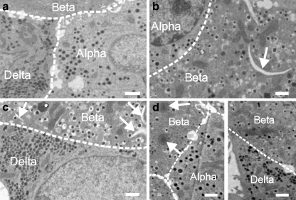
Methods: Mouse pancreatic islets were cultured for 72 h at 4.5 or 15 mmol/l glucose with or without 0.5 mmol/l oleate or palmitate. The release of glucagon and somatostatin during subsequent 1 h incubations at 1 or 20 mmol/l glucose as well as the islet content of the two hormones were determined. Lipid-induced changes in islet cell ultrastructure were assessed by electron microscopy.
Results: Culture at 15 mmol/l glucose increased islet glucagon content by approximately 50% relative to that observed following culture at 4.5 mmol/l glucose. Inclusion of oleate or palmitate reduced islet glucagon content by 25% (at 4.5 mmol/l glucose) to 50% (at 15 mmol/l glucose). Long-term exposure to the NEFA increased glucagon secretion at 1 mmol/l glucose by 50% (when islets had been cultured at 15 mmol/l glucose) to 100% (with 4.5 mmol/l glucose in the culture medium) and abolished the inhibitory effect of 20 mmol/l glucose on glucagon secretion. Somatostatin content was unaffected by glucose and lipids, but glucose-induced somatostatin secretion was reduced by approximately 50% following long-term exposure to either of the NEFA, regardless of whether the culture medium contained 4.5 or 15 mmol/l glucose. Ultrastructural evidence of lipid deposition was seen in <10% of non-beta cells but in >80% of the beta cells.
Conclusions/interpretation: Long-term exposure to high glucose and/or NEFA affects the release of somatostatin and glucagon. The effects on glucagon secretion are very pronounced and in type 2 diabetes in vivo may aggravate the hyperglycaemic effects due to lack of insulin.
Figures

Similar articles
-
Effects of gastric inhibitory polypeptide, vasoactive intestinal polypeptide and peptide histidine isoleucine on the secretion of hormones by isolated mouse pancreatic islets.J Endocrinol. 1990 Jun;125(3):375-9. doi: 10.1677/joe.0.1250375. J Endocrinol. 1990. PMID: 1973701
-
Loss of a priming effect of glucose on A and D cell secretion in perfused pancreases from alloxan-diabetic rats: role of insulin and alloxan.Diabetologia. 1983 Jan;24(1):47-51. doi: 10.1007/BF00275947. Diabetologia. 1983. PMID: 6131006
-
Long-term exposure to glucose and lipids inhibits glucose-induced insulin secretion downstream of granule fusion with plasma membrane.Diabetes. 2007 Jul;56(7):1888-97. doi: 10.2337/db06-1150. Epub 2007 Apr 24. Diabetes. 2007. PMID: 17456851
-
Regulatory Role of Fatty Acid Metabolism on Glucose-Induced Changes in Insulin and Glucagon Secretion by Pancreatic Islet Cells.Int J Mol Sci. 2024 May 31;25(11):6052. doi: 10.3390/ijms25116052. Int J Mol Sci. 2024. PMID: 38892240 Free PMC article. Review.
-
Gap junction coupling and islet delta-cell function in health and disease.Peptides. 2022 Jan;147:170704. doi: 10.1016/j.peptides.2021.170704. Epub 2021 Nov 23. Peptides. 2022. PMID: 34826505 Review.
Cited by
-
Structural basis for delta cell paracrine regulation in pancreatic islets.Nat Commun. 2019 Aug 16;10(1):3700. doi: 10.1038/s41467-019-11517-x. Nat Commun. 2019. PMID: 31420552 Free PMC article.
-
Revisiting the role of glucagon in health, diabetes mellitus and other metabolic diseases.Nat Rev Endocrinol. 2023 Jun;19(6):321-335. doi: 10.1038/s41574-023-00817-4. Epub 2023 Mar 17. Nat Rev Endocrinol. 2023. PMID: 36932176 Review.
-
The mitochondrial β-oxidation enzyme HADHA restrains hepatic glucagon response by promoting β-hydroxybutyrate production.Nat Commun. 2022 Jan 19;13(1):386. doi: 10.1038/s41467-022-28044-x. Nat Commun. 2022. PMID: 35046401 Free PMC article.
-
Chronic Exposure to Palmitic Acid Down-Regulates AKT in Beta-Cells through Activation of mTOR.Am J Pathol. 2022 Jan;192(1):130-145. doi: 10.1016/j.ajpath.2021.09.008. Epub 2021 Oct 5. Am J Pathol. 2022. PMID: 34619135 Free PMC article.
-
The Human and Mouse Islet Peptidome: Effects of Obesity and Type 2 Diabetes, and Assessment of Intraislet Production of Glucagon-like Peptide-1.J Proteome Res. 2021 Sep 3;20(9):4507-4517. doi: 10.1021/acs.jproteome.1c00463. Epub 2021 Aug 23. J Proteome Res. 2021. PMID: 34423991 Free PMC article.
References
Publication types
MeSH terms
Substances
Grants and funding
LinkOut - more resources
Full Text Sources
