Review
. 2008 Nov;392(6):1019-30.
doi: 10.1007/s00216-008-2244-0. Epub 2008 Jul 13.

Substrate binding to cytochromes P450

Affiliations
Review

Substrate binding to cytochromes P450

Emre M Isin et al. Anal Bioanal Chem. 2008 Nov.

Abstract

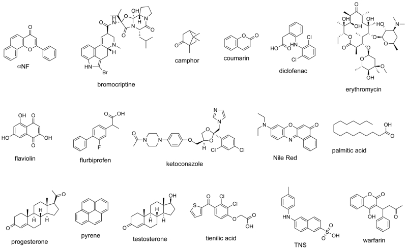
P450s have attracted tremendous attention owing to not only their involvement in the metabolism of drug molecules and endogenous substrates but also the unusual nature of the reaction they catalyze, namely, the oxidation of unactivated C-H bonds. The binding of substrates to P450s, which is usually viewed as the first step in the catalytic cycle, has been studied extensively via a variety of biochemical and biophysical approaches. These studies were directed towards answering different questions related to P450s, including mechanism of oxidation, substrate properties, unusual substrate oxidation kinetics, function, and active-site features. Some of the substrate binding studies extending over a period of more than 40 years of dedicated work have been summarized in this review and categorized by the techniques employed in the binding studies.


Figures

Fig. 1
Fig. 1. Generalized P450 catalytic cycle
Fig. 2
Fig. 2. Structures of selected ligands described in the text
Fig. 3
Fig. 3. Spectra of P450 2A6 complexes
A, Spectra were recorded in 50 mM potassium phosphate buffer (pH 7.4) with 5.2 μM P450 2A6, either without (—) or with (—) 50 μM coumarin. B, Difference spectrum obtained by mathematically subtracting the spectrum of the unbound P450 from the bound. Reprinted with permission from [20].
Fig. 4
Fig. 4. Steady-state kinetics of oxidations catalyzed by P450 1A2
Pyrene 1-hydroxylation; data points are set to the equation v = kcat·Sn (S50n + Sn)−1, with kcat = 3.0 ± 0.1 min−1, n = 3.6 ± 0.6 and S50 = 9.9 ± 0.5 μM. Reprinted with permission from [41].
Fig. 5
Fig. 5. A, Scheme depicting proposed events in ligand binding to ferric P450 1A2
See text and original reference [41] for more discussion. Step 1: The ligand L first interacts with P450 1A2 at a peripheral site. Step 2: L is translocated to the interior of the protein, Step 3a: A conformational change in the P450 occurs. Step 3b: If L is small enough for two molecules (of L) to occupy the active site, a second molecule of L can enter the active site. Step 4: Conformational change of the P450. B, Model and rate constants used in fitting. E: P450 1A2, S: pyrene, P: 1-hydroxypyrene, Q: dihydroxypyrene products (1,5-, 1,6-, and 1,8-). The kinetic scheme involves sequential binding of two pyrene molecules (k1, k−1, k2, k−2),a conformational change (k3, k−3),, oxidation of pyrene only in the complex with two pyrenes (k4, k−4), release of 1-hydroxypyrene (k5, k−5), and conversion of 1-hydroxypyrene to dihydroxypyrene(s) from the binary complex (k6, k−6). Reprinted with permission from [41].

References

    1. Guengerich FP. Toxicol Lett. 1994;70:133–138. - PubMed
    1. Ortiz de Montellano PR, editor. Cytochrome P450: Structure, Mechanism, and Biochemistry. 3rd KluwerAcademic/Plenum Publishers; New York: 2005.
    1. Denisov IG, Makris TM, Sligar SG, Schlichting I. Chem Rev. 2005;105:2253–2277. - PubMed
    1. Guengerich FP. Chem Res Toxicol. 2008;21:70–83. - PubMed
    1. Lewis DFV, Ito Y, Goldfarb PS. Drug Develop Res. 2006;66:19–24.

Publication types

LinkOut - more resources