. 2009 Feb;126(2):233-45.
doi: 10.1111/j.1365-2567.2008.02892.x. Epub 2008 Jun 23.

Fatty acid-induced induction of Toll-like receptor-4/nuclear factor-kappaB pathway in adipocytes links nutritional signalling with innate immunity

Affiliations

Fatty acid-induced induction of Toll-like receptor-4/nuclear factor-kappaB pathway in adipocytes links nutritional signalling with innate immunity

Andreas Schaeffler et al. Immunology. 2009 Feb.

Abstract

To study the effects of fatty acids and the involvement of the Toll-like receptor-4/nuclear factor-kappaB (TLR-4/NF-kappaB) pathway with respect to the secretion of adipokines from adipocytes 3T3-L1 adipocytes were stimulated with increasing doses of fatty acids. The secretion of adiponectin, resistin and monocyte chemoattractant protein-1 (MCP-1) was measured by enzyme-linked immunosorbent assay. The NF-kappaB p65 nuclear translocation and TLR-4 expression were investigated by Western blot. The effects mediated by NF-kappaB were tested using a specific NF-kappaB-inhibitor and TLR-4-induced effects were analysed with a neutralizing TLR-4 antibody. Binding of (14)C-labelled fatty acids to TLR-4/MD-2 was investigated using a FLAG-tagged extracellular part of TLR-4 fused to full-length MD-2 via a linker (lipopolysaccharide-Trap). The messenger RNA (mRNA) expression of adipokines in abdominal adipose tissue of rats fed a standard chow or a high-fat diet was investigated by reverse transcription-polymerase chain reaction. The TLR-4 is induced during adipocyte differentiation and its expression is enhanced following fatty acid stimulation. The stimulatory effects of stearic and palmitic acids on MCP-1 secretion and of palmitoleic acid on resistin secretion are mediated via NF-kappaB. The stimulatory effects of stearic, palmitic and palmitoleic acids on resistin secretion and the stimulatory effect of stearic acid on MCP-1 secretion are mediated via TLR-4. Fatty acid-mediated effects are caused by an endogenous ligand because fatty acids were shown not to bind directly to TLR-4/MD-2. Adipose tissue mRNA expression and serum levels of adipokines did not differ in rats fed a high-fat diet. These data provide a new molecular mechanism by which fatty acids can link nutrition with innate immunity.


Figures

Figure 1
Figure 1
Effects of fatty acids on the secretion of adiponectin from differentiated 3T3-L1 adipocytes. Six wells were used for each experimental group. After data normalization for total protein content, the secretion of adiponectin is given in ng/ml/24 hr. Box plots show median (thick black line), lower and upper quartiles (box) and lower and upper extreme values (thin vertical lines). The dose effects of palmitic acid (a), stearic acid (b), oleic acid (c), palmitoleic acid (d) and linoleic acid (e) were investigated.
Figure 2
Figure 2
Effects of fatty acids on the secretion of resistin from differentiated 3T3-L1 adipocytes. Six wells were used for each experimental group. After data normalization for total protein content, the secretion of adiponectin is given in ng/ml/24 hr. Box plots show median (thick black line), lower and upper quartiles (box) and lower and upper extreme values (thin vertical lines). The dose effects of palmitic acid (a), stearic acid (b), oleic acid (c), palmitoleic acid (d) and linoleic acid (e) were investigated.
Figure 3
Figure 3
Effects of fatty acids on the secretion of monocyte chemoattractant protein-1 (MCP-1) from differentiated 3T3-L1 adipocytes. Six wells were used for each experimental group. After data normalization for total protein content, the secretion of MCP-1 (n= 6) is given in pg/ml/24 hr. Box plots show median (thick black line), lower and upper quartiles (box) and lower and upper extreme values (thin vertical lines). The dose effects of palmitic acid, stearic acid (a), oleic acid, palmitoleic acid (b) and linoleic acid (c) were investigated.
Figure 4
Figure 4
The role of nuclear factor-κB (NF-κB) in the fatty acid-induced secretion of resistin and monocyte chemoattractant protein-1 (MCP-1) from differentiated 3T3-L1 adipocytes. (a) Effects of NF-κB inhibition on the basal and palmitoleic acid-induced secretion of resistin. Three wells were used for each experimental group. After data normalization for total protein content, the secretion of resistin (n= 3) is given in pg/ml/24 hr. Box plots show median (thick black line), lower and upper quartiles (box) and lower and upper extreme values (thin vertical lines). (b) Effects of NF-κB inhibition on the basal and stearic acid-induced and palmitic acid-induced secretion of MCP-1. Three wells were used for each experimental group. After data normalization for total protein content, the secretion of MCP-1 (n= 3) is given in pg/ml/24 hr. Box plots show median (thick black line), lower and upper quartiles (box) and lower and upper extreme values (thin vertical lines). (c) Western blot analysis using nuclear extracts prepared from 3T3-L1 adipocytes. When compared to controls (absence of fatty acids), stearic acid, palmitic acid and palmitoleic acid increase NF-κB p65 nuclear translocation. As a loading control, the Coomassie stained gel is shown.
Figure 5
Figure 5
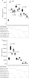
Effects of antibody-mediated Toll-like receptor-4 (TLR-4) blockade on the fatty acid-induced secretion of resistin and monocyte chemoattractant protein-1 (MCP-1) from differentiated 3T3-L1 adipocytes. Three wells were used for each experimental group. After data normalization for total protein content, the secretion of resistin and MCP-1 (n= 3 wells/group) is given in ng/ml/24 hr and pg/ml/24 hr, respectively. Box plots show median (thick black line), lower and upper quartiles (box) and lower and upper extreme values (thin vertical lines). Three doses (0·1, 1 and 5 μg per well) of a neutralizing TLR-4 antibody (°) were used to block TLR-4-induced signalling. (a) Effects of antibody-mediated TLR-4 blockade on the fatty acid-induced secretion of resistin. *Significant when compared to control; °Significant when compared to fatty acid stimulation without TLR-4 blockade. (b) Effects of antibody-mediated TLR-4 blockade on the fatty acid-induced effects on the secretion of MCP-1. *Significant when compared to control; °Significant when compared to fatty acid stimulation without TLR-4 blockade.
Figure 6
Figure 6
Analysis of Toll-like receptor-4 (TLR-4) expression by Western blot using extracts prepared from 3T3-L1 adipocytes during differentiation and after stimulation with fatty acids. The TLR-4 is almost absent in preadipocytes and is induced during adipocyte differentiation. Fatty acids slightly increase the amount of TLR-4 with palmitoleic acid showing the most pronounced effect. After stimulation with palmitoleic acid, adipocytes have about equal amounts of TLR-4 compared to monocytic THP-1 cells, which were used as a positive control. Lane 1 = 3T3-L1 preadipocytes, lane 2 = adipocytes after 4 days of differentiation, lane 3 = adipocytes after 7 days of differentiation, lane 4 = mature adipocytes after 9 days of differentiation, lane 5 = mature adipocytes stimulated with 100 μm stearic acid (24 h), lane 6 = mature adipocytes stimulated with 100 μm palmitic acid (24 hr), lane 7 = mature adipocytes stimulated with 1 μm palmitoleic acid (24 hr), lane 8 = THP-1 cells (monocytes), lane 9 = negative control (phosphate-buffered saline).
Figure 7
Figure 7
Investigation of fatty acid binding to Toll-like receptor-4 (TLR-4)/MD-2 by using a FLAG-tagged extracellular part of TLR-4 fused to full-length MD-2 [lipopolysaccharide (LPS)-Trap]. The binding of 14C-labelled stearic and oleic acids to a soluble fusion complex consisting of the FLAG-tagged extracellular part of TLR-4 fused to full-length MD-2 via a flexible linker (LPS-Trap) was investigated by immunoprecipitation (upper panel) and by fatty acid binding studies (lower panel). The LPS-Trap was incubated with an excess of C14-labelled fatty acids or with LPS alone. The complex was immunoprecipitated with anti-FLAG antibody M2. LPS-Trap was incubated with biotin-labelled LPS with and without a 100 × molar excess of stearic or oleic acid, a dose that should compete effectively with LPS if specific fatty acid binding occurs. The LPS/LPS-Trap complex was then pulled down with streptavidin–agarose, Western-blotted, and the blot was probed with anti-FLAG antibody. Upper panel: Neither stearic acid nor oleic acid can bind to the TLR-4/MD-2 heterodimer. Lower panel: There was no specific binding of C14-labelled fatty acids to the TLR-4/MD-2 complex. IP= immunoprecipitation, WB= Western blotting.

References

    1. Weisberg SP, McCann D, Desai M, Rosenbaum M, Leibel RL, Ferrante AW Jr. Obesity is associated with macrophage accumulation in adipose tissue. J Clin Invest. 2003;112:1796–808. - PMC - PubMed
    1. Xu H, Barnes GT, Yang Q, et al. Chronic inflammation in fat plays a crucial role in the development of obesity-related insulin resistance. J Clin Invest. 2003;112:1821–30. - PMC - PubMed
    1. Whitehead JP, Richards AA, Hickman IJ, Macdonald GA, Prins JB. Adiponectin – a key adipokine in the metabolic syndrome. Diabetes Obes Metab. 2006;8:264–80. - PubMed
    1. Okamoto Y, Kihara S, Funahashi T, Matsuzawa Y, Libby P. Adiponectin: a key adipocytokine in metabolic syndrome. Clin Sci (Lond) 2006;110:267–78. - PubMed
    1. Vasseur F. Adiponectin and its receptors: partners contributing to the “vicious circle” leading to the metabolic syndrome? Pharmacol Res. 2006;53:478–81. - PubMed

Publication types

MeSH terms