Review
. 2008 Dec;12(6A):2263-80.
doi: 10.1111/j.1582-4934.2008.00402.x. Epub 2008 Jun 27.

Neuronal cell death in neurodegenerative diseases: recurring themes around protein handling

Affiliations
Review

Neuronal cell death in neurodegenerative diseases: recurring themes around protein handling

Adrienne M Gorman. J Cell Mol Med. 2008 Dec.

Abstract

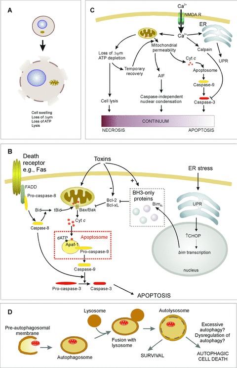
Neuronal cell death plays a role in many chronic neurodegenerative diseases with the loss of particular subsets of neurons. The loss of the neurons occurs during a period of many years, which can make the mode(s) of cell death and the initiating factors difficult to determine. In vitro and in vivo models have proved invaluable in this regard, yielding insight into cell death pathways. This review describes the main mechanisms of neuronal cell death, particularly apoptosis, necrosis, excitotoxicity and autophagic cell death, and their role in neurodegenerative diseases such as ischaemia, Alzheimer's, Parkinson's and Huntington's diseases. Crosstalk between these death mechanisms is also discussed. The link between cell death and protein mishandling, including misfolded proteins, impairment of protein degradation, protein aggregation is described and finally, some pro-survival strategies are discussed.


Figures

Figure 1
Figure 1
Mechanisms of cell death. See text for details. (A) Necrosis is characterized by loss of ATR which leads to cell lysis and release of cellular contents into the extracellular environment. (B) Apoptosis induction involves different pathways leading to caspase activation. In the extrinsic pathway ligand binding to death receptors {e.g. Fas) induces their trimerization and recruitment of caspase-8 through death domain-containing adaptor proteins (e.g. FADD). This leads to auto-activation of caspase-8, which can directly activate effector caspases. Alternatively in some cells Bid cleavage by caspase-8 is required for death. Truncated Bid translocates to the mitochondria and activates the intrinsic pathway. The intrinsic pathway is activated by toxins or conditions that cause stress to the mitochondria or endoplasmic reticulum (ER). Such stresses often alter the levels of, or cause post-translational modification of, Bcl-2 family proteins, initiating the release of cytochrome c from the mitochondria. For example, ER stress up-regulates the unfolded protein response (UPR), which increases expression of BimEL, a BH3-only protein that antagonizes Bcl-2 and Bcl-xL, permitting Bax-mediated cytochrome c release. Once in the cytosol, cytochrome c stimulates formation of the apoptosome complex, leading to caspase-9 activation. Initiator caspases (e.g. caspase-8 and -9) are shown in yellow, downstream effector caspases (e.g. caspase-3) are shown in red. (C) Excitotoxicity is caused by the influx of Ca2+ through the NMDA receptor. This can result in apoptosis, necrosis, or cell death that exhibits apoptotic nuclear morphology without caspase activation, suggesting a continuum of death patterns. (D) Autophagic cell death may be due to excessive autophagic digestion, as a result of dysregulation of the pathway by unknown mechanisms.
Figure 2
Figure 2
Crosstalk between apoptosis, autophagy and autophagic cell death. The schematic depicts some points of crosstalk between these pathways (see text for details).
Figure 3
Figure 3
Protein metabolism in neurons. Misfolded proteins can occur as a result of mutations, oxidative damage, erroneous proteolysis or other post-translational modifications. These can be refolded by chaperones such as Hsps, or targeted for proteasomal degradation by the addition of ubiqui-tin (Ub). Impairment of UPS-mediated degradation can lead to the formation of protein aggregates, which may be degraded via autophagy. The accumulation of misfolded proteins or protein aggregates leads to neuronal cell death by an unknown mechanism.

References

    1. Bredesen DE, Rao RV, Mehlen P. Cell death in the nervous system. Nature. 2006;443:796–802. - PMC - PubMed
    1. Yankner BA, Lu T, Loerch P. The aging brain. Annu Rev Pathol. 2008;3:41–66. - PubMed
    1. Stroh C, Schulze-Osthoff K. Death by a thousand cuts: an ever increasing list of caspase substrates. Cell Death Differ. 1998;5:997–1000. - PubMed
    1. Li P, Nijhawan D, Budihardjo I, Srinivasula SM, Ahmad M, Alnemri ES, Wang X. Cytochrome c and dATP-depend-ent formation of Apaf-1/caspase-9 complex initiates an apoptotic protease cascade. Cell. 1997;91:479–89. - PubMed
    1. Zou H, Li Y, Liu X, Wang X. An APAF-1.cytochrome c multimeric complex is a functional apoptosome that activates pro-caspase-9. J Biol Chem. 1999;274:11549–56. - PubMed

Publication types

MeSH terms