Review
. 2008 Oct;12(5B):1848-62.
doi: 10.1111/j.1582-4934.2008.00411.x. Epub 2008 Jul 10.

Parenchymal and vascular Abeta-deposition and its effects on the degeneration of neurons and cognition in Alzheimer's disease

Affiliations
Review

Parenchymal and vascular Abeta-deposition and its effects on the degeneration of neurons and cognition in Alzheimer's disease

Dietmar Rudolf Thal et al. J Cell Mol Med. 2008 Oct.

Abstract

The deposition of the amyloid beta-protein (Abeta) is one of the pathological hallmarks of Alzheimer's disease (AD). Abeta-deposits show the morphology of senile plaques and cerebral amyloid angiopathy (CAA). Senile plaques and vascular Abeta-deposits occur first in neocorti-cal areas. Then, they expand hierarchically into further brain regions. The distribution of Abeta plaques throughout the entire brain, thereby correlates with the clinical status of the patients. Imaging techniques for Abeta make use of the hierarchical distribution of Abeta to distinguish AD patients from non-AD patients. However, pathology seen in AD patients represents a late stage of a pathological process starting 10-30 years earlier in cognitively normal individuals. In addition to the fibrillar amyloid of senile plaques, oligomeric and monomeric Abeta is found in the brain. Recent studies revealed that oligomeric Abeta is presumably the most toxic Abeta-aggregate, which interacts with glutamatergic synapses. In doing so, dendrites are presumed to be the primary target for Abeta-toxicity. In addition, vascular Abeta-deposits can lead to capillary occlusion and blood flow disturbances presumably contributing to the alteration of neurons in addition to the direct neurotoxic effects of Abeta. All these findings point to an important role of Abeta and its aggregates in the neurodegenerative process of AD. Since there is already significant neuron loss in AD patients, treatment strategies aimed at reducing the amyloid load will presumably not cure the symptoms of dementia but they may stop disease progression. Therefore, it seems to be necessary to protect the brain from Abeta-toxicity already in stages of the disease with minor neuron loss before the onset of cognitive symptoms.


Figures
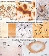
Fig 1
Fig 1
(A–C) Senile plaques in the temporal neocortex of AD cases. All layers are occupied by senile plaques, which contain Aβ as demonstrated by immunolabelling with an antibody raised against Aβ17–24 (4G8). In the higher magnification, the fibrillar nature of the Aβ deposits can be seen (arrows in B). The ‘needle-like’ appearance characterized even diffuse Aβ-deposits (arrows in B; [152]). C shows a cored neuritic plaques stained in a combination of a Gallyas-silver staining for neurofibrillary material (black) and anti-Aβ8–17 (6F3D) immunohistochemistry (brown). The amyloid core (*) is seen in the center of the plaques surrounded by a halo of diffuse Aβ-deposits (arrows). Here dystrophic neurites occur, which contain argyrophilic neurofibrillary material (arrowheads) indicative for neuritic plaques [35]. Adjacent to the cored neuritic plaques there is a NFT (t). (D, E) NFTs and NTs in the temporal cortex of an AD case. NFTs are most prominent in the pyramidal cell layers III and V. NFTs and neuropil threads contain fibrillar material detected be the Gallyas silver methods (D). These fibrils consist of aggregates of abnormally phosphorylated τ-protein (E). With the antibody against abnormal τ-protein not only Gallyas-positive fibrils are marked but also non-aggregated abnormal τ-protein (E; [8]). (F G) NFTs (arrows) and NTs (arrowheads) in the subicu-lum/ CA1 region at the higher magnification level. Both structures are stained with the Gallyas silver method and an antibody directed against abnormal τ-protein (AT-8). The neuron indicated with the open arrow shows accumulation of abnormal τ-protein in the pre-tangle status. (H, I) Cerebral amyloid angiopathy in the parietal cortex of an AD patient. There are Aβ-deposits in leptomeningeal and cortical vessels (arrows in H). The higher magnification shows the destruction of the vessel wall by Aβ-deposits that replace smooth muscle cells of the media (arrows in I). The numbers I–VI indicate the cortical layers in A and D. Calibration bar in I valid for: A = 300 μm; B = 30 μm; C, I = 15 μm; D, H = 150 μm; E = 370 μm; F, G = 50 μm.
Fig 2
Fig 2
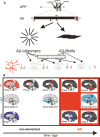
Schematic representation of Aβ generation, aggregation, deposition in the brain and its relation to neuronal changes. (A) Aβ is the cleavage product of β- and γ-secretase cleavage of the amyloid precursor protein (APP) [14]. It is a 39–43 amino acid protein. Aβ40 and Aβ42 are the major forms [40]. Aβ forms oligomers [64, 70] and fibrils [13, 86]. It is not clear whether oligomeric Aβ can form fibrils. However, the hypothesis that a conformational switch of Aβ is decisive for either fibril formation or oligomer formation has been supported by a recent study [153]. Fibrillar and oligomeric Aβ alter neurons [, , , , –74, 154]. An interaction between Aβ-oligomers with glutamatergic synapses has been demonstrated [–67]. Moreover, neurons with a prominent, highly ramified dendritic tree are more vulnerable than neurons exhibiting only single dendrites indicating a selective vulnerability of different types of neurons depending on the dendritic tree anatomy [51]. Neurons in grey represent degenerated neurons whereas those painted in colour are intact. (B) The hierarchical expansion of Aβ-deposits throughout the brain follows five phases [17] (areas marked in red are newly involved in Aβ-plaque deposition, areas marked in black are not newly involved in Aβ-plaque pathology but exhibit Aβ-plaques): First Aβ-plaques occur in the neocortex (phase 1). Then they expand into allocortical regions (phase 2), the basal ganglia and the dien-cephalon (phase 3), and into the midbrain and the medulla oblongata (phase 4). In the fifth and final phase the pons and the cerebellum also exhibit Aβ-deposits. The regions of the medial temporal lobe exhibit a similar sequence of Aβ-plaque deposition in its subfields that strongly correlates with these phases [17, 18] (Table 1). The expansion of Aβ-plaque pathology goes along with that of NFTs as indicated by the Braak-stages [19] (Braak-stages: areas marked in light blue are newly involved in NFT pathology, areas marked in dark blue are not newly involved in NFT pathology but exhibit NFTs). End stage Aβ- and NFT-pathology (Aβ-phase 4, 5; Braak stage V, VI) is associated with the clinical picture of AD whereas early stages (Aβ-phase 1–2; Braak stage I-III) of the disease are usually not clinically apparent [17, 125]. Phase 3 and Braak-stage IV are often associated with AD but are also found in non-demented cases [17]. Parallel with the deposition of Aβ-plaques and the generation of NFTs CAA develops (CAA-stages: areas marked in scarab blue are newly involved in vascular Aβ deposition, areas marked in black are not newly involved in vascular Aβ pathology but exhibit CAA). First vascular Aβ-deposits occur in the first stage of CAA in leptomeningeal and parenchymal vessels of neocortical regions. In the second stage, allocortical regions, the midbrain and the cerebellum become involved (Table 1). In stage 3, CAA is also seen in the pons, the medulla oblongata, the basal ganglia and the thalamus [20]. AD cases most frequently exhibit late stage CAA, i.e. CAA-stages 2 and 3 as well (see also Fig. 3C). Animal experiments indicated that the phases of Aβ-deposition represent a time course of the development of this pathology [33]. Together with the time-dependent degeneration of distinct types of neurons these data strongly suggest that Aβ triggers the process of AD-related neurodegeneration. This hypothesis is strongly supported by the finding that Aβ-triggers τ-pathology in APP-τ-transgenic mice [155, 156] and after injection into the brain of τ-sin-gle transgenic mice [157].
Fig 3
Fig 3
Expansion of Aβ-plaque pathology in the medial temporal lobe (Phase of Aβ-deposition) [18] (A), NFTs (Braak stage [19]) (B), and CAA (CAA-stage) [20] (C) in the brain of non-demented and demented patients. The degree of dementia is given by the CDR-score. AD cases (cases with CDR-scores of 1–3; other causes of dementia were excluded) showed more widely distributed Aβ-plaques (A; n = 214 cases; Student's t-test P < 0.001), NFTs (B; n = 214 cases; Student's t-test P< 0.001), and CAA (C; n = 67 cases; Student's t-test P< 0.001) than non-demented cases with CDR-scores of 0. MCI patients with a CDR-score of 0.5 [158, 159] showed intermediate stages. (D) The Aβ-load (obtained as described earlier [44]) in the temporal neocortex was also higher in AD cases than in non-demented individuals with a CDR-score of 0 (n = 177 cases; Student's t-test P< 0.001). Mean values are presented and the standard deviation is indicated by the bars.
Fig 4
Fig 4
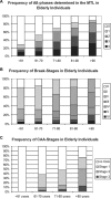
(A) The frequency of patients with Aβ-deposits increases with age. Only 11% of the patients older than 90 years of age were free of Aβ-deposits in a sample of 506 autopsy cases. Accordingly, the prevalence of higher phases of Aβ-deposition as observed in the medial temporal lobe [18] also increases with age. (B) Similar to the deposition of Aβ NFTs occur in most individuals older than 90 years. In our sample, there was no one free of NFTs at this age. The percentage of cases with NFTs in cases younger than 71 years of age was strikingly higher than that of those with Aβ-plaques. This result is in line with previously published samples [48]. (C) In parallel with the increasing frequency of Aβ-deposits and NFTs in elderly people CAA occurs more often in advanced ages and the prevalence of higher stages increases when compared with younger age groups. This is demonstrated in a sample of 88 autopsy cases (reproduced with kind permission from [83]).
Fig 5
Fig 5
Prevalence of CAA and its subtypes in AD and age-matched non-demented control cases. In non-demented controls, most individuals were free of CAA (60.4%). Controls with CAA most frequently exhibited CAA-type 2 lacking capillary involvement (28.2%). Only 11.4% of the controls showed capillary CAA (CAA-type 1). On the other hand, capillary CAA (CAA-type 1) was found in 51% of the AD cases. Only 40% of the AD cases exhibited CAA-type 2 and 9% were free of CAA. These data confirm that capillary CAA is frequent in AD [96].

References

    1. Alzheimer A. Ueber eine eigenartige Erkrankung der Hirnrinde. Allg Zschr Psych. 1907;64:146–8.
    1. DeKosky ST, Scheff SW. Synapse loss in frontal cortex biopsies in Alzheimer's disease: correlation with cognitive severity. Ann Neurol. 1990;27:457–64. - PubMed
    1. Masliah E, Mallory M, Hansen L, DeTeresa R, Alford M, Terry R. Synaptic and neuritic alterations during the progression of Alzheimer's disease. Neurosci Lett. 1994;174:67–72. - PubMed
    1. Terry RD, Peck A, DeTeresa R, Schechter R, Horoupian DS. Some morphometric aspects of the brain in senile dementia of the Alzheimer type. Ann Neurol. 1981;10:184–92. - PubMed
    1. Duyckaerts C, Dickson DW. Neuro -pathology of Alzheimer's disease. In: Dickson D, editor. Neurodegeneration: the molecular pathology of dementia and movement disorders. ISN Neuropath Press; 2003. pp. 47–65. In:, editor.. Basel:.

Publication types

Substances