. 2008 Sep 12;381(4):1012-24.
doi: 10.1016/j.jmb.2008.06.082. Epub 2008 Jul 3.

The crystal structure of CHIR-AB1: a primordial avian classical Fc receptor

Affiliations

The crystal structure of CHIR-AB1: a primordial avian classical Fc receptor

Tal I Arnon et al. J Mol Biol. .

Abstract

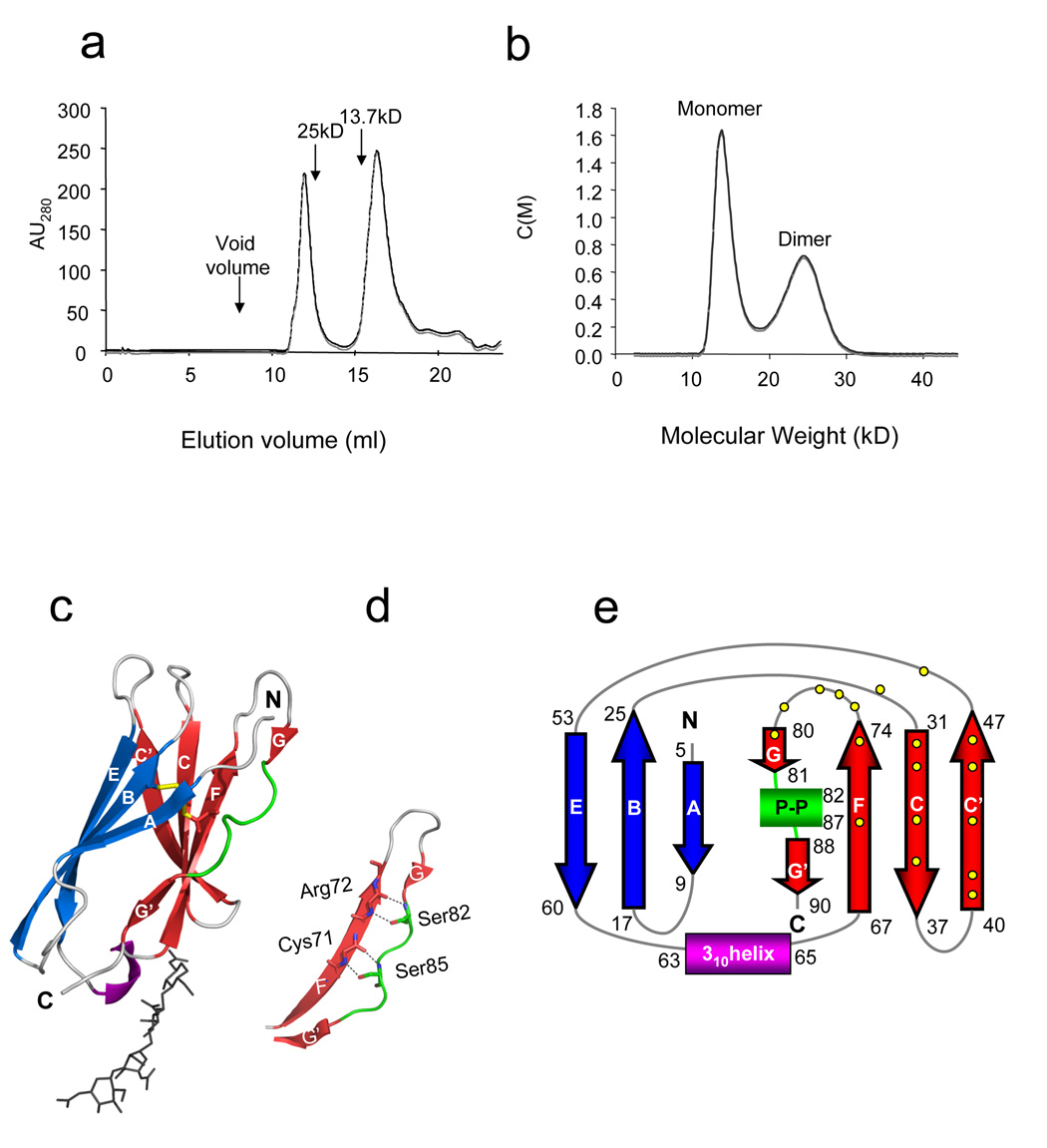
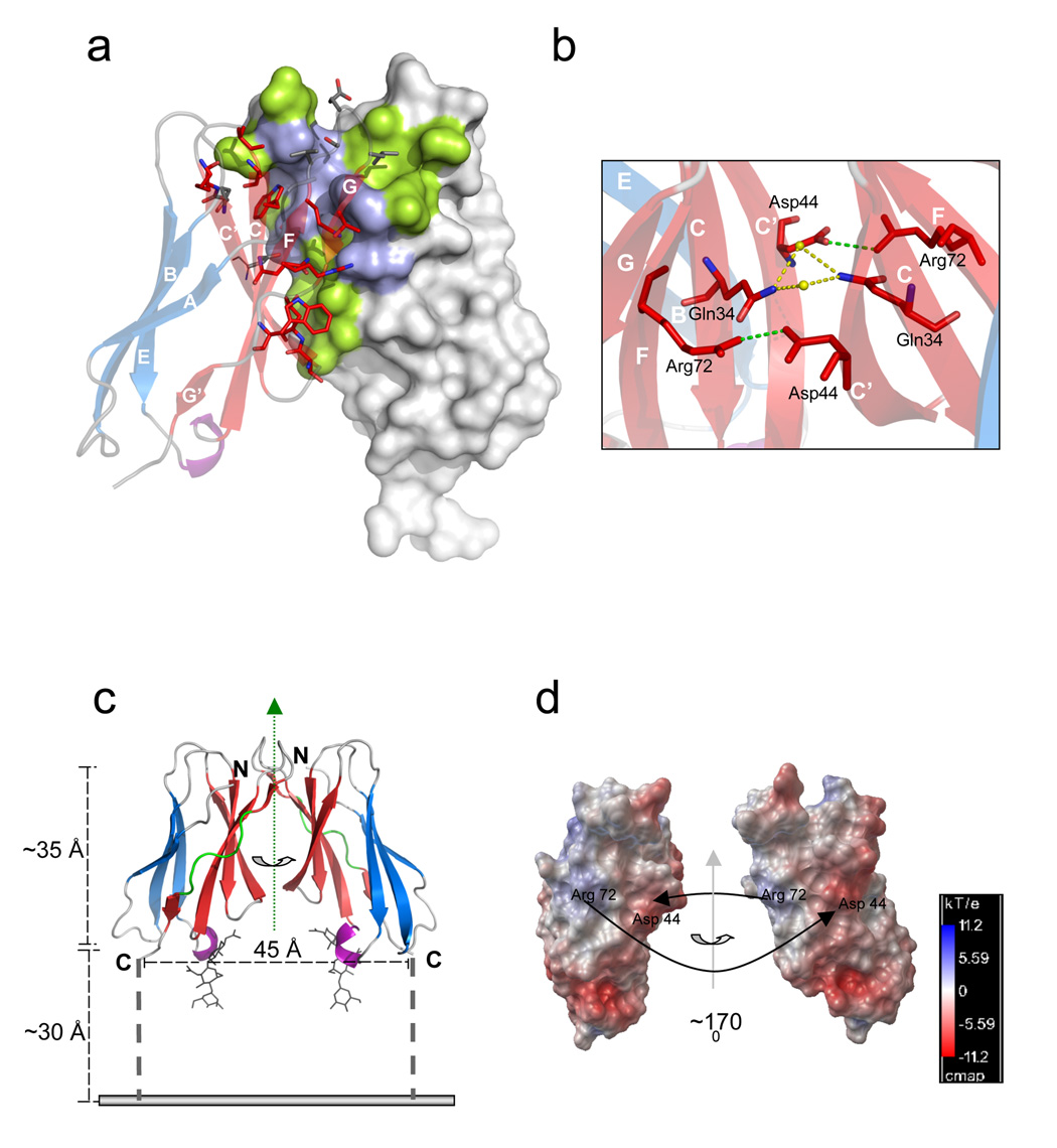
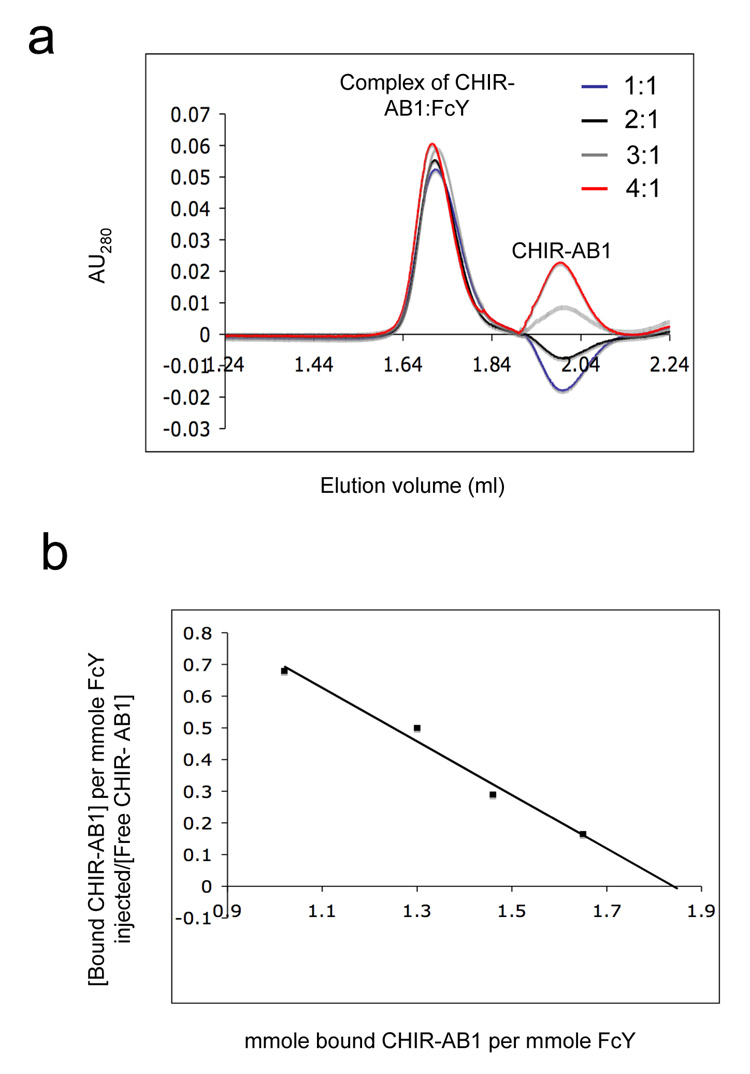
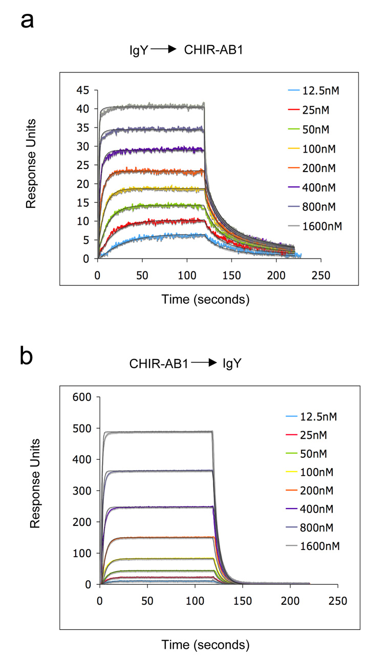
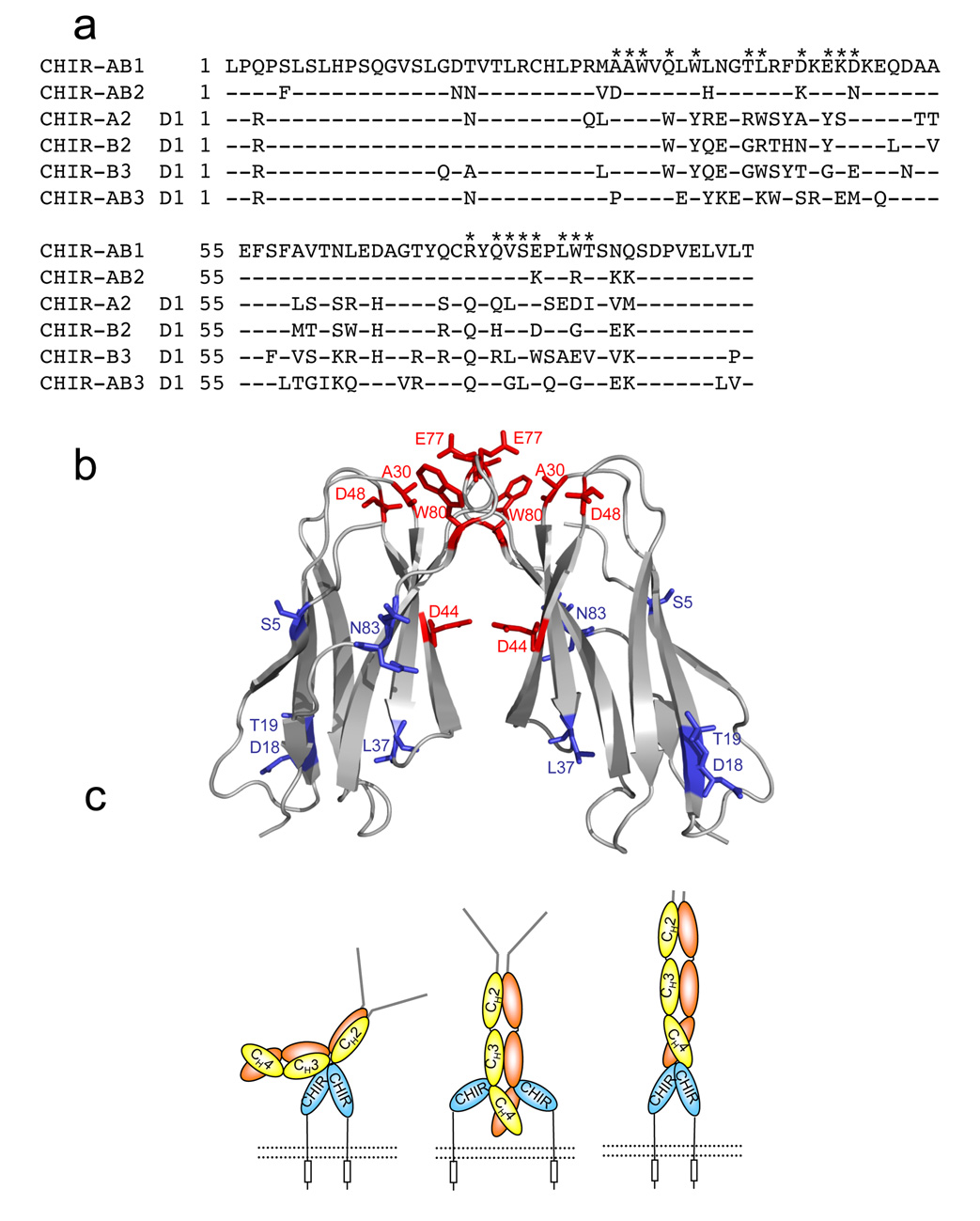
CHIR-AB1 is a newly identified avian immunoglobulin (Ig) receptor that includes both activating and inhibitory motifs and was therefore classified as a potentially bifunctional receptor. Recently, CHIR-AB1 was shown to bind the Fc region of chicken IgY and to induce calcium mobilization via association with the common gamma-chain, a subunit that transmits signals upon ligation of many different immunoreceptors. Here we describe the 1.8-A-resolution crystal structure of the CHIR-AB1 ectodomain. The receptor ectodomain consists of a single C2-type Ig domain resembling the Ig-like domains found in mammalian Fc receptors such as FcgammaRs and FcalphaRI. Unlike these receptors and other monomeric Ig superfamily members, CHIR-AB1 crystallized as a 2-fold symmetrical homodimer that bears no resemblance to variable or constant region dimers in an antibody. Analytical ultracentrifugation demonstrated that CHIR-AB1 exists as a mixture of monomers and dimers in solution, and equilibrium gel filtration revealed a 2:1 receptor/ligand binding stoichiometry. Measurement of the 1:1 CHIR-AB1/IgY interaction affinity indicates a relatively low affinity complex, but a 2:1 CHIR-AB1/IgY interaction allows an increase in apparent affinity due to avidity effects when the receptor is tethered to a surface. Taken together, these results add to the structural understanding of Fc receptors and their functional mechanisms.


Figures

Figure 1
Figure 1. Structure of CHIR-AB1
(a). Elution profile of purified CHIR-AB1 on a S200 Superdex gel filtration column. The elution volumes of molecular weight standards are indicated by arrows. (b) Velocity sedimentation analytical ultracentrifugation analysis of purified CHIR-AB1 showing the existence of monomeric (14.3kD) and dimeric (25.0kD) forms. Data are displayed at the 95% confidence level. (c) Ribbon diagram of the CHIR-AB1 structure. β-strands A, B, and E are shown in blue, strands CC’FGG’ in red, a 310 helix in purple and a polyproline II helix in green. The disulfide bond is yellow and carbohydrate residues are shown in stick representation. (d) Close-up view of the region of the polyproline II helix showing hydrogen bonds between the side chains of Ser 82 and Ser 85 and main chain atoms of residues Arg72 and Cys71. Side chains are shown as sticks using atom-based color code (oxygen, red; nitrogen, blue). (e) Topology diagram of CHIR-AB1. β-strands, 310 helix and the polyproline II helix (P-P) are colored as above. Contact resides involved in dimerization (Figure 2 and Table 2) are represented by yellow dots.
Figure 2
Figure 2. The dimer interface
(a) Dimerization interface region with one subunit as a surface representation (hydrophilic contact residues in light blue and hydrophobic contact residues in green) and the other subunit as transparent ribbons diagram colored as in Figure 1b (β-strands A, B, and E are shown in blue, strands CC’FGG’ in red, a 310 helix in purple and loops are colored grey). Strands are labeled in white and contact residues are shown as sticks. (b) Cross section of the dimer interface illustrating the two symmetrical inter-subunit salt bridges (green lines) between Asp44 and Arg72. Water molecules (yellow spheres) that mediate a hydrogen bond network (yellow dotted line) between the Gln34 residues of each subunit are also shown. Strands are labeled in white and colored as above (c) Proposed orientation of a CHIR-AB1 dimer on a membrane (grey line). Monomers are colored as in Figure 1b. The two-fold symmetry axis relating the monomers is indicated by a vertical green dotted arrow in the plane of the page. The distance between the C-terminus of each ectodomain (45Å) and the approximate length of the ectodomain (~35Å) are indicated (black dashed lines). The 8-residue stem region connecting each ectodomain subunit to the transmembrane region is shown as a broken black bar with a maximum theoretical length of ~30Å (calculated assuming 3.8Å per residue). (d) Molecular surfaces of the two subunits in a CHIR-AB1 dimer with colors highlighting the electrostatic potential calculated with APBS tools . Electrostatic potential is plotted from −11.2 kT/e (electronegative; red) to +11.2 kT/e (electropositive; blue) with white indicating electroneutrality. The right monomer is related to the left monomer by a rotation about the indicated axis of ~170°. Black arrows point toward contact partner residues.
Figure 3
Figure 3. CHIR-AB1-FcY binding stoichiometry
(a) Equilibrium gel filtration analysis of the CHIR-AB1-FcY complex. The column was equilibrated with and run in a buffer containing 2.5 µM CHIR-AB1. Complexes were prepared by incubating 2.5 µM FcY with varying amounts of CHIR-AB1 at receptor:FcY molar ratios of 1:1, 2:1, 3:1 and 4:1. Similar experiments were conducted using columns equilibrated with 1.5µM, 5µM and 10 µM of CHIR-AB1 (data not shown). (b) Scatchard plot including data from equilibration buffers containing 1.5µM, 2.5µM, 5µM and 10µM CHIR-AB1. The best fit line to the data yields a KD of 840 nM and an x-intercept of 1.83.
Figure 4
Figure 4. Biosensor analyses of CHIR-AB1/IgY interactions
Representative sensorgrams describing the binding response of increasing concentrations of IgY injected over immobilized CHIR-AB1 (58RUs) (a) or CHIR-AB1 injected over immobilized IgY (2000RUs) (b). Colored lines show the observed response overlaid with the calculated response (black line) based on a 2:1 binding model. Fitting of the same data with a 1:1 binding model resulted with KD values of 17 ± 9nM (a) and 800nM (b). Similar results were obtained when CHIR-AB1 was coupled to the chip at different densities (140RU, 320RU and 560RU; data not shown).
Figure 5
Figure 5. Models for the CHIR-AB1/FcY interaction
(a) Sequence alignment of the extracellular domains of CHIR-AB1 and CHIR AB2 with the D1 domains of CHIR-A2, CHIR-B2, and CHIR-AB3 (GenBank accession numbers AJ745094, AJ745095, AJ745093, AJ639837 and AJ879909, respectively). Residues at the dimer interface of CHIR-AB1 are indicated by asterisks above the sequence. (b) Ribbon diagram of a CHIR-AB1 dimer with residues that differ from CHIR-AB2 highlighted as red (dimer interface residues) or blue (all others) sticks. The highlighted amino acids are labeled (c) Potential models for binding between CHIR-AB1 and IgY. The CHIR-AB1 ectodomain is blue, the ITIM motif in the cytoplasmic tail is represented by a rectangle, and the cell membrane is shown as a dotted black line. IgY is shown with a yellow and orange Fc region and grey Fabs. (Left) A dimer of CHIR-AB1 is bound asymmetrically to the lower hinge region between the CH2 and CH3 domains of FcY, analogous to the binding of FcγRs and FcεRI to Fcs. (Middle) CHIR-AB1 monomers bind to the CH3-CH4 interdomain interface to create a symmetrical 2:1 complex, analogous to the binding of FcαRI to Fc. (Right) The two-fold symmetry axis of a CHIR-AB1 dimer aligns with the two-fold symmetry axis of FcY to form a symmetric 2:1 complex in which each CHIR-AB1 monomer binds to the bottom of a FcY CH4 domain.

References

    1. Nimmerjahn F, Ravetch JV. Fc-receptors as regulators of immunity. Adv Immunol. 2007;96:179–204. - PubMed
    1. Nimmerjahn F, Ravetch JV. The antiinflammatory activity of IgG: the intravenous IgG paradox. J Exp Med. 2007;204:11–15. - PMC - PubMed
    1. Nimmerjahn F, Ravetch JV. Fcgamma receptors as regulators of immune responses. Nat Rev Immunol. 2008;8:34–47. - PubMed
    1. Woof JM, Burton DR. Human antibody-Fc receptor interactions illuminated by crystal structures. Nat Rev Immunol. 2004;4:89–99. - PubMed
    1. Garman SC, Kinet JP, Jardetzky TS. Crystal structure of the human high-affinity IgE receptor. Cell. 1998;95:951–961. - PubMed

Publication types

Associated data