Review
. 2009;56 Suppl 1(Suppl 1):149-59.
doi: 10.1016/j.neuropharm.2008.06.028. Epub 2008 Jun 25.

Dissecting motivational circuitry to understand substance abuse

Affiliations
Review

Dissecting motivational circuitry to understand substance abuse

Robert A Wheeler et al. Neuropharmacology. 2009.

Abstract

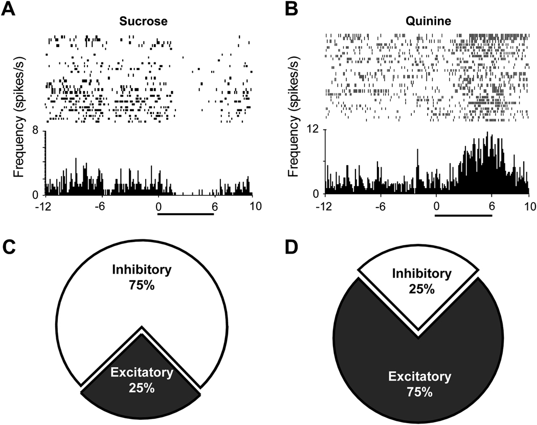
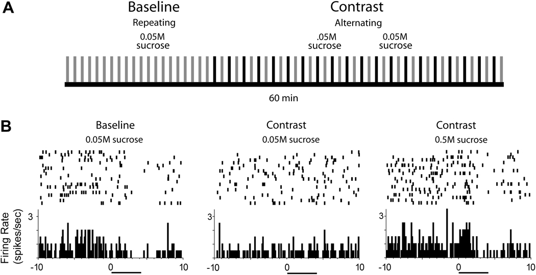
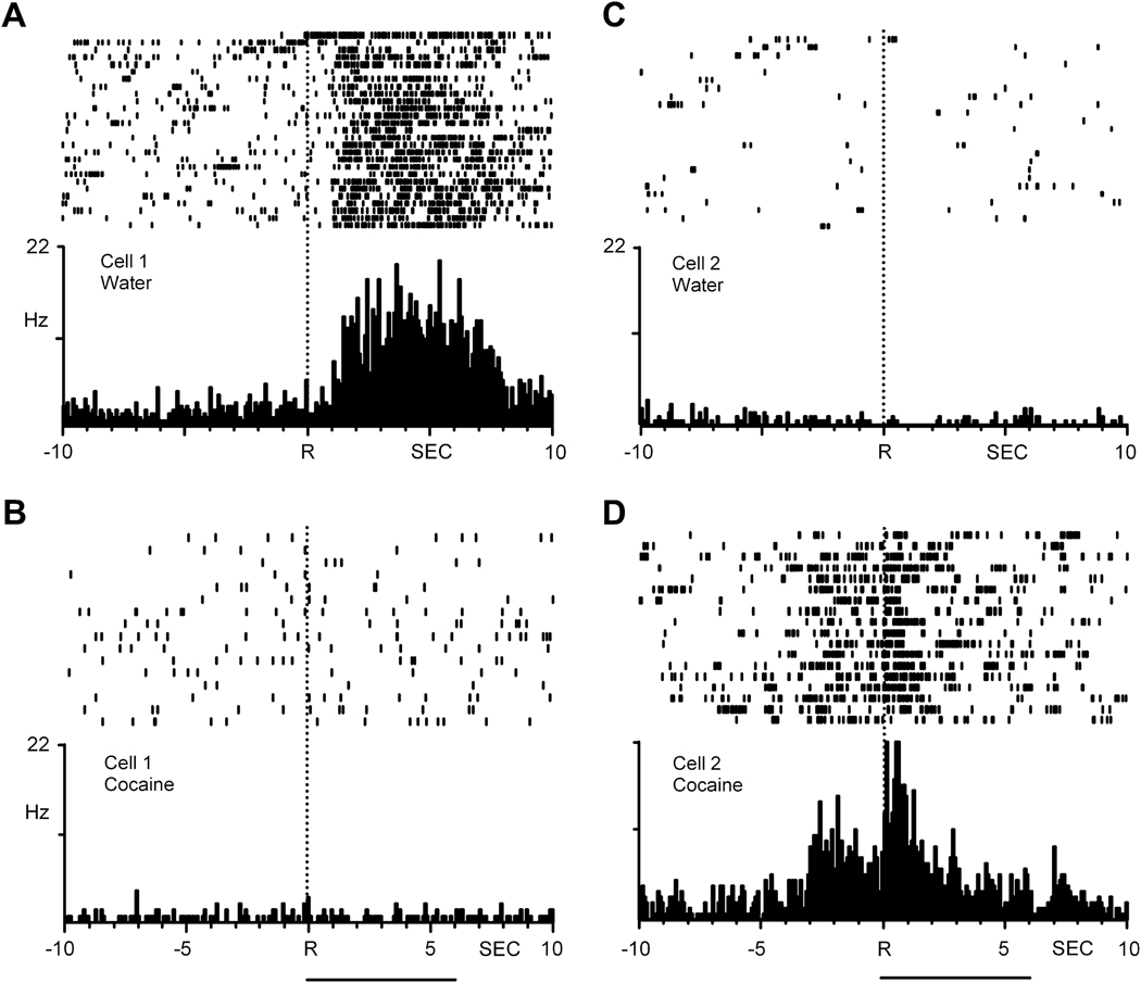
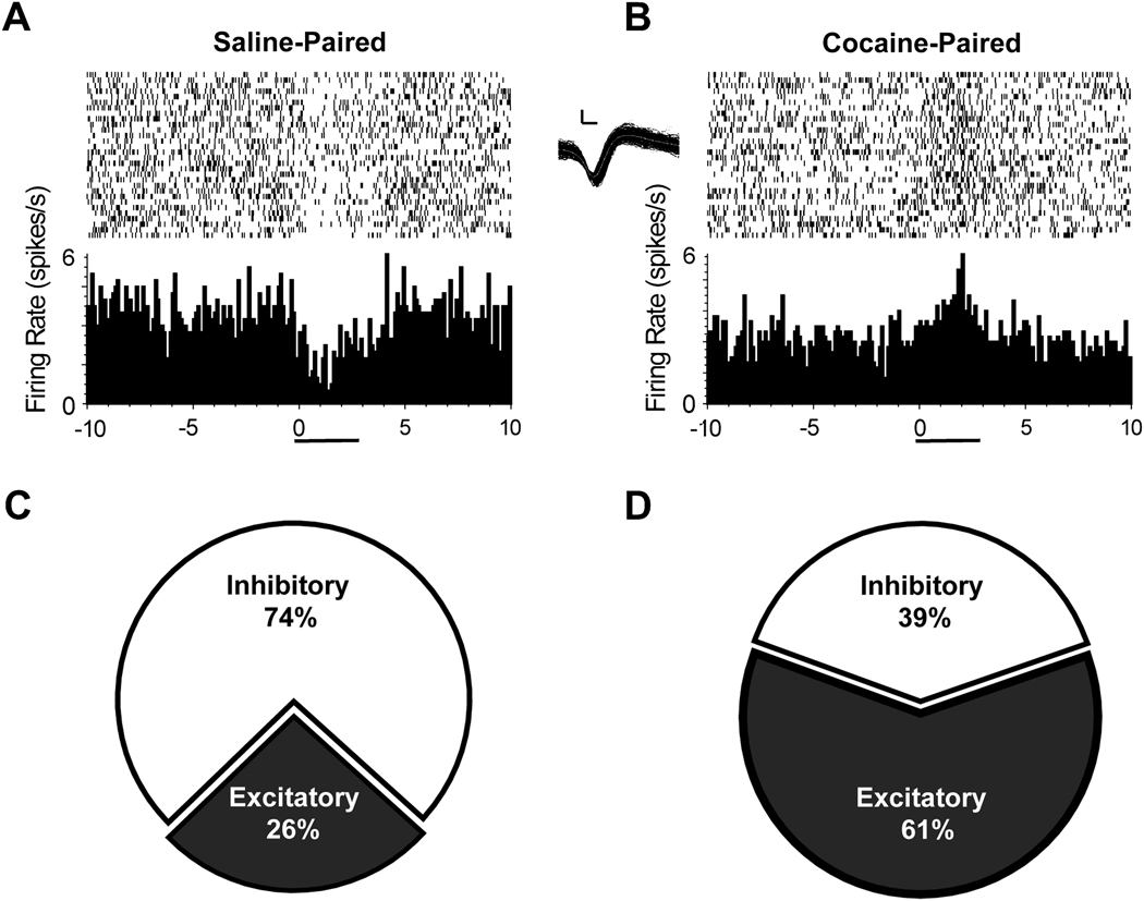
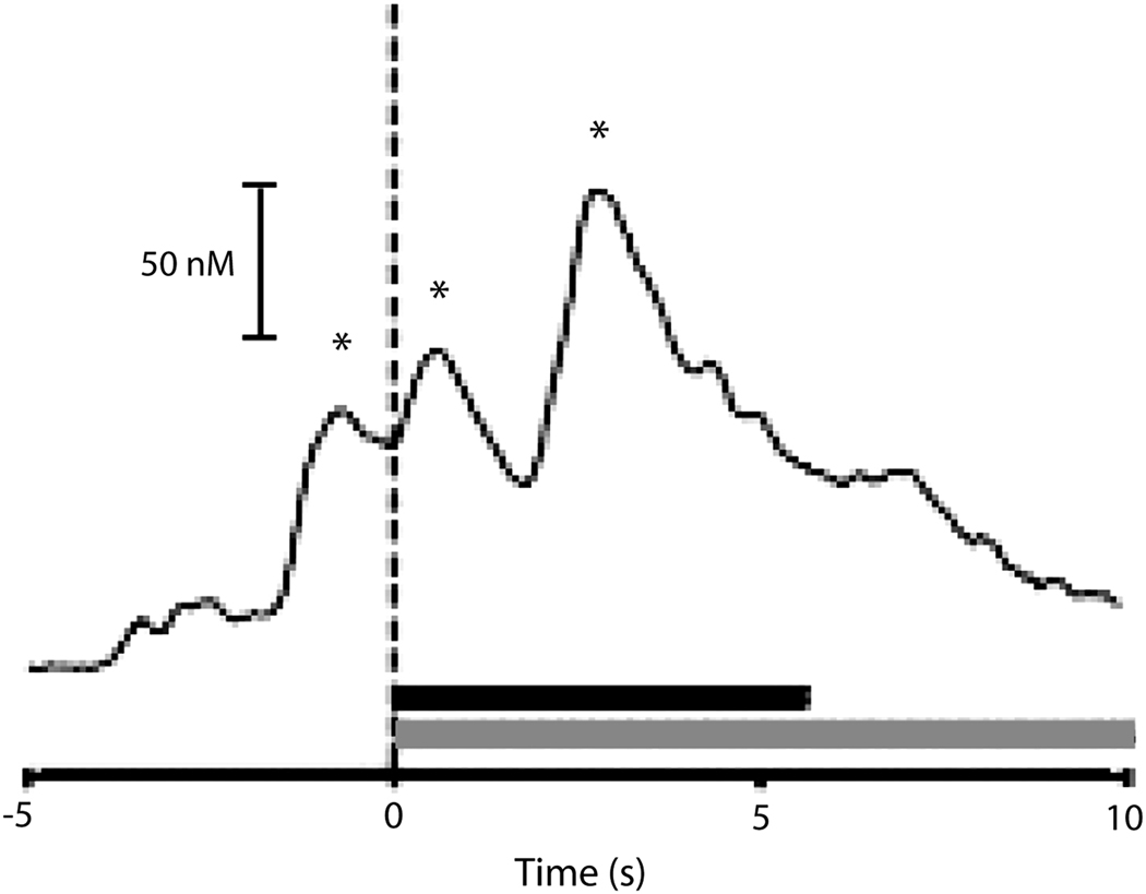
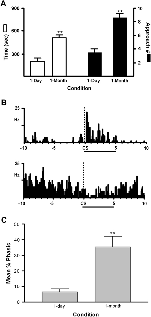
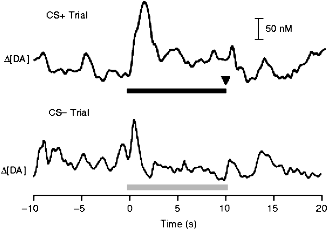
An important goal of cocaine addiction research is to understand the neurobiological mechanisms underlying this disease state. Here, we review studies from our laboratory that examined nucleus accumbens (NAc) cell firing and rapid dopamine signaling using electrophysiological and electrochemical recordings in behaving rodents. A major advantage of these techniques is that they allow for the characterization of NAc activity and rapid dopamine release during specific phases of motivated behavior. Moreover, each approach enables an examination of the dynamic nature of NAc signaling as a function of factors such as hedonics and associative learning. We show that NAc neurons differentially respond to rewarding and aversive stimuli and their predictors in a bivalent manner. This differential responding is modifiable and can be altered by the presentation of other natural rewards or cocaine. Likewise, the dynamic nature of NAc cell firing is also reflected in the differential activation of distinct populations of NAc neurons during goal-directed behaviors for natural versus drug rewards, and the heightened activation of some NAc neurons following cocaine abstinence. Our electrochemical data also show that rapid dopamine signaling in the NAc reflects primary rewards and their predictors and appears to modulate specific NAc neuronal responses. In some cases, these influences are observed in a regionally specific manner that matches previous pharmacological manipulations. Collectively, these findings provide critical insight into the functional organization of the NAc that can be used to guide additional studies aimed at dissecting the neural code underlying compulsive drug-seeking behavior.


Figures

Figure 1
Figure 1
NAc neurons exhibit opposite firing patterns during intraoral infusion of rewarding versus aversive tastants. Top: The activity of a representative NAc cell that exhibited a decrease in firing rate during infusion of a sweet sucrose solution (A), and another NAc neuron that showed a clear increase in activity during intraoral infusions of a bitter quinine solution (B). Pump onset at time 0 in PEH; horizontal black bar indicates intraoral infusion duration. The piecharts show the percentage of all recorded NAc neurons that displayed either excitatory or inhibitory responses to intraoral infusions of sucrose (C) or quinine (D). Figure modified from Roitman et al. (2005) with permission from Elsevier.
Figure 2
Figure 2
Dynamic modulation of NAc cell firing during reward comparison involving two natural rewards. A. Design of the behavioral task. A low concentration of sucrose (0.05 M) was repeatedly delivered via intraoral infusion during the baseline period. Next, the same concentration of sucrose was delivered, but in alternation with a higher, more palatable solution (0.5 M). Each tick mark represents an infusion of the low (grey) or high sucrose concentration (black). B. The baseline response of one cell to infusions of the 0.05 M sucrose was a pronounced reduction in firing rate (left). However, the same cell exhibited no change in activity to the same sucrose solution (middle) when given in an alternating manner with the more palatable solution (right). Time 0 indicates infusion onset; horizontal black bar below each PEH indicates the duration of the intraoral infusion.
Figure 3
Figure 3
NAc neurons exhibit differential, nonoverlapping firing patterns during goal-directed behaviors for natural (water) versus cocaine reward. Left: PEHs show the activity of a single neuron that exhibited increases in firing rate immediately following lever press responding for water reinforcement (A) but no changes in activity (i.e., nonphasic firing) relative to the reinforced response for intravenous cocaine during the self-administration phase of the multiple schedule. (B). Right: Another NAc neuron recorded in a second animal exhibited nonphasic firing during the water-reinforcement phase and a shift to patterned firing during self-administration. Drug infusion indicated by horizontal lines below bottom PEHs. R is reinforced response. Figure modified from Carelli et al. (2003). Copyright 2003 by the Society for Neuroscience.
Figure 4
Figure 4
NAc neurons track a learned aversion to saccharin following its repeated pairing with cocaine. Top: A: PEH of a representative NAc neuron that exhibited a decrease in firing rate during intraoral infusion of a distinctly flavored saccharin infusion that predicted availability to self-administer saline. B: The same neuron shifted its firing profile to an excitatory response during infusion of a differently flavored saccharin solution of the same concentration that was repeatedly paired with cocaine self-administration. (Inset) Scale calibration: 26 mV, 100 ms. Bottom; The piecharts show that the majority of NAc neurons exhibited inhibitory responses to the tastant paired with saline self-administration (C), but shifted to predominately excitatory activity during infusion of the tastant paired with cocaine self-administration (D). Figure modified from Wheeler et al. (2008) with permission from Elsevier.
Figure 5
Figure 5
Rapid dopamine release is evident within seconds of reinforced responding for cocaine during self-administration sessions. Dopamine significantly increased within seconds preceding and immediately following the cocaine reinforced lever press response (dashed line, time 0). One representative trial shown. Black bar indicates the duration of cocaine infusion (0.33 mg/inf); shaded bar indicates the presentation of the drug-associated cues. Asterisks indicate confirmed dopamine events. Figure modified from Stuber et al. (2005) with permission from Elsevier.
Figure 6
Figure 6
One-month abstinence from cocaine self-administration increases the percentage of NAc neurons that respond to cocaine-associated cues (CS). A. CS-induced behavioral activation (time in, and number of approaches to, cocaine-associated lever quadrant). B. PEHs show examples of representative neurons exhibiting excitatory (top) or inhibitory (bottom) responses to the CS (5 s duration; onset indicated by dashed line labeled ‘CS’ in each PEH). C. Percentage of NAc neurons that were significantly activated by the CS for 1 d and 1 month cocaine abstinent rats. **p < 0.01. Figure from Hollander et al. (2007). Copyright 2007 by the Society for Neuroscience.
Figure 7
Figure 7
After training, rapid dopamine release events in the NAc shift from the onset of primary (sucrose) reward to conditioned stimuli (CS) associated with reward delivery. Top: Representative changes in dopamine signaling during a trial in which the CS+ (top) signaled reward delivery (at inverted arrow). The horizontal black bar indicates the duration of the CS+. Bottom: Smaller dopamine release event during a trial in which the CS− signaled no reward delivery. The horizontal grey bar indicates the duration of the CS−. Figure modified from Day et al. (2007) with permission from Nature Publishing Group.
Figure 8
Figure 8
Combined electrophysiological and electrochemical measurements reveal similar temporal dynamics of rapid dopamine release and NAc cell firing during intracranial self-stimulation. The overlaid trace (grey) shows the time course of extracellular dopamine concentration changes simultaneously measured at the same location where a NAc neuron was recorded. Note that changes in rapid dopamine release events occurred during the period at which the NAc neuron exhibited a phasic inhibition in cell firing. Cue onset at time 0; lever extension at 1s; gray symbols in raster represent lever presses; stimulation delivery indicated by symbols under double arrow in raster display. Figure modified from Cheer et al. (2008) with permission from Elsevier.

References

    1. Ahmed SH, Kenny PJ, Koob GF, Markou A. Neurobiological evidence for hedonic allostasis associated with escalating cocaine use. Nat Neurosci. 2002;5:625–626. - PubMed
    1. Ahmed SH, Koob GF. Transition from moderate to excessive drug intake: change in hedonic set point. Science. 1998;282:298–300. - PubMed
    1. Arbuthnott GW, Wickens J. Space, time and dopamine. Trends Neurosci. 2007;30:62–69. - PubMed
    1. Avena NM, Bocarsly ME, Rada P, Kim A, Hoebel BG. After daily bingeing on a sucrose solution, food deprivation induces anxiety and accumbens dopamine/acetylcholine imbalance. Physiol Behav. 2008 - PMC - PubMed
    1. Baldo BA, Kelley AE. Discrete neurochemical coding of distinguishable motivational processes: insights from nucleus accumbens control of feeding. Psychopharmacology (Berl) 2007;191:439–459. - PubMed

Publication types