. 2008 Sep 12;283(37):25364-25371.
doi: 10.1074/jbc.M804966200. Epub 2008 Jul 14.

cAMP-dependent protein kinase A (PKA) signaling induces TNFR1 exosome-like vesicle release via anchoring of PKA regulatory subunit RIIbeta to BIG2

Affiliations

cAMP-dependent protein kinase A (PKA) signaling induces TNFR1 exosome-like vesicle release via anchoring of PKA regulatory subunit RIIbeta to BIG2

Aminul Islam et al. J Biol Chem. .

Abstract

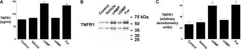
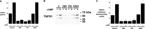
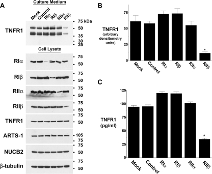
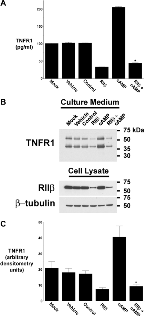
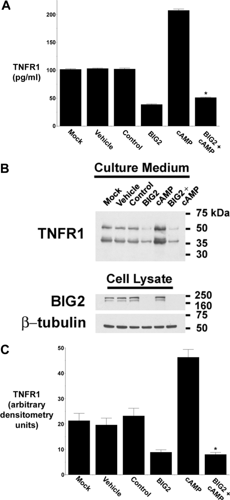
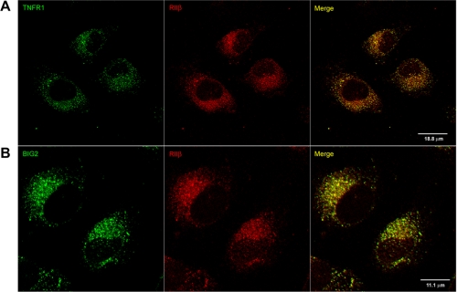
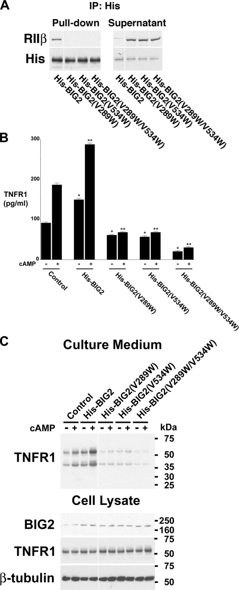
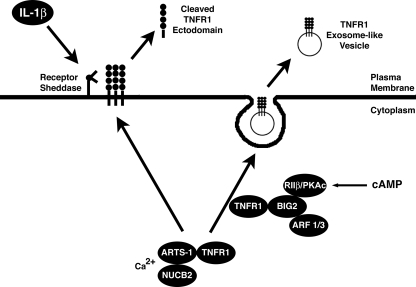
The 55-kDa TNFR1 (type I tumor necrosis factor receptor) can be released to the extracellular space by two mechanisms, the proteolytic cleavage and shedding of soluble receptor ectodomains and the release of full-length receptors within exosome-like vesicles. We have shown that the brefeldin A-inhibited guanine nucleotide exchange protein BIG2 associates with TNFR1 and selectively modulates the release of TNFR1 exosome-like vesicles via an ARF1- and ARF3-dependent mechanism. Here, we assessed the role of BIG2 A kinase-anchoring protein (AKAP) domains in the regulation of TNFR1 exosome-like vesicle release from human vascular endothelial cells. We show that 8-bromo-cyclic AMP induced the release of full-length, 55-kDa TNFR1 within exosome-like vesicles via a protein kinase A (PKA)-dependent mechanism. Using RNA interference to decrease specifically the levels of individual PKA regulatory subunits, we demonstrate that RIIbeta modulates both the constitutive and cAMP-induced release of TNFR1 exosome-like vesicles. Consistent with its AKAP function, BIG2 was required for the cAMP-induced PKA-dependent release of TNFR1 exosome-like vesicles via a mechanism that involved the binding of RIIbeta to BIG2 AKAP domains B and C. We conclude that both the constitutive and cAMP-induced release of TNFR1 exosome-like vesicles occur via PKA-dependent pathways that are regulated by the anchoring of RIIbeta to BIG2 via AKAP domains B and C. Thus, BIG2 regulates TNFR1 exosome-like vesicle release by two distinct mechanisms, as a guanine nucleotide exchange protein that activates class I ADP-ribosylation factors and as an AKAP for RIIbeta that localizes PKA signaling within cellular TNFR1 trafficking pathways.


Figures

FIGURE 1.
FIGURE 1.
Effect of cAMP and forskolin on the release of TNFR1 exosome-like vesicles. HUVEC were incubated without or with 1 mm 8-Br-cAMP (cAMP), 1 mm 8-Br-cGMP (cGMP), or 50 μm forskolin (For) for 24 h. A, TNFR1 concentration in medium quantified by ELISA. *, significant difference (n = 6) between cells treated with vehicle (DMSO) and those treated with 8-Br-cAMP (p < 10-12) or forskolin (p < 10-7). B, Western blot of TNFR1 in medium from one of five experiments that demonstrated similar results. C, densitometry was performed on the Western blots from B, and the quantity of the 55-kDa TNFR1 that was released into conditioned medium is presented as arbitrary densitometry units. *, significant difference (n = 5) between cells treated with vehicle and those treated with 8-Br-cAMP (p < 0.006) or forskolin (p < 0.003).
FIGURE 2.
FIGURE 2.
Constitutive and cAMP-mediated TNFR1 exosome-like vesicle release are PKA-dependent. HUVEC were incubated for 24 h without or with 1 mm 8-Br-cAMP (cAMP) and 10 μm H-89, 50 μm Myr-PKI, or 50 μm ERKI. A, TNFR1 concentration in medium quantified by ELISA. *, significant reduction (n = 6) in constitutive release of TNFR1 between cells treated with vehicle (DMSO) and those treated with H-89 (p < 10-9) or Myr-PKI (p < 10-9). **, significant reduction (n = 6) in cAMP-mediated release of TNFR1 from cells treated with vehicle (DMSO) and those treated with H-89 (p < 10-14) or Myr-PKI (p < 10-14). B, Western blot of TNFR1 in medium from one of five experiments that demonstrated similar results. C, densitometry was performed on the Western blots from B, and the quantity of the 55-kDa TNFR1 that was released into conditioned medium is presented as arbitrary densitometry units. *, significant reduction (n = 5) in the constitutive release of TNFR1 into culture medium from cells treated with vehicle (DMSO) and those treated with H-89 (p < 0.015) or Myr-PKI (p < 0.015). **, significant reduction (n = 6) in cAMP-mediated release of TNFR1 from cells treated with vehicle (DMSO) and those treated with H-89 (p < 10-3) or Myr-PKI (p < 0.004).
FIGURE 3.
FIGURE 3.
The constitutive release of TNFR1 exosome-like vesicles is regulated by PKA RIIβ. HUVEC were transfected with vehicle (Mock); 100 nm control, nontargeting siRNA (Control); or 100 nm siRNA targeting RIα, RIβ, RIIα, or RIIβ for 3 days prior to the addition of fresh, exosome-depleted medium for 24 h. A, representative Western blot, from one of three experiments, showing TNFR1 in medium and cell lysates and showing RIα, RIβ, RIIα, RIIβ, ARTS-1, NUCB2, and β-tubulin in cell lysates. B, densitometry was performed on the Western blots from A, and the quantity of the 55-kDa TNFR1 that was constitutively released into conditioned medium is presented as arbitrary densitometry units. *, a significant decrease in the quantity of the 55-kDa TNFR1 present in conditioned medium from cells transfected with siRNA targeting RIIβ as compared with those transfected with control, nontargeting siRNA (p < 0.002, n = 3). C, TNFR1 in medium was quantified by ELISA. *, significant difference from cells transfected with siRNA targeting RIIβ as compared with those transfected with control, nontargeting siRNA (p < 10-7, n = 6).
FIGURE 4.
FIGURE 4.
cAMP-mediated TNFR1 exosome-like vesicle release is regulated by PKA RIIβ. HUVEC were transfected with vehicle (Mock), DMSO (Vehicle); 100 nm control, nontargeting siRNA (Control); or 100 nm siRNA targeting RIIβ, without or with 1 mm cAMP for 3 days prior to the addition of fresh, exosome-depleted medium for 24 h. A, TNFR1 concentration in medium was quantified by ELISA. *, significant reduction in the quantity of TNFR1 present in medium from cells treated with cAMP and siRNA targeting RIIβ as compared with those treated with cAMP alone (p < 10-9, n = 6). B, representative Western blot, from one of five experiments, showing TNFR1 in medium and cell lysates, and RIIβ and β-tubulin in cell lysates. C, densitometry was performed on the Western blots from B, and the quantity of the 55-kDa TNFR1 that was constitutively released into conditioned medium is presented as arbitrary densitometry units. *, significant decrease in the quantity of the 55-kDa TNFR1 present in conditioned medium from those transfected with siRNA targeting RIIβ as compared with cells treated with cAMP alone (p < 0.003, n = 5).
FIGURE 5.
FIGURE 5.
cAMP-mediated TNFR1 exosome-like vesicle release requires BIG2. HUVEC were transfected with vehicle (Mock); DMSO (Vehicle), 100 nm control, nontargeting siRNA (Control); or 100 nm siRNA targeting BIG2, without or with 1 mm cAMP for 3 days prior to the addition of fresh, exosome-depleted medium for 24 h. A, TNFR1 concentration in medium was quantified by ELISA. *, significant reduction in the quantity of TNFR1 present in medium from cells treated with cAMP and siRNA targeting BIG2 as compared with those treated with cAMP alone (p < 10-13, n = 6). B, representative Western blot, from one of three experiments, showing TNFR1 in medium and cell lysates and showing BIG2 and β-tubulin in cell lysates. C, densitometry was performed on the Western blots from B, and the quantity of the 55-kDa TNFR1 that was released into conditioned medium is presented as arbitrary densitometry units. *, significant decrease in the quantity of the 55-kDa TNFR1 present in conditioned medium from cells transfected with siRNA targeting BIG2 as compared with those treated with cAMP alone (p = 0.006, n = 3).
FIGURE 6.
FIGURE 6.
Constitutive association between PKA RIIβ and TNFR1 in HUVEC. HUVEC were grown on collagen I-coated slides and reacted with antibodies against TNFR1 and RIIβ (A) or BIG2 and RIIβ (B) and secondary antibodies conjugated with Alexa Fluor 488 (green) or Alexa Fluor 568 (red), respectively, before immunofluorescence confocal laser-scanning microscopy.
FIGURE 7.
FIGURE 7.
Constitutive and cAMP-mediated TNFR1 exosome-like vesicle release requires the anchoring of RIIβ to BIG2 AKAP domains B and C. HUVEC were transfected with plasmids encoding either His-tagged wild-type BIG2 (His-BIG2) or BIG2 mutants that contained single amino acid substitutions in AKAP domain B (His-BIG2(V289W)), domain C (His-BIG2(V534W)), or domains B and C (His-BIG2(V289W/V534W)) or empty plasmid (Control), for 2 days before the addition of fresh, exosome-depleted medium for 24 h, without or with 1 mm 8-Br-cAMP (cAMP). A, proteins immunoprecipitated (IP) with the anti-His antibody (Pull-down) or remaining in the supernatant were immunoblotted with antibodies against RIIβ or the His tag. This blot is representative of three individual experiments. B, TNFR1 concentration in medium was quantified by ELISA. *, significant increase (His-BIG2, p < 10-3) or decrease (His-BIG2(V289W), p < 10-3; His-BIG2(V534W), p < 0.002; His-BIG2(V289W/V534W), p < 10-3) in the quantity of TNFR1 constitutively released into the medium as compared with control cells transfected with the empty plasmid (n = 6). **, significant increase (His-BIG2, p < 0.012) or decrease (His-BIG2(V289W), p < 10-4; His-BIG2(V534W), p < 10-4; His-BIG2(V289W/V534W), p < 10-4) in the quantity of TNFR1 released into the medium following stimulation with 1 mm 8-Br-cAMP for 24 h as compared with control cells transfected with the empty plasmid (n = 6). The quantity of constitutive and 8-Br-cAMP-mediated TNFR1 release from cells transfected with the domain B/C double mutant (His-BIG2(V289W/V534W)) was significantly decreased as compared with those transfected with His-BIG2(V289W) (p < 10-3) or His-BIG2(V534W) (p < 10-3), respectively (n = 6). C, representative Western blot, from one of three experiments, showing TNFR1 in medium and cell lysates, and BIG2 and β-tubulin in cell lysates. Images were grouped from adjacent parts of the same gel in A and C.
FIGURE 8.
FIGURE 8.
Proposed model by which cAMP-dependent PKA signaling induces TNFR1 exosome-like vesicle release via anchoring of PKA regulatory subunit RIIβ to BIG2. The calcium-dependent intracellular NUCB2-ARTS-1 complex associates with TNFR1 before divergence of the pathways that lead either to the inducible proteolytic cleavage of TNFR1 ectodomains, shown here in response to IL-1β stimulation, or the constitutive release of TNFR1 exosome-like vesicles. The association of TNFR1 with BIG2 occurs after its interaction with ARTS-1 and NUCB2 and is related selectively to the extracellular release of TNFR1 exosome-like vesicles. BIG2, via AKAP domains B and C, spatiotemporally anchors constitutive and cAMP-mediated PKA catalytic activity (PKAc) via regulatory subunit RIIβ and thereby regulates the release of TNFR1 exosome-like vesicles to the extracellular compartment. Consistent with the ability of BIG2 to activate class I ARFs, the constitutive release of TNFR1 exosome-like vesicles also requires the nonredundant actions of both ARF1 and ARF3 (31).

References

    1. Chen, G., and Goeddel, D. V. (2002) Science 296 1634-1635 - PubMed
    1. Locksley, R. M., Killeen, N., and Lenardo, M. J. (2001) Cell 104 487-501 - PubMed
    1. Wajant, H., Pfizenmaier, K., and Scheurich, P. (2003) Cell Death Differ. 10 45-65 - PubMed
    1. Wallach, D., Varfolomeev, E. E., Malinin, N. L., Goltsev, Y. V., Kovalenko, A. V., and Boldin, M. P. (1999) Annu. Rev. Immunol. 17 331-367 - PubMed
    1. Engelmann, H., Aderka, D., Rubinstein, M., Rotman, D., and Wallach, D. (1989) J. Biol. Chem. 264 11974-11980 - PubMed

Publication types

MeSH terms

Substances