. 2008 Sep;76(9):4259-68.
doi: 10.1128/IAI.00422-08. Epub 2008 Jul 14.

Characteristics of biofilm formation by Streptococcus mutans in the presence of saliva

Affiliations

Characteristics of biofilm formation by Streptococcus mutans in the presence of saliva

Sug-Joon Ahn et al. Infect Immun. 2008 Sep.

Abstract

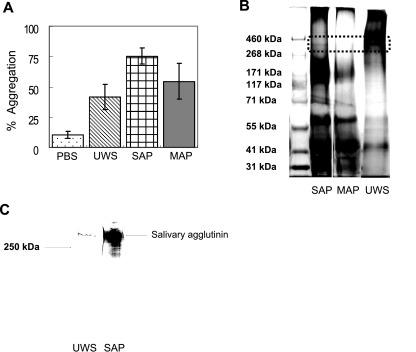
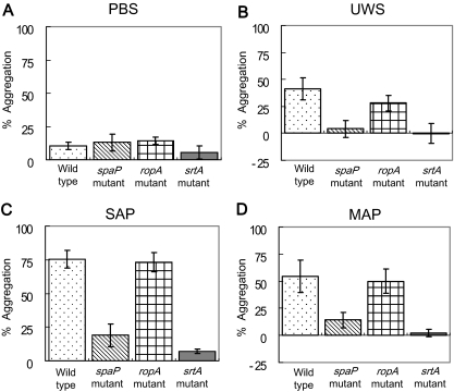
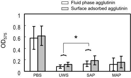
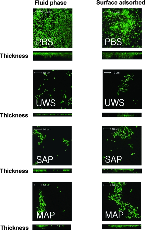
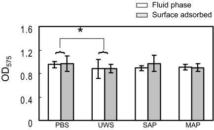
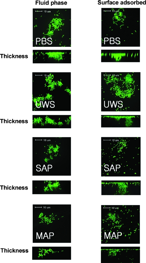
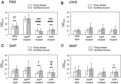
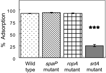
Interactions between salivary agglutinin and the adhesin P1 of Streptococcus mutans contribute to bacterial aggregation and mediate sucrose-independent adherence to tooth surfaces. We have examined biofilm formation by S. mutans UA159, and derivative strains carrying mutations affecting the localization or expression of P1, in the presence of fluid-phase or adsorbed saliva or salivary agglutinin preparations. Whole saliva- and salivary agglutinin-induced aggregation of S. mutans was adversely affected by the loss of P1 and sortase (SrtA) but not by the loss of trigger factor (RopA). Fluid-phase salivary agglutinin and, to a lesser extent, immobilized agglutinin inhibited biofilm development by S. mutans in the absence of sucrose, and whole saliva was more effective at decreasing biofilm formation than salivary agglutinin. Inhibition of biofilm development by salivary agglutinin was differently influenced by particular mutations, with the P1-deficient strain displaying a greater inhibition of biofilm development than the SrtA- or RopA-deficient strains. As expected, biofilm-forming capacities of all strains in the presence of salivary preparations were markedly enhanced in the presence of sucrose, although biofilm formation by the mutants was less efficient than that by the parental strain. Aeration strongly inhibited biofilm development, and the presence of salivary components did not restore biofilm formation in aerated conditions. The results disclose a potent ability of salivary constituents to moderate biofilm formation by S. mutans through P1-dependent and P1-independent pathways.


Figures

FIG. 1.
FIG. 1.
(A) Bacterial aggregation profiles of S. mutans UA159. SAP and MAP were prepared from S. mutans NG8, and 100 μl of UWS, SAP, or MAP was added to 900 μl of bacterial suspensions. Aggregation was calculated as the percent reduction in OD after 120 min relative to that of the initial cell suspension. Aggregation data were repeated at least five times, and error bars represent standard deviations. There were statistically significant differences in bacterial aggregation capacity among salivary preparations (SAP > MAP > UWS > PBS; P < 0.01). (B) Tris-acetate gel (3 to 8%) electrophoresis of high-molecular-weight markers (lane 1), SAP (lane 2), MAP (lane 3), and UWS (lane 4). Following electrophoresis, proteins were stained with both a glycoprotein detection kit and a silver-staining kit. The dotted rectangle indicates the position of salivary agglutinin migration, which stains as a band of approximately 340 kDa in UWS and SAP but is absent in MAP. (C) Western blot analysis of UWS (lane 1) and SAP (lane 2) using an antibody to gp340 (Affinity BioReagents, Golden, CO).
FIG. 2.
FIG. 2.
Bacterial aggregation by wild-type Streptococcus mutans UA159 (dots) and its spaP (diagonal lines), ropA (hatching), and srtA (solid) mutants in the presence of PBS (A), UWS (B), SAP (C), and MAP (D). Salivary preparations and aggregation assays were conducted as described in the methods section and in the legend for Fig. 1. Aggregation data were repeated at least five times, and the data were analyzed using a two-way ANOVA with respect to bacterial strain and salivary preparations. The error bars represent standard deviations. The wild-type and RopA-deficient strains aggregated more effectively than the P1- and SrtA-deficient strains (P < 0.001). SAP aggregated S. mutans strains better than UWS and MAP (P < 0.001). In addition, a significant interaction was detected between strains and salivary preparations. Salivary preparations aggregated the parental strain and the ropA mutant better than the spaP and srtA mutants (P < 0.001).
FIG. 3.
FIG. 3.
Comparisons of biofilm development by Streptococcus mutans UA159 when fluid-phase (white bars) and surface-phase (gray bars) salivary preparations were utilized. The cultures were grown in BM medium supplemented with 20 mM glucose. Fluid-phase salivary preparations were added directly to each well with the cell suspensions, whereas wells were coated with salivary preparations before inoculation with cell suspensions in the case of adsorbed salivary preparations. Biofilm formation was assayed on polystyrene microtiter plates after staining with crystal violet. Data shown are obtained from at least five independent experiments performed in quadruplicate. The error bars represent standard deviations. Biofilm development was inhibited more by fluid-phase salivary preparations than by surface-bound salivary preparations (P < 0.05), except for PBS, which showed no difference. Among salivary preparations, SAP allowed biofilm formation better than UWS (*, P < 0.05), while MAP allowed biofilm development that was intermediate to SAP and UWS.
FIG. 4.
FIG. 4.
Confocal microscopic images of S. mutans UA159 glucose biofilms. Biofilms of S. mutans UA159 were grown in BM supplemented with 20 mM glucose in the presence of fluid-phase PBS, UWS, salivary agglutinin preparations (SAG), and mock agglutinin preparations (MAG) and stained with SYTO 13. Data presented are representative of three independent experiments.
FIG. 5.
FIG. 5.
Comparison of biofilm development by Streptococcus mutans UA159 when fluid-phase (white bars) and surface-phase (gray bars) salivary preparations were utilized. The cultures were grown in BM medium supplemented with 20 mM sucrose. Salivary treatment and biofilm quantification were as described in Materials and Methods and in the legend for Fig. 3. Data were obtained from at least five independent experiments performed in quadruplicate. Error bars represent standard deviations. There were no significant differences in biofilm development between fluid-phase and adsorbed salivary preparations. Significant differences in biofilm development were detected only between PBS and UWS. *, P < 0.05; two-way ANOVA.
FIG. 6.
FIG. 6.
Confocal microscopic images of S. mutans UA159 sucrose biofilms. Biofilms of S. mutans UA159 were formed in BM supplemented with 20 mM sucrose in the presence of fluid-phase PBS, UWS, salivary agglutinin preparations (SAG), and mock agglutinin preparations (MAG), and stained with SYTO 13. There was no significant difference in microscopic images between adsorbed and fluid-phase salivary preparations. Data presented here are representative of three independent experiments.
FIG. 7.
FIG. 7.
Results of biofilm formation of Streptococcus mutans UA159 and its derivatives in BM supplemented with 20 mM glucose, with respect to PBS (A), UWS (B), SAP (C), and MAP (D). Biofilm formation assays were performed in the following two different ways: salivary preparations were added to each well with the cell suspensions (fluid-phase salivary preparations) (white bars), or wells were first coated with salivary preparations before inoculation with cell suspensions (surface-phase salivary preparations) (gray bars). Biofilm assays and data analysis were as described in Materials and Methods and in the legend for Fig. 6. Biofilm development was inhibited more by fluid-phase salivary preparations than by surface-bound salivary preparations (P < 0.05). The differences in biofilm development between the P1-defective strain and other strains are marked by a cross, and those between the wild type and other strains are marked by an asterisk. †, P < 0.05; ††, P < 0.01; †††, P < 0.001; *, P < 0.05; ***, P < 0.001. See the text for more details.
FIG. 8.
FIG. 8.
Cell surface hydrophobicity of Streptococcus mutans UA159 strains. The bacterial suspensions (1.2 ml) were mixed with 0.6 ml of hexadecane. Adsorption was calculated as the percent loss in OD of the aqueous phase relative to that of the initial cell suspension. Error bars represent standard deviations. ***, P < 0.001; one-way ANOVA.

References

    1. Ahn, S. J., and R. A. Burne. 2007. Effects of oxygen on biofilm formation and the AtlA autolysin of Streptococcus mutans. J. Bacteriol. 1896293-6302. - PMC - PubMed
    1. Ahn, S. J., Z. T. Wen, and R. A. Burne. 2006. Multilevel control of competence development and stress tolerance in Streptococcus mutans UA159. Infect. Immun. 741631-1642. - PMC - PubMed
    1. Babu, J. P., E. H. Beachey, D. L. Hasty, and W. A. Simpson. 1986. Isolation and characterization of a 60-kilodalton salivary glycoprotein with agglutinating activity against strains of Streptococcus mutans. Infect. Immun. 51405-413. - PMC - PubMed
    1. Brady, L. J., D. A. Piacentini, P. J. Crowley, P. C. F. Oyston, and A. S. Bleiweis. 1992. Differentiation of salivary agglutinin-mediated adherence and aggregation of mutans streptococci by use of monoclonal antibodies against the major surface adhesin P1. Infect. Immun. 601008-1017. - PMC - PubMed
    1. Burne, R. A., R. G. Quivey, Jr., and R. E. Marquis. 1999. Physiologic homeostasis and stress responses in oral biofilms. Methods Enzymol. 310441-460. - PubMed

Publication types