

Review
doi: 10.1513/pats.200804-037DW.
Stem cells and cell therapies in lung biology and lung diseases
Affiliations
- PMID: 18625757
- PMCID: PMC2645238
- DOI: 10.1513/pats.200804-037DW
Item in Clipboard
Review
Stem cells and cell therapies in lung biology and lung diseases
Proc Am Thorac Soc.
.
No abstract available
Figures

Human β2-microglobulin–positive cells can be detected in NOD-SCID (nonobese diabetic/severe combined immunodeficient) mouse airways for up to 3 months after systemic administration of human cord blood–derived mesenchymal stem cells (CB-MSCs) (arrowheads). NOD-SCID lung sections 3 months following CB-MSC administration. Blue = DAPI (4′-6-diamidino-2-phenylindole) nuclear stain, green = pancytokeratin, red = β2-microglobulin, white = colocalization of pancytokeratin and β2-microglobulin. Original magnifications, ×40, ×200. Adapted by permission from Reference .
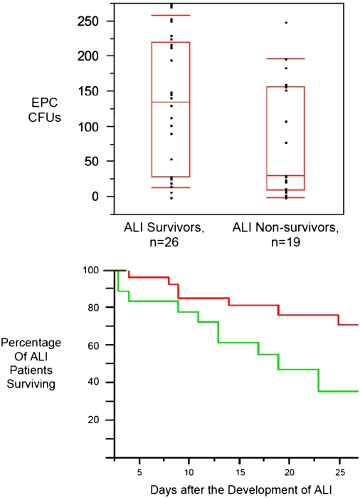
Patients with higher numbers of circulating endothelial progenitor cells (EPCs) during the first 72 hours of acute lung injury (ALI) had improved survival. (Top) Boxes represent the median, 25th, and 75th percentiles. Tenth and 90th percentiles are signified by floating red bars. (Bottom) Kaplan-Meier curve of patients with ALI stratified by EPC cfu counts of ⩾35 (red line) or <35 (green line). Patients with EPC CFU counts ⩾35 had survival benefit (70 vs. 35% survival at 28 d). Adapted by permission from Reference .

Levels of circulating CD45+ Col1+ cells are increased in blood from patients with usual interstitial pneumonitis (UIP) or fibrotic nonspecific interstitial pneumonitis (NSIP). *P < 0.05 compared with normal volunteers (n = 5 per group). Adapted by permission from Reference .
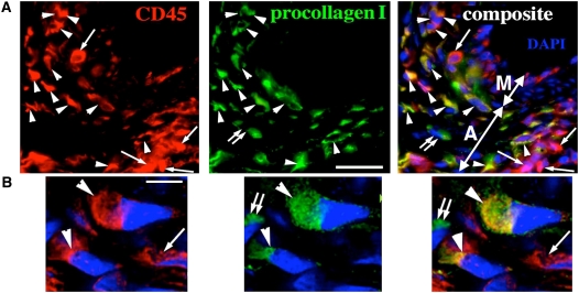
Fibrocytes (CD45+/procollagen+) comprise almost 40% of leukocytes in the remodeled pulmonary artery adventitia of neonatal calves with severe hypoxic pulmonary hypertension. (A, B) Triple-labeled immunofluorescent staining (CD45, red; procollagen, green; cell nuclei [DAPI], blue) demonstrates numerous fibrocytes (CD45+/procollagen+; marked by arrowheads) in the remodeled adventitia. Leukocytes (CD45+/procollagen−) are marked by single arrows, and fibroblasts (CD45−/procollagen+) by double arrows. Pulmonary artery adventitia (A) and media (M) are marked by double-headed arrows in (A). Scale bars, 20 mm (A); 5 mm (B). (B) Deconvolution confocal microscopy with a high magnification of a field as shown in (A). Adapted by permission from Reference .

Mice receiving intratracheal administration of mesenchymal stem cells (MSCs) demonstrated decreased histologic injury and bronchoalveolar lavage (BAL) fluid levels of proinflammatory cytokines in response to endotoxin-induced lung injury. (A) Hematoxylin-and-eosin–stained lung sections 48 hours after instillation of endotoxin + phosphate-buffered saline (PBS) (A) or endotoxin + MSCs (B). Original magnification, 20× (B). BAL fluid levels of macrophage inflammatory protein (MIP)-2 (C) and tumor necrosis factor (TNF)-α (D) were decreased 48 hours after instillation of either PBS or MSCs in endotoxin-induced lung injury. Adapted by permission from Reference .
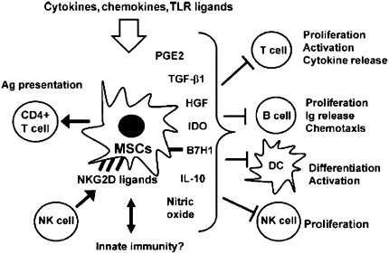
Schematic illustrating the range of in vitro immune-modulating effects described for mesenchymal stem cells (MSCs). Ag = antigen; DC = dendritic cell; HGF = hepatocyte growth factor; NK = natural killer; PGE = prostaglandin E; TGF = transforming growth factor; TLR = Toll-like receptor. Adapted by permission from Reference .
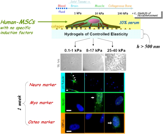
Schematic demonstrating inherent matrix stiffness of different tissues and that culture of human mesenchymal stem cells (MSCs) in hydrogels of increasing stiffness directs differentiation along different pathways. Adapted by permission from Reference .

Expression of epithelial and endothelial markers in gelfoam sponges implanted into rat lung. Sixty days after injection of gelfoam sponges impregnated with fetal rat lungs cells into adult rat lungs, expression of various markers of mature lung cells was detected in the sponge. Original magnification, ×100. pro-SPC = pro–surfactant protein C; vWF = von Willebrand factor. Adapted by permission from Reference .

Schematic of endogenous progenitor cells. D = dorsal; V = ventral. Adapted by permission from Reference .

Human embryonic stem cells (hES E.9) transduced with a pro-surfactant protein C (pro-SPC)/Neo.74 vector and selected in G418 express surfactant proteins A, B, and C (red stain). Nuclei are counterstained with DAPI (blue). ATII = type II alveolar epithelial cells. Original magnification, ×400. Adapted by permission from Reference .
References
-
- Weiss DJ, Berberich MA, Borok Z, Gail DB, Kolls JK, Penland C, Prockop DJ. Adult stem cells, lung biology, and lung disease. NHLBI/Cystic Fibrosis Foundation Workshop. Proc Am Thorac Soc 2006;3:193–207. - PubMed
-
- Brown JK, Hogan BLM, Randell SH, Stripp B, Weiss DJ. Human embryonic stem cell research: an official ATS research policy statement. Am J Respir Crit Care Med 2006;173:1–3.
-
- Fisher KA, Summer RS. 4. Stem and progenitor cells in the formation of the pulmonary vasculature. Curr Top Dev Biol 2006;74:117–131. - PubMed
Publication types
MeSH terms
Grants and funding
LinkOut - more resources
Full Text Sources
Other Literature Sources
Medical