. 2008 Jul 14;182(1):19-26.
doi: 10.1083/jcb.200711040.

Nucleophosmin and its AML-associated mutant regulate c-Myc turnover through Fbw7 gamma

Affiliations

Nucleophosmin and its AML-associated mutant regulate c-Myc turnover through Fbw7 gamma

Paola Bonetti et al. J Cell Biol. .

Abstract

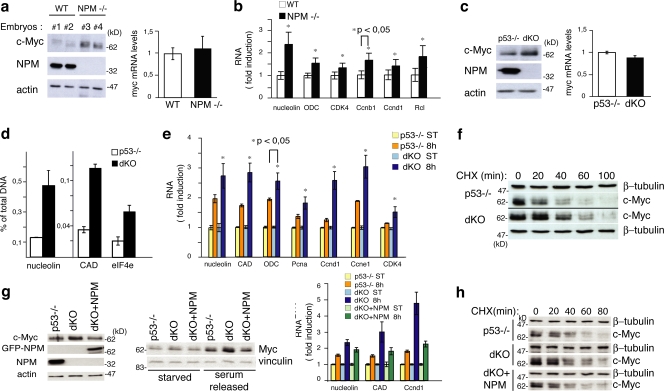
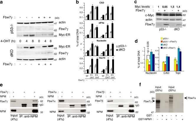
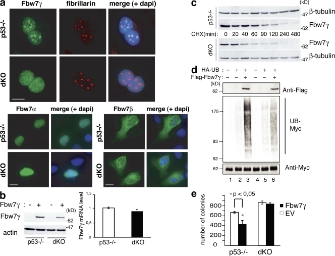
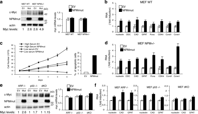
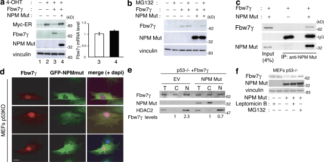
Mutations leading to aberrant cytoplasmic localization of nucleophosmin (NPM) are the most frequent genetic alteration in acute myelogenous leukemia (AML). NPM binds the Arf tumor suppressor and protects it from degradation. The AML-associated NPM mutant (NPMmut) also binds p19Arf but is unable to protect it from degradation, which suggests that inactivation of p19Arf contributes to leukemogenesis in AMLs. We report here that NPM regulates turnover of the c-Myc oncoprotein by acting on the F-box protein Fbw7gamma, a component of the E3 ligase complex involved in the ubiquitination and proteasome degradation of c-Myc. NPM was required for nucleolar localization and stabilization of Fbw7gamma. As a consequence, c-Myc was stabilized in cells lacking NPM. Expression of NPMmut also led to c-Myc stabilization because of its ability to interact with Fbw7gamma and delocalize it to the cytoplasm, where it is degraded. Because Fbw7 induces degradation of other growth-promoting proteins, the NPM-Fbw7 interaction emerges as a central tumor suppressor mechanism in human cancer.


Figures

Figure 1.
Figure 1.
NPM regulates c-Myc protein stability. (a, left) WB analysis of two NPM WT (Nos. 1 and 2) and knockout (KO; Nos. 2 and 3) embryos at 10.5 d post coitum (Colombo et al., 2005). (right) c-Myc mRNA levels in the same embryos evaluated by QPCR (results are normalized against WT samples). (b) Expression of c-Myc target genes analyzed by QPCR using mRNA from WT and NPM KO embryos. Expression was standardized with ubiquitin and normalized against the control WT RNAs. (c) c-Myc protein levels (left) and c-Myc mRNA levels (right) in p53−/− and dKO MEFs. (d) QPCR of anti c-Myc ChIP. The percentage of DNA bound to c-Myc was calculated as described previously (Frank et al., 2001). (e) QPCR analysis of c-Myc target genes at 8 h after serum treatment. Results analyzed as in panel b. (f) c-Myc protein level in p53−/− and dKO MEFs. Cells were treated with CHX and harvested at the indicated time points. (g, left) c-Myc protein levels in p53−/− and dKO MEFs infected with retroviruses expressing the GFP-NPM1 protein (+NPM) or control retroviruses. (middle) WB analysis of the same cells during serum starvation and upon serum release. (right) QPCR analysis of c-Myc target gene expression after serum treatment (8 h) as in panel d. (h) c-Myc protein level in p53−/−, dKO, and dKO + GFP-NPM MEFs as described in panel f. Data represent the mean of three determinations ± SEM.
Figure 2.
Figure 2.
NPM is required for Fbw7γ activity. (a) WB analysis in p53−/− and dKO MEFs infected with Myc-ER and, where indicated (+), HA-Fbw7γ–expressing retroviruses. Cells were treated with 4-OHT for the indicated hours or left untreated (0 h). (b) QPCR of anti–c-Myc ChIP on target promoters (as indicated) after 4-OHT treatment of the same cells as in panel a. (c) WB analysis in p53−/− and dKO MEFs infected with retroviruses expressing HA-Fbw7γ (+) or control retroviruses (−) as indicated. c-Myc levels have been calculated by densimetric analysis, normalizing band intensity to actin and control p53−/− cells. (d) QPCR analysis of anti c-Myc ChIP on target promoters (as indicated) of the same cells as in panel c. (e) dKO MEFs were cotransfected with plasmids expressing NPM1 and flag-Fbw7γ (left), flag-Fbw7α (middle), and flag-Fbw7β (right). Total lysates and immunoprecipitates were blotted with antibodies against NPM (NPMa) or anti-flag. (f) GST pull-down assay using in vitro translated 35S-labeled Fbw7γ or Fbw7α and equal amounts of GST or GST-NPM proteins. Data represent the mean of three determinations ± SEM.
Figure 3.
Figure 3.
NPM is required Fbw7γ nucleolar localization and stability. (a) IF analysis of Fbw7γ (top), Fbw7α (bottom left), and Fbw7β (bottom right) localization in p53−/− and dKO MEFs transiently transfected with the corresponding flag-Fbw7 constructs (green staining). Anti-fibrillarin staining (red) is shown as a marker for nucleoli. Bars, 10 μm. (b, left) WB analysis in p53−/− and dKO MEFs infected with retroviruses expressing HA-Fbw7γ (+) or control retroviruses (−) as indicated. (right) Fbw7γ mRNA levels in p53−/− and dKO MEFs. (c) HA-Fbw7γ protein stability in p53−/− and dKO MEFs. Cells were treated with CHX and harvested at the indicated time points. (d) Effects of Fbw7γ expression on Myc ubiquitination in p53−/− and dKO cells. p53−/− (lanes 1–3) and dKO (lanes 4–6) MEFs were transfected with expression vectors for HA-tagged ubiquitin and flag-tagged Fbw7γ, as indicated. Cell lysates were IPed with an anti-Myc antibody and blotted with anti-HA (to identify ubiquitinated myc; UB-myc) and anti-Myc (as control) antibodies. Levels of flag-Fbw7γ in the input were analyzed with an anti-flag antibody. (e) Methylcellulose colony assay of p53−/− and dKO MEFs expressing Myc-ER protein and infected with control (EV) or HA-Fbw7γ–expressing retroviruses. 4-OHT was added every 3 d; colonies were counted after 15 d. Data represent the mean of three determinations ± SEM. *, P < 0.045.
Figure 4.
Figure 4.
Mutant NPM stabilizes the c-Myc protein. (a, left) WB analysis in WT and NPM± MEFs infected with retroviruses expressing NPMmut (Mut) or control (EV) retroviruses. (right) c-Myc mRNA levels in the same cell. (b) QPCR analysis of the indicated c-Myc target genes. (c, left) Growth curves of NPM± MEFs infected with retroviruses expressing NPMmut or control (EV) retroviruses. 104 cells were plated in presence of 10% serum (high serum) or 0.5% serum (low serum) as indicated. (right) The percentage of apoptosis in the same cells maintained in low serum culture conditions for 24 h. (d) QPCR analysis of the indicated c-Myc target genes in NPM± MEFs infected with EV or NPMmut retroviruses. (e, left) WB analysis in ARF−/−, p53−/−, and dKO MEFs infected with retroviruses expressing NPMmut (mut) or control (EV) retroviruses. (right) c-Myc mRNA levels in the same samples. (f) QPCR analysis of the indicated c-Myc target genes in the same cells as in panel e. Data represent the mean of three determinations ± SEM.
Figure 5.
Figure 5.
Mutant NPM delocalizes and destabilizes Fbw7γ. (a, left) WB analysis in p53−/−;Myc-ER cells infected with empty (No. 2) or HA-Fbw7γ-expressing (No. 4) retroviruses. HA-Fbw7γ–infected cells were reinfected with empty or NPMmut-expressing lentiviruses (also expressing GFP as marker). Cells were treated for 2 h with 4-OHT (Nos. 2–4). (right) QPCR analysis of Fbw7γ mRNA levels in samples Nos. 3 and 4. Data represent the mean of three determinations ± SEM. (b) WB analysis in the same cells as in panel a. Cells were or were not treated with MG132 for 2 h, as indicated. (c) dKO MEFs were cotransfected with plasmids expressing flag-Fbw7γ and NPMmut or the empty vector, as indicated. Total lysates and IPs were blotted with antibodies against NPMmut or the flag epitope. (d) IF analysis of p53−/− MEFs infected with GFP-NPMmut– and flag-Fbw7γ (red staining)-expressing constructs. A merge of the NPMmut, Fbw7γ, and DAPI staining is also shown. Bar, 10 μm. (e) Nucleus–cytoplasm fractionation in p53−/− MEFs expressing flag-Fbw7γ infected with GFP-NPMmut or control (EV) retroviruses. Total cell lysates (T) or cellular fractions (C, cytoplasm; N, nucleus) were analyzed by WB as indicated. (f) The same cells as in panel e were or were not treated with 1 μM LMB or MG132 for 3 h, as indicated. Expression of Fbw7γ was analyzed by WB on total cells lysates using anti-flag antibodies.

Comment in

References

    1. Adhikary, S., and M. Eilers. 2005. Transcriptional regulation and transformation by Myc proteins. Nat. Rev. Mol. Cell Biol. 6:635–645. - PubMed
    1. Bartkova, J., Z. Horejsi, K. Koed, A. Kramer, F. Tort, K. Zieger, P. Guldberg, M. Sehested, J.M. Nesland, C. Lukas, et al. 2005. DNA damage response as a candidate anti-cancer barrier in early human tumorigenesis. Nature. 434:864–870. - PubMed
    1. Borer, R.A., C.F. Lehner, H.M. Eppenberger, and E.A. Nigg. 1989. Major nucleolar proteins shuttle between nucleus and cytoplasm. Cell. 56:379–390. - PubMed
    1. Colombo, E., J.C. Marine, D. Danovi, B. Falini, and P.G. Pelicci. 2002. Nucleophosmin regulates the stability and transcriptional activity of p53. Nat. Cell Biol. 4:529–533. - PubMed
    1. Colombo, E., P. Bonetti, E. Lazzerini Denchi, P. Martinelli, R. Zamponi, J.C. Marine, K. Helin, B. Falini, and P.G. Pelicci. 2005. Nucleophosmin is required for DNA integrity and p19Arf protein stability. Mol. Cell. Biol. 25:8874–8886. - PMC - PubMed

Publication types