. 2008 Jul 14;182(1):27-33.
doi: 10.1083/jcb.200712080.

Mars promotes dTACC dephosphorylation on mitotic spindles to ensure spindle stability

Affiliations

Mars promotes dTACC dephosphorylation on mitotic spindles to ensure spindle stability

Shengjiang Tan et al. J Cell Biol. .

Abstract

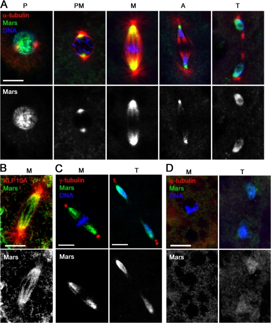
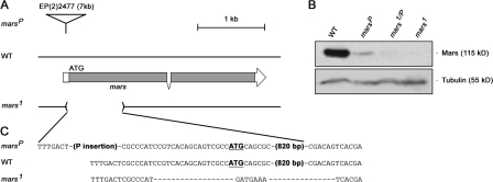
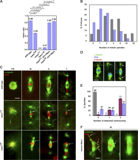
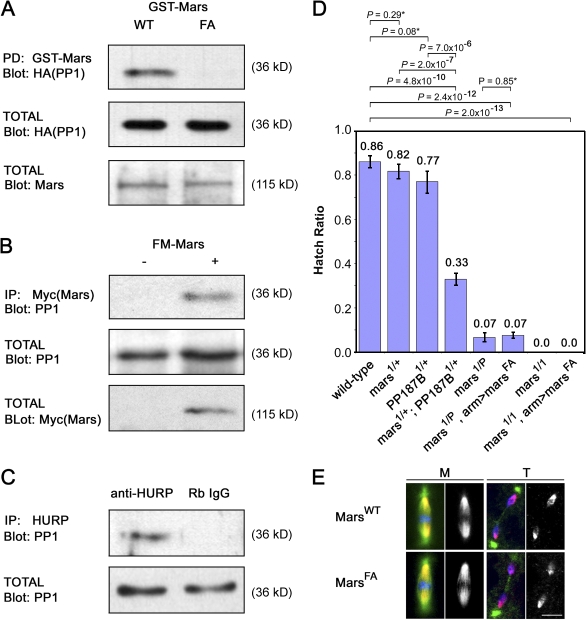
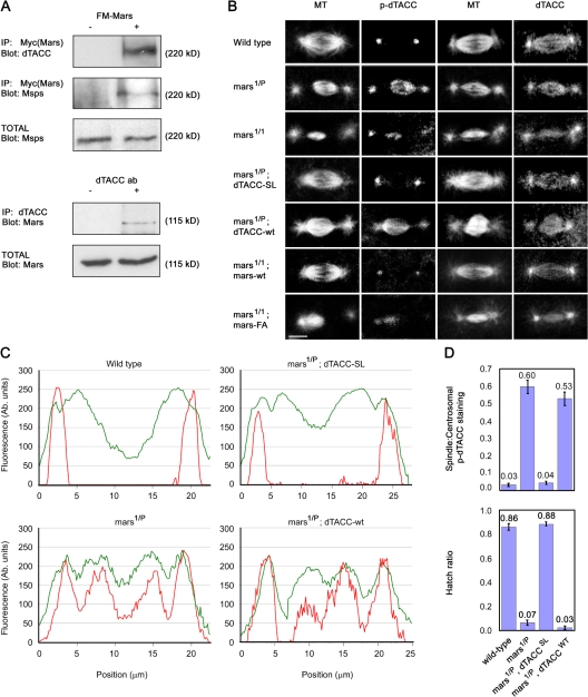
Microtubule-associated proteins (MAPs) ensure the fidelity of chromosome segregation by controlling microtubule (MT) dynamics and mitotic spindle stability. However, many aspects of MAP function and regulation are poorly understood in a developmental context. We show that mars, which encodes a Drosophila melanogaster member of the hepatoma up-regulated protein family of MAPs, is essential for MT stabilization during early embryogenesis. As well as associating with spindle MTs in vivo, Mars binds directly to protein phosphatase 1 (PP1) and coimmunoprecipitates from embryo extracts with minispindles and Drosophila transforming acidic coiled-coil (dTACC), two MAPs that function as spindle assembly factors. Disruption of binding to PP1 or loss of mars function results in elevated levels of phosphorylated dTACC on spindles. A nonphosphorylatable form of dTACC is capable of rescuing the lethality of mars mutants. We propose that Mars mediates spatially controlled dephosphorylation of dTACC, which is critical for spindle stabilization.


Figures

Figure 1.
Figure 1.
Mars localizes to spindle MTs. (A) Fixed wild-type embryos stained to reveal the distribution of Mars (green), α-tubulin (red), and DNA (blue) during mitosis. (B and C) Mars is concentrated at MT minus ends. Fixed wild-type embryos stained to reveal distribution of Mars (green) and either KLP10A (red; B) or γ-tubulin (red) and DNA (blue; C). (D) An embryo treated with 500 μg/ml colchicine before fixation to depolymerize MTs. Under these conditions, Mars staining (green) disappears from the spindle during metaphase but can be seen on the nuclear envelope at telophase. P, prophase; PM, prometaphase; M, metaphase; A, anaphase; T, telophase. Bars, 10 μm.
Figure 2.
Figure 2.
Molecular defects in mars mutants. (A) mars genomic region in marsP, wild type, and mars1. The coding region of mars is represented by shading. marsP contains a P element insertion, EP(2)2477, in the mars untranslated region. mars1 has a 0.84-kb deletion that removes the mars initiation codon and part of the coding region. (B) Mars protein produced in embryos from wild-type, marsP, mars1/P, and mars1 flies. Total protein was analyzed by immunoblotting using Mars antibody. marsP produced full-length Mars protein in a much lower amount, whereas mars1 failed to produce any detectable protein. Blotting with α-tubulin antibodies controlled for loading. (C) Genomic sequence of wild-type and mars mutant strains. The initiation codon (ATG) of mars is bold and underlined.
Figure 3.
Figure 3.
mars is essential for spindle stability. (A) Hatch ratios, plotted as the mean ± standard error of five experiments, showing greatly reduced ability of embryos laid by mars mutant females to hatch compared with wild type. Ectopic expression of UASP-FM-marsWT under control of the arm-GAL4 driver (arm>Mars) rescues this phenotype. P-values determined from student's t tests are shown above the graph. (B) Graph showing significant reduction in mean number of mitotic spindles in 15–45-min embryos laid by mars1 mutant mothers (blue; n = 144 embryos) compared with wild-type mothers (gray; n = 185 embryos; P = 2.9 × 10⁻¹⁰). 81.9% of mars1 mutant embryos progress through meiosis to form one or more mitotic figure. (C) Images of mitotic spindles from embryos laid by wild-type, mars1, and mars1/P mothers showing small spindles in mars mutants (arrows) and loss of centrosome attachment to the spindle body. Loss of chromosome attachment to monopolar MTs is apparent in polar bodies. PB, polar body; M, metaphase; A, anaphase; T, telophase. (D) Images of spindles from mutant embryos, showing detached centrosomes (arrows). (E) Bar graph showing percentage of spindles with detached centrosomes from wild type (gray), mars1 (red), and mars1/P (blue), plotted as the mean ± standard error of three experiments. (F) Representative images showing effects of strong ectopic expression of marsWT with nanos-GAL4VP16. Compared with the wild type (C, top), these embryos display abnormally robust spindles, which often have aberrantly aligned MT fibers at metaphase (arrow). Bars, 10 μm.
Figure 4.
Figure 4.
Binding to PP1 is essential for Mars function. (A) Mars binds to PP1α87B in a GST pulldown (PD) assay. Binding requires Phe839 of Mars. arm-GAL4 UAS-HA-PP1α87B fly extracts were incubated with lysates from Escherichia coli expressing GST-MarsWT or MarsF839A. Mars complexes were purified on glutathione-Sepharose and immunoblotted with HA to test for coprecipitation of PP1α87B. Blots of total extracts confirmed equal levels of HA-PP1 and GST-Mars in these experiments. (B) Mars coprecipitates PP1 from D. melanogaster embryonic nuclear extracts. arm-GAL4 (−) and arm-GAL4 UASP-FM-mars (+) embryo extracts were subjected to immunoprecipitation (IP) with Myc antibodies followed by immunoblotting with PP1 antibodies. Blots of total extracts confirmed levels of PP1 and FM-tagged Mars. (C) HURP coprecipitates PP1 from HeLa cell nuclear extracts. Immunoprecipitation of extracts with rabbit HURP antibody or with rabbit random IgG (Rb IgG) was followed by immunoblotting with PP1 antibodies to test for binding. (D) Hatch ratios, plotted as the mean ± standard error of seven experiments. Loss of one copy of PP1α87B enhances mars1/+ to semilethality. Ectopic marsFA fails to rescue lethality of embryos laid by mars mutant mothers. P-values indicated by an asterisk are not statistically significant. (E) Ectopic MarsWT and MarsF839A have identical distributions on mitotic spindles. Fixed embryos from arm>marsWT or arm>marsF839A mothers were stained to reveal the distribution of α-tubulin (green), FLAG-tagged Mars (red and greyscale), and DNA (blue) during mitosis. M, metaphase; T, telophase.
Figure 5.
Figure 5.
An essential role of mars is to promote dTACC dephosphorylation. (A) Mars coprecipitates dTACC and Msps from D. melanogaster embryonic nuclear extracts. Immunoprecipitation of arm-GAL4 (−) and arm>marsWT (+) embryo extracts with Myc antibody, followed by immunoblotting with dTACC and Msps antibodies, showed binding to FM-tagged Mars. Immunoprecipitation with dTACC antibody followed by immunoblotting with Mars antibody confirmed dTACC binding. (B) Distribution of dTACC and p-dTACC on mitotic spindles in embryos from mothers of different genotypes, as indicated. In wild type, p-dTACC is only found at the centrosome but in mars mutants, p-dTACC abnormally accumulates on mitotic spindles. Total dTACC staining is largely unaffected in mars mutants. A nonphosphorylatable mutant form of dTACC (dTACCSL), but not wild-type dTACC (dTACCWT), restores spindle structure and normal distribution of p-dTACC in a mars1/P mutant background. Similarly, arm>marsWT, but not arm>marsFA, restores normal spindle structure and p-dTACC staining in a mars1 background. Bar, 10 μm. (C) Linescans of fluorescence intensity (arbitrary units) across spindles from embryos of different genotypes, as indicated. The distribution of p-dTACC (red trace) is shown relative to α-tubulin (green trace). (D) Top graph shows quantification of ratio of spindle/centrosomal p-dTACC staining. dTACCSL, but not dTACCWT, restores a normal p-dTACC ratio in embryos from mars1/P mothers. Bottom graph shows that dTACCSL, but not dTACCWT, rescues lethality of embryos laid by mars1/P mothers. Hatch ratios are plotted as the mean ± standard error from n = 5 experiments.

References

    1. Baksa, K., H. Morawietz, V. Dombrádi, M. Axton, H. Taubert, G. Szabo, I. Torok, A. Udvardy, H. Gyurkovics, and B. Szöor. 1993. Mutations in the protein phosphatase 1 gene at 87B can differentially affect suppression of position-effect variegation and mitosis in Drosophila melanogaster.Genetics. 135:117–125. - PMC - PubMed
    1. Barros, T.P., K. Kinoshita, A.A. Hyman, and J.W. Raff. 2005. Aurora A activates D-TACC–Msps complexes exclusively at centrosomes to stabilize centrosomal microtubules. J. Cell Biol. 170:1039–1046. - PMC - PubMed
    1. Bennett, D., and L. Alphey. 2004. Cloning and expression of mars, a novel member of the guanylate kinase associated protein family in Drosophila. Gene Expr. Patterns. 4:529–535. - PubMed
    1. Bennett, D., E. Lyulcheva, and L. Alphey. 2006. Towards a comprehensive analysis of the protein phosphatase 1 interactome in Drosophila. J. Mol. Biol. 364:196–212. - PubMed
    1. Brittle, A.L., and H. Ohkura. 2005. Centrosome maturation: Aurora lights the way to the poles. Curr. Biol. 15:R880–R882. - PubMed

Publication types

MeSH terms