. 2008 Jul 2;3(7):e2571.
doi: 10.1371/journal.pone.0002571.

In-cell biochemistry using NMR spectroscopy

Affiliations

In-cell biochemistry using NMR spectroscopy

David S Burz et al. PLoS One. .

Abstract

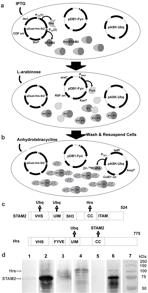
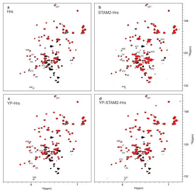
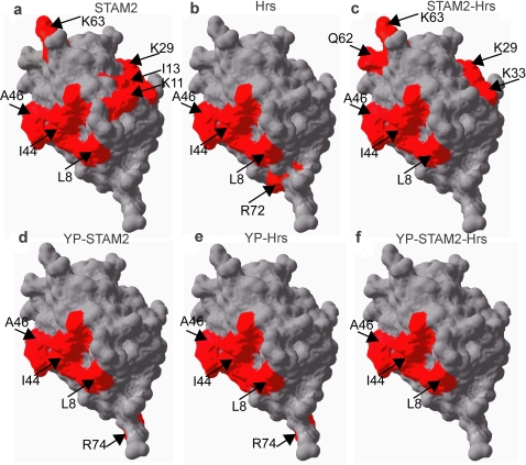
Biochemistry and structural biology are undergoing a dramatic revolution. Until now, mostly in vitro techniques have been used to study subtle and complex biological processes under conditions usually remote from those existing in the cell. We developed a novel in-cell methodology to post-translationally modify interactor proteins and identify the amino acids that comprise the interaction surface of a target protein when bound to the post-translationally modified interactors. Modifying the interactor proteins causes structural changes that manifest themselves on the interacting surface of the target protein and these changes are monitored using in-cell NMR. We show how Ubiquitin interacts with phosphorylated and non-phosphorylated components of the receptor tyrosine kinase (RTK) endocytic sorting machinery: STAM2 (Signal-transducing adaptor molecule), Hrs (Hepatocyte growth factor regulated substrate) and the STAM2-Hrs heterodimer. Ubiquitin binding mediates the processivity of a large network of interactions required for proper functioning of the RTK sorting machinery. The results are consistent with a weakening of the network of interactions when the interactor proteins are phosphorylated. The methodology can be applied to any stable target molecule and may be extended to include other post-translational modifications such as ubiquitination or sumoylation, thus providing a long-awaited leap to high resolution in cell biochemistry.


Conflict of interest statement

Competing Interests: The authors have declared that no competing interests exist.

Figures

Figure 1
Figure 1. Sequential overexpression and in-cell post-translational modification of interacting proteins for STINT-NMR.
E. coli are transformed with up to three compatible plasmids and grown overnight in LB-glucose medium containing antibiotics. The cells are washed and resuspended in label-free medium. a) IPTG is used to induce overexpression of interactor proteins, STAM2 and Hrs, which form heterodimers. L-arabinose is then used to induce overexpression of Fyn kinase, which phosphorylates tyrosine residues (PY) on the interactors. b) The cells are washed and resuspended in labeling medium. Anhydrotetracycline is used to induce overexpression of uniformly labeled [U-15N] Ubiquitin target protein, which binds to the interactors. Samples are taken as the concentration of target increases. Changes in the target protein structure are monitored using in-cell NMR spectroscopy. A sample of labeled target containing no interactor is prepared separately as a reference. N.b. The experiment can be performed using a single interactor protein and without post-translational modification. The protocol can also be reversed, overexpressing labeled target first, followed by interactor(s) and post-translational modification (see Materials and Methods). c) Domain structure of STAM2 and Hrs. VHS (Vps27-Hrs-Stam domain); UIM (ubiquitin interacting motif); SH3 (src homology domain 3); CC (coiled coil domain); FYVE (FYVE-finger domain); up arrows indicate Ubiquitin, STAM2 or Hrs binding domain. d) Western blot of overexpressed interactor proteins from whole cells in the absence and presence of overexpressed Fyn kinase, probed with anti-tyrosine phosphate HRP-conjugate antibody. Lane 1: STAM2; lane 2: STAM2 & Fyn kinase; lane 3: Hrs; lane 4: Hrs & Fyn kinase; lane 5: STAM2 & Hrs; lane 6: STAM2, Hrs & Fyn kinase; Lane 7: MW.
Figure 2
Figure 2. NMR-spectra of Ubiquitin-ligand complexes.
1H{15N}HSQC spectra of E. coli after 3-h of [15N]-Ubiquitin overexpression (black), overlaid with spectra (red) obtained from E. coli after 2-h of [15N]-Ubiquitin overexpression and: a) 4-h of Hrs overexpression; b) 4-h of STAM2 & Hrs co-overexpression; c) 4-h of Hrs and 2-h of Fyn kinase co-overexpression; d) 4-h of Hrs & STAM2 and 2-h of Fyn kinase co-overexpression. Individual peaks exhibiting either a chemical shift change >0.1 ppm or significant differential broadening (>30% change in intensity) are labeled with corresponding assignments. The strong peaks in the spectra between 8.5 and 7.8 ppm correspond to various metabolites of [U-15N] ammonium ion. NMR experiments were acquired at T = 298 K on Bruker Avance 700 MHz NMR spectrometer equipped with a cryoprobe. 1H{15N}-edited HSQC data were recorded with 16 transients as 512{128} complex points, apodized with a squared cosine-bell window function and zero-filled to 1k{256) points prior to Fourier transformation. The corresponding sweep widths were 12 and 35 ppm in the 1H and 15N dimensions, respectively. The Q49 peak is obscured by peaks from the [U-15N] ammonium ion metabolites and is not labeled. Ubiquitin ligands are indicated in each panel.
Figure 3
Figure 3. Interaction surface maps of Ubiquitin-ligand complexes.
Interaction surface of Ubiquitin mapped onto the three-dimensional structure of Ubiquitin (PDB code 1D3Z). Individual residues exhibiting either a chemical shift change >0.05 ppm or significant differential broadening are indicated in red. All perturbed residues lie on the Ubiquitin surface and, therefore, reflect changes in the interaction surface of the molecule rather than changes in tertiary or quaternary structure. a) STAM2-Ubq interaction; b) Hrs-Ubq interaction; c) STAM2-Hrs-Ubq interaction; d) phosphorylated STAM2-Ubq interaction (YP-STAM2); e) phosphorylated Hrs-Ubq interaction (YP-Hrs); f) phosphorylated STAM2-Hrs-Ubq interaction (YP-STAM2-Hrs). Ubiquitin ligands are indicated in each panel.

References

    1. Seet BT, Dikic I, Zhou MM, Pawson T. Reading protein modifications with interaction domains. Nat Rev Mol Cell Biol. 2006;7:473–483. - PubMed
    1. Haglund K, Di Fiore PP, Dikic I. Distinct monoubiquitin signals in receptor endocytosis. Trends Biochem Sci. 2003;28:598–603. - PubMed
    1. Clague MJ, Urbé S. The interface of receptor trafficking and signalling. J Cell Sci. 2001;114:3075–3081. - PubMed
    1. Marmor MD, Yarden Y. Role of protein ubiquitylation in regulating endocytosis of receptor tyrosine kinases. Oncogene. 2004;23:2057–2070. - PubMed
    1. Burz DS, Dutta K, Cowburn D, Shekhtman A. Mapping structural interactions using in-cell NMR spectroscopy (STINT-NMR). Nat Methods. 2006;3:91–93. - PMC - PubMed

Publication types

MeSH terms

Substances

LinkOut - more resources