. 2008 Jul;64(1):71-87.
doi: 10.1002/ana.21408.

Dok-7 myasthenia: phenotypic and molecular genetic studies in 16 patients

Affiliations

Dok-7 myasthenia: phenotypic and molecular genetic studies in 16 patients

Duygu Selcen et al. Ann Neurol. 2008 Jul.

Abstract

Objective: Detailed analysis of phenotypic and molecular genetic aspects of Dok-7 myasthenia in 16 patients.

Methods: We assessed our patients by clinical and electromyographic studies, by intercostal muscle biopsies for in vitro microelectrode analysis of neuromuscular transmission and quantitative electron microscopy EM of 409 end plates (EPs), and by mutation analysis, and expression studies of the mutants.

Results: The clinical spectrum varied from mild static limb-girdle weakness to severe generalized progressive disease. The synaptic contacts were single or multiple, and some, but not all, were small. In vitro microelectrode studies indicated variable decreases of the number of released quanta and of the synaptic response to acetylcholine; acetylcholine receptor (AChR) channel kinetics were normal. EM analysis demonstrated widespread and previously unrecognized destruction and remodeling of the EPs. Each patient carries 2 or more heteroallelic mutations: 11 in genomic DNA, 7 of which are novel; and 6 identifiable only in complementary DNA or cloned complementary DNA, 3 of which are novel. The pathogenicity of the mutations was confirmed by expression studies. Although the functions of Dok-7 include AChR beta-subunit phosphorylation and maintaining AChR site density, patient EPs showed normal AChR beta-subunit phosphorylation, and the AChR density on the remaining junctional folds appeared normal.

Interpretation: First, the clinical features of Dok-7 myasthenia are highly variable. Second, some mutations are complex and identifiable only in cloned complementary DNA. Third, Dok-7 is essential for maintaining not only the size but also the structural integrity of the EP. Fourth, the profound structural alterations at the EPs likely contribute importantly to the reduced safety margin of neuromuscular transmission.


Figures
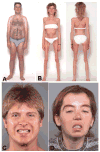
Fig 1
Fig 1
Phenotypic variability of Dok-7 myasthenia. Patient 10 has mild weakness and atrophy of limb girdle muscles, and mild eyelid ptosis (A). Patient 6 has severe diffuse weakness and atrophy of limb and axial muscles (B). Patient 1 shows mild asymmetric ptosis with slight facial weakness (C). Patient 13 shows marked eyelid ptosis and severe facial weakness (D).
Fig 2
Fig 2
Synaptic contact areas visualized with the cholinesterase reaction. Single small (B), multiple small (D–F), and perforated (A, C) contact areas were observed. Nerve sprouts are recognizable (D, asterisk) as faint brown lines connecting contact areas. Scale bar = 50μm.
Fig 3
Fig 3
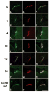
End-plate (EP) localization of Dok-7 (green signal, left column), acetylcholine receptor (AChR; red signal, center column except T), acetylcholinesterase (AChE; T), and merge (right column) in control subject (C), in Patients 1, 4, 10, 12, and 14, and EP AChR deficiency caused by low-expressor AChR ε subunit mutations. Apotome optics, 0.43μm slice distance. Scale bar = 20μm.
Fig 4
Fig 4
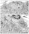
Electron micrographs of normal (A) and degenerating (B) neuromuscular junction in same patient. (B) Most junctional folds are replaced by globular debris (asterisk), causing widening of the synaptic space. This predicts a decreased synaptic response to acetylcholine (ACh) caused by loss of acetylcholine receptor (AChR) from tips of the destroyed folds, loss of ACh by diffusion from the widened synaptic space, and decreased input resistance of the remaining simple folds. Scale bars = 1μm.
Fig 5
Fig 5
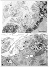
End plates (EPs) with presynaptic and postsynaptic abnormalities. (A) A highly abnormal EP region devoid of nerve terminal. Some junctional folds are degenerating (asterisk). The subsynaptic sarcoplasm harbors large myeloid structures. Nerve sprouts (s) surrounded by Schwann cell (SC) appear above the junction. EP region in (B) is denuded of its nerve terminal and shows marked degeneration of its junctional folds (asterisks) and subsynaptic organelles. Schwann cell process (SC) is present amid relics of the folds. Two nerve sprouts (s) appear at top left. Bars = 1μm.
Fig 6
Fig 6
Acetylcholine receptor (AChR) localization with peroxidase-labeled α-bungarotoxin (α-bgt). (A) Junctional folds are preserved and show a normal density and distribution of AChR. (B) Folds at bottom left react strongly for AChR but are capped by a degenerating nerve terminal. The folds at top right are degenerate, do not react for AChR, and are not covered by nerve terminal. nt = nerve terminal. Bars = 1μm.
Fig 7
Fig 7
Genomic structure of DOK7 and identified rearrangements in 16 patients. (inset) Intron 1 retention (thick horizontal line).
Fig 8
Fig 8
Scaled linear models of wild-type DOK7 and predicted peptides of mutant transcripts. White and shaded regions represent wild-type and missense residues. Solid circle shows location of missense mutation. The pleckstrin homology (PH) and phosphotyrosine-binding (PTB) domains, and the four important tyrosine residues in exon 7 are marked in wild type. Novel rearrangements are indicated in boldface type. NFS = no frameshift; NBP = nonbenign polymorphism.
Fig 9
Fig 9
Immunoblot demonstrating expression of wild-type (WT) and mutant DOK7 transcripts in human embryonic kidney (HEK) cells. Expression levels are normalized for cotransfected β-galactosidase. Bars indicate means and standard errors of four transfections.
Fig 10
Fig 10
Immunoblot demonstrating MuSK and phosphorylated MuSK in affinity-purified extracts of human embryonic kidney (HEK) cells transfected with MUSK and wild-type or exon-skipped constructs of DOK7. Products of transcripts lacking exons 3, 3 to 4, or 3 to 6 do not phosphorylate MuSK.
Fig 11
Fig 11
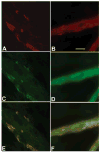
Localization of acetylcholine receptor (AChR; red; A, B), Flag-Dok-7 (green; C, D), merge (E, F) in C2C12 myotubes transfected with Flag-tagged wild-type (A, C, E) and flag-tagged 1139_1141delinsA-DOK7 complementary DNA (B, D, F). Apotome optics, 0.43μm slice distance. Scale bar = 20μm.

References

    1. Engel AG, Sine SM. Current understanding of congenital myasthenic syndromes. Curr Opin Pharmacol. 2005;5:306–321. - PubMed
    1. Okada K, Inoue A, Okada M, et al. The muscle protein Dok-7 is essential for neuromuscular synaptogenesis. Science. 2006;312:1802–1805. - PubMed
    1. Beeson D, Higuchi O, Palace J, et al. Dok-7 mutations underlie a neuromuscular junction synaptopathy. Science. 2006;313:1975–1978. - PubMed
    1. Palace J, Lashley D, Newsom-Davis J, et al. Clinical features of the DOK7 neuromuscular junction synaptopathy. Brain. 2007;130:1507–1515. - PubMed
    1. Muller JS, Herczegfalvi A, Vilchez JJ, et al. Phenotypical spectrum of DOK7 mutations in congenital myasthenic syndromes. Brain. 2007;130:1497–1506. - PubMed

Publication types

MeSH terms