. 2008 Jul;20(7):1760-74.
doi: 10.1105/tpc.107.057570. Epub 2008 Jul 15.

Auxin regulates Arabidopsis anther dehiscence, pollen maturation, and filament elongation

Affiliations

Auxin regulates Arabidopsis anther dehiscence, pollen maturation, and filament elongation

Valentina Cecchetti et al. Plant Cell. 2008 Jul.

Abstract

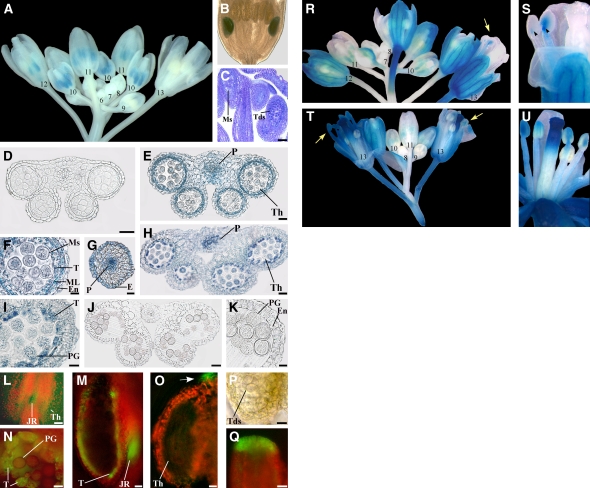
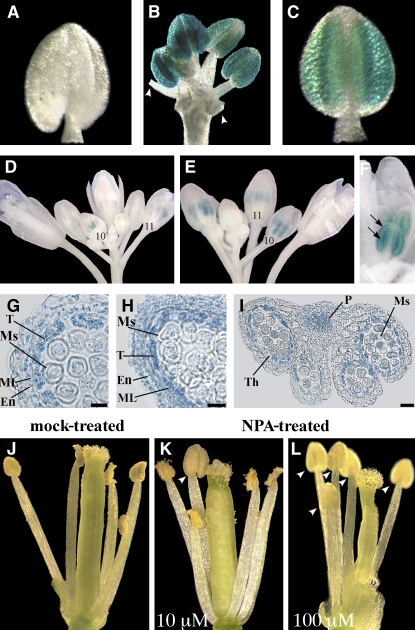
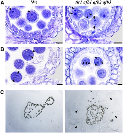
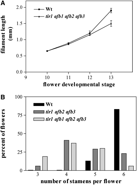
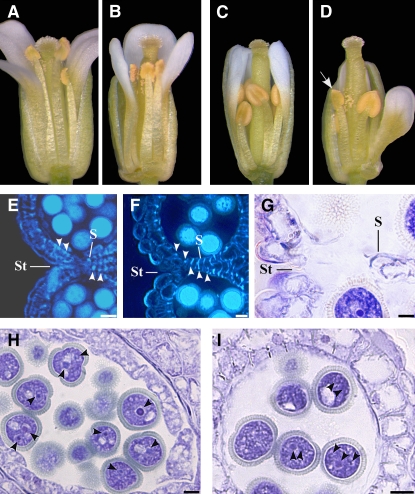
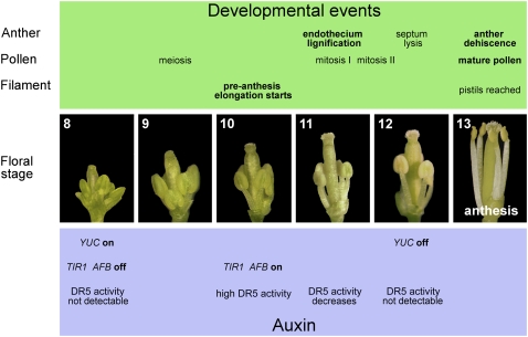
We provide evidence on the localization, synthesis, transport, and effects of auxin on the processes occurring late in Arabidopsis thaliana stamen development: anther dehiscence, pollen maturation, and preanthesis filament elongation. Expression of auxin-sensitive reporter constructs suggests that auxin effects begin in anthers between the end of meiosis and the bilocular stage in the somatic tissues involved in the first step of dehiscence as well as in the microspores and in the junction region between anther and filament. In situ hybridizations of the auxin biosynthetic genes YUC2 and YUC6 suggest that auxin is synthesized in anthers. In agreement with the timing of auxin effects, the TIR1, AFB1, AFB2, and AFB3 auxin receptor-encoding genes are transcribed in anthers only during late stages of development starting at the end of meiosis. We found that in tir1 afb triple and quadruple mutants, anther dehiscence and pollen maturation occur earlier than in the wild type, causing the release of mature pollen grains before the completion of filament elongation. We also assessed the contribution of auxin transport to late stamen developmental processes. Our results suggest that auxin synthesized in anthers plays a major role in coordinating anther dehiscence and pollen maturation, while auxin transport contributes to the independent regulation of preanthesis filament elongation.


Figures

Figure 1.
Figure 1.
Auxin-Responsive Reporter Genes Are Transcribed in Anthers before the Onset of Late Developmental Programs and Are Induced by Exogenous Auxin. (A) to (C) Expression of the DR5:GUS auxin-responsive reporter in flowers at different developmental stages. (A) DR5:GUS inflorescence showing no GUS staining in flower buds at early stages of development (6 to 9), intense GUS staining (blue) in anthers at stages 10 and 11, GUS staining at stage 12 only in short stamens, and no GUS staining at stage 13. Numbers indicate flower developmental stages. (B) DR5:GUS flower bud at early stage 12: GUS staining is observed in the anthers of short stamens. (C) Longitudinal section of a DR5:GUS flower bud at stage 10: microspores are visible in the anthers of long stamens, whereas tetrads are visible in those of short stamens. Bar = 30 μm. (D) to (K) Histochemical analysis of anthers in transection of DR5:GUS stamens. (D) Anther at stage 9. GUS staining is absent at the onset of meiosis. Bar = 20 μm. (E) Anther at late stage 10. Strong GUS staining at the end of meiosis in the theca and procambium. Bar = 20 μm. (F) Detail of (E). Strong GUS staining in the tapetum, middle layer, endothecium, and in microspores. Bar = 10 μm. (G) Upper stamen filament at late stage 10: transverse section at the end of meiosis showing intense GUS staining in the procambium and epidermal cells. Bar = 20 μm. (H) Anther at stage 11. GUS staining is observed in the theca and procambium. Bar = 20 μm. (I) Detail of (H). GUS signal is localized in tapetum degenerating cells and in pollen grains. Bar = 10 μm. (J) Anther at stage 12. GUS staining is absent after septum lysis (bilocular anther). Bar = 30 μm. (K) Detail of (J). GUS signal is absent in the endothecium and pollen grains. Bar = 10 μm. (L) to (Q) Fluorescence and bright-light images of anthers and pistils of DR5:GFP auxin-responsive reporter at different developmental stages. (L) Stamen at late stage 10. GFP fluorescence (green) is visible in the theca and in the junction region between anther and filament. Bar = 80 μm. (M) Stamen at late stage 11. Hand-cut longitudinal section showing intense GFP fluorescence in the remnants of the tapetum and in the junction region between anther and filament. Bar = 20 μm. (N) Anther theca at late stage 11. Hand-cut transverse section showing intense GFP fluorescence in tapetum degenerating cells and faint fluorescence in pollen grains. Bar = 10 μm. (O) to (Q) Anther and pistil at late stage 9. (O) Anther theca in longitudinal section showing GFP fluorescence at the tip of the anther (arrow) but not in the theca. Bar = 10 μm. (P) Detail of (O) in bright light showing tetrads. Bar = 10 μm. (Q) Detail of the apical region of the pistil. GFP fluorescence is intense in cells differentiating the papillae. Bar = 20 μm. (R) to (U) Expression of the DR5:GUS reporter after 10 mM NAA treatment of in planta inflorescences ([R] and [S]) or excised inflorescences ([T] and [U]). (R) DR5:GUS inflorescence showing no GUS staining in flower buds at early stages of development (7 and 8), intense GUS staining in anthers at stages 10 and 11, and GUS staining in stamens at stage 13 (arrow). Numbers indicate flower developmental stages. (S) Detail of (R) flower at stage 13: GUS signal is observed in filaments and in the junction region between anther and filament (arrowheads). (T) DR5:GUS inflorescence showing no GUS staining in flower buds at early stages of development (8 and 9), intense GUS staining in anthers at stages 10 and 11, and intense GUS staining in stamens at stage 13 (arrows). Numbers indicate flower developmental stages. (U) DR5:GUS flower at late stage 12: GUS signal is observed in filaments and anthers. E, epidermal cells; En, endothecium; JR, junction region; ML, middle layer; Ms, microspores; P, procambium; PG, pollen grains; T, tapetum; Tds, tetrads; Th, theca.
Figure 2.
Figure 2.
Analysis of the Expression Profiles of YUC2 and YUC6 during Stamen Development. (A) to (G) RNA in situ hybridization of YUC2. Longitudinal and transverse sections of stamens and anthers. (A) Stamen at stage 8. A strong signal is present in the theca. Bar = 20 μm. (B) Anther at early stage 9. A strong signal is present in the tissues surrounding the theca and meiocytes. The signal is also present in the procambium. Bar = 10 μm. (C) Anther at late stage 9. A strong signal is present in the endothecium, tapetum, tetrads, and in the procambium. Bar = 10 μm. (D) Upper stamen filament at late stage 9. A signal is present in the procambium and epidermal cells. Bar = 20 μm. (E) Anther at stage 10. A weak signal is present in the tissues surrounding the theca and in microspores. Bar = 10 μm. (F) Anther at late stage 11. A signal is present in the procambium, absent in the endothecium, and faint in pollen grains. Bar = 20 μm. (G) Anther at stage 12. Signal is absent. Bar = 30 μm. (H) to (K) RNA in situ hybridization of YUC6. (H) Stamen at stage 8. A strong signal is present in the theca. Bar = 20 μm. (I) Anther at late stage 9. A strong signal is present in the tissues surrounding the theca, tetrads, and in the procambium. Bar = 10 μm. (J) Anther at stage 10. A weak signal is present in the tissues surrounding the theca and microspores. Bar = 10 μm. (K) Anther at stage 12. Signal is absent. Bar = 20 μm. Control hybridizations with sense probes for YUC2 and YUC6 are shown in Supplemental Figure 2 online. E, epidermal cells; En, endothecium; Mc, meiocytes; Ms, microspores; P, procambium; PG, pollen grains; T, tapetum; Tds, tetrads; Th, theca.
Figure 3.
Figure 3.
Effects of Blocking Auxin Transport on Auxin Reporter DR5:GUS Expression and on Stamen Development. (A) to (C) GUS staining of DR5:GUS flower buds and severed anthers cultured in vitro. (A) Severed anther at time 0. Staining is absent. (B) Flower bud after 24 h of culture. Strong staining is observed in all anthers. Arrowheads indicate filaments of severed anthers. (C) Severed anther after 24 h of culture. Strong staining is observed in the whole anther. (D) to (F) Expression of the DR5:GUS reporter in flower buds treated with water (D) or 10 mM NPA ([E] and [F]). (D) Water-treated DR5:GUS inflorescence showing GUS staining in anthers at stages 10 and 11. Numbers indicate flower developmental stages. (E) NPA-treated DR5:GUS inflorescence showing GUS staining in anthers of stages late 10 and 11. Numbers indicate flower developmental stages. (F) Detail of (E) NPA-treated DR5:GUS inflorescence at late stage 10: GUS signal is observed in the theca and in the tissues surrounding the theca (arrows). (G) to (I) Histochemical analysis of anther transverse sections of DR5:GUS flower buds treated with water (G) or 100 μM NPA ([H] and [I]). (G) Theca of a water-treated anther at late stage 10. GUS staining is observed in tapetum and middle layer cells, in the endothecium, and in microspores. Bar = 10 μm. (H) Theca of an NPA-treated anther at late stage 10. GUS staining is observed in tapetum and middle layer cells, in the endothecium, and in microspores. Bar = 10 μm. (I) NPA-treated anther at late stage 11. Intense GUS staining is observed in the theca, in microspores, and in the procambium. Bar = 30 μm. (J) to (L) Phenotype of stamens in mature flowers from DR5:GUS inflorescences treated with water (J), 10 μM NPA (K), or 100 μM NPA (L). (J) Water-treated flower at stage 13. All the anthers are dehiscent. (K) Flower treated with 10 μM NPA at stage 13. One anther is still indehiscent (arrowhead). (L) Flower treated with 100 μM NPA at stage 13. All anthers are indehiscent (arrowheads). En, endothecium; ML, middle layer; Ms, microspores; P, procambium; T, tapetum; Th, theca.
Figure 4.
Figure 4.
Activity of TIR1, AFB1, AFB2, and AFB3 Promoters in Developing Stamens and Pistils. (A) TIR1:GUS flower buds at different developmental stages: GUS staining is absent in the stamens and intense in the pistil at late stage 10 (A1). Staining is intense in the junction region between anther and filament (arrowheads), in the vascular tissue of the anthers (arrowheads), and in the apical region of the pistil at stages 11 (A2) and 12 (A3). Staining is intense in the anthers and filaments and very intense in the apical region of the filaments (arrowheads) at stage 13 (A4). (B) AFB1:GUS flower buds at different developmental stages: GUS staining is absent in the stamens and intense in the pistil at late stage 10 (B1). Staining is detectable in the junction region between anther and filament (arrowhead) and in the apical region of the pistil at early stage 11 (B2). Staining is intense in the whole anthers and in the apical region of the pistil at stage 11 (B3). Staining is intense in the junction region between anther and filament (arrowhead), in the filaments, and in the apical region of the pistil at stage 12 (B4). (C) AFB2:GUS flower buds at different developmental stages: GUS staining is absent in the stamens and faint in the pistil at stage 9 (C1). Staining is absent in the stamens and intense in the apical region of the pistil at stages 10 (C2) and 11 (C3). Staining is strong in the anthers, in the filaments, and in the apical region of the pistil at stage 13 (C4). (D) AFB3:GUS flower buds at different developmental stages: GUS staining is absent at stage 9 (D1). Staining is intense in the anthers at stages 10 (D2) and 11 (D3). Staining is strong in the anthers, in the filaments, in pollen grains, and in the apical region of the pistil at stage 13 (D4).
Figure 5.
Figure 5.
Early Anther Dehiscence Due to Premature Endothecium Lignification in tir1 afb2 afb3 Triple and tir1 afb1 afb2 afb3 Quadruple Mutant Flowers. (A) Percentage of dehiscent anthers in wild-type, tir1 afb2 afb3, and tir1 afb1 afb2 afb3 flowers at different developmental stages. Seven plants of each genotype were analyzed. Means ± se from 50 flowers are reported. (B) to (D) Wild-type and tir1 afb1 afb2 afb3 flowers at different stages of development. (B) Wild-type and tir1 afb1 afb2 afb3 flowers at stage 11: nondehiscent anthers in wild-type flowers, whereas partially dehiscent anthers are visible in tir1 afb1 afb2 afb3 flowers (arrows). (C) Wild-type and tir1 afb1 afb2 afb3 flowers at stage 12: nondehiscent anthers in wild-type flowers and fully dehiscent anthers are visible in tir1 afb1 afb2 afb3 flowers (arrows). (D) Wild-type and tir1 afb1 afb2 afb3 flowers at stage 13: fully dehiscent anthers are visible in the wild type and in tir1 afb1 afb2 afb3 flowers. (E) to (I) Histological analysis of wild-type and tir1 afb1 afb2 afb3 anthers at different developmental stages. Representative transverse sections are shown. (E) and (F) Anthers visualized by fluorescence microscopy. (E) Wild-type and tir1 afb1 afb2 afb3 anthers at stage 10: lignification is absent in wild-type endothecium but abundant in the endothecium of tir1 afb1 afb2 afb3 anthers (arrowheads). Bars = 20 μm. (F) Wild-type and tir1 afb1 afb2 afb3 anthers at early stage 12: endothecium lignification is completed in both wild-type and tir1 afb1 afb2 afb3 anthers (arrowheads). Note the locules bent inwards in the tir1 afb1 afb2 afb3 anthers. Bars = 20 μm. (G) to (I) Anthers stained with Toluidine blue and visualized under light microscopy. (G) Wild-type and tir1 afb1 afb2 afb3 anthers at stage 12: lysis of the septum occurred and stomium cells are unbroken in wild-type anthers, whereas concomitant lysis of the stomium occurred in tir1 afb1 afb2 afb3 anthers. Bars = 20 μm. (H) Wild-type anther at early stage 12: detail of the septum region during septum lysis. Stomium cells are unbroken. Bar = 10 μm. (I) tir1 afb1 afb2 afb3 anther at early stage 12: detail of the septum region during septum lysis. The locules are bent inwards and stomium cells are parted. Bar = 10 μm. S, septum; St, stomium; T, tapetum.
Figure 6.
Figure 6.
Premature Pollen Maturation in tir1 afb1 afb2 afb3 Flowers. (A) and (B) Histological analysis of pollen maturation of wild-type and tir1 afb1 afb2 afb3 anthers at different developmental stages. Anthers were stained with Toluidine blue and visualized in bright field. (A) Wild-type and tir1 afb1 afb2 afb3 anthers at early stage 11: one nucleus is observed in pollen grains of wild-type anthers (arrowhead), whereas two nuclei are observed in pollen grains of tir1 afb1 afb2 afb3 anthers (arrowhead). Bars = 10 μm. (B) Wild-type and tir1 afb1 afb2 afb3 anthers at late stage 11: two nuclei (arrowheads) are observed in pollen grains of wild-type anthers, whereas three nuclei (arrowheads) are observed in pollen grains of tir1 afb1 afb2 afb3 anthers. Bars = 10 μm. (C) In vitro germination assay of pollen grains from wild-type and tir1 afb1 afb2 afb3 early-dehiscing anthers. Pollen grains from wild-type nondehiscent anthers at stage 11 are shown on the left. No pollen tube formation is observed after 24 h of culture. Pollen grains from early-dehiscing anthers of tir1 afb1 afb2 afb3 flowers at stage 11 are shown on the right. Pollen tube formation (arrowheads) is observed in many pollen grains after 24 h of culture.
Figure 7.
Figure 7.
Reduction in Preanthesis Filament Elongation and Number of Stamens in tir1 afb Mutant Flowers. (A) Stamen filament elongation rate in wild-type and tir1 afb1 afb2 afb3 flowers. Filaments from four wild-type and four mutant plants were analyzed. Bars represent sd from the mean (n = 12). (B) Percentage of flowers with a given stamen number at anthesis in the wild type, tir1 afb2 afb3, and tir1 afb1 afb2 afb3. One hundred flowers from four plants of each genotype were analyzed.
Figure 8.
Figure 8.
Analysis of Late Stamen Development in mdr1 pgp1 Mutant Flowers Defective in Auxin Transport. (A) to (D) Wild-type and mdr1 pgp1 flowers at different stages of development. (A) and (B) Wild-type and mdr1 pgp1 flowers at stage 13: stamen filaments reached the pistil in the wild-type flower (A), whereas they are shortened in the mdr1 pgp1 flower (B). (C) and (D) Wild-type and mdr1 pgp1 flowers at stage 12: nondehiscent anthers in the wild-type flower (C) and one dehiscent anther (arrow) in the mdr1 pgp1 flower (D). (E) to (I) Histological analysis of wild-type and mdr1 pgp1 anthers at different developmental stages. Representative transverse sections are shown. (E) and (F) Anthers visualized by fluorescence microscopy. Wild-type (E) and mdr1 pgp1 anthers (F) at late stage 11. Endothecium lignification (arrowheads) is observed in wild-type anthers, whereas it is increased in mdr1 pgp1 anthers. Bars = 10 μm. (G) to (I) Anthers were stained with Toluidine blue and visualized under light microscopy. (G) mdr1 pgp1 anther at stage 12: detail of the septum region during septum lysis. Stomium cells are parted. Bar = 10 μm. (H) and (I) Hystological analysis of pollen maturation in mdr1 pgp1 anthers. (H) mdr1 pgp1 anther ar early stage 11: one or two nuclei (arrowheads) are observed in pollen grains. Bar = 10 μm. (I) mdr1 pgp1 anther at late stage 11: two or three nuclei (arrowheads) are observed in pollen grains. Bar = 10 μm. S, septum; St, stomium.
Figure 9.
Figure 9.
Landmarks in Late Stamen Development. Main events in the development of anther, pollen, and filament (green panel) and events related to auxin (blue panel), occurring in stamens during late flower development (central panels), as inferred from our data, from stages 8 to 13 (according to Bowman, 1994).

Comment in

References

    1. Aloni, R., Langhans, M., Aloni, E., and Ullrich, C.I. (2006). Role of auxin in regulating Arabidopsis flower development. Planta 223 315–328. - PubMed
    1. Blilou, I., Xu, J., Wildwater, M., Willemsen, V., Paponov, I., Friml, J., Heidstra, R., Aida, M., Palme, K., and Scheres, B. (2005). The PIN auxin efflux facilitator network controls growth and patterning in Arabidopsis roots. Nature 433 39–44. - PubMed
    1. Bonner, L.J., and Dickinson, H.G. (1989). Anther dehiscence in Lycopersicon esculentum Mill. I. Structural aspects. New Phytol. 113 97–115. - PubMed
    1. Bowman, J. (1994). Arabidopsis: An Atlas of Morphology and Development. (New York: Springer-Verlag).
    1. Cecchetti, V., Altamura, M.M., Serino, G., Falasca, G., Costantino, P., and Cardarelli, M. (2007). ROX1, a gene induced by rolB, is involved in procambial cell proliferation and xylem differentiation in tobacco stamen. Plant J. 49 27–37. - PubMed

Publication types

MeSH terms