. 2008 Jul 15;22(14):1865-70.
doi: 10.1101/gad.1673508.

Control of differentiation in a self-renewing mammalian tissue by the histone demethylase JMJD3

Affiliations

Control of differentiation in a self-renewing mammalian tissue by the histone demethylase JMJD3

George L Sen et al. Genes Dev. .

Abstract

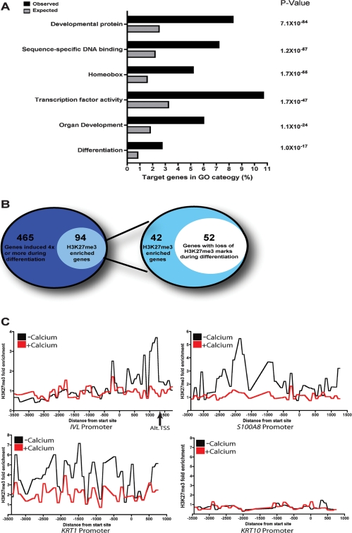
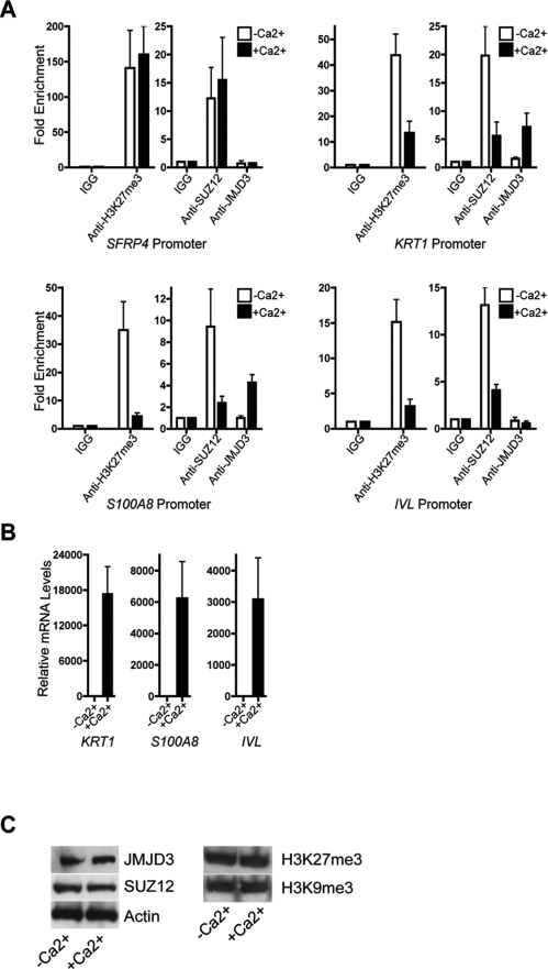
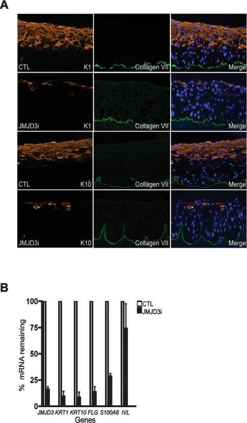
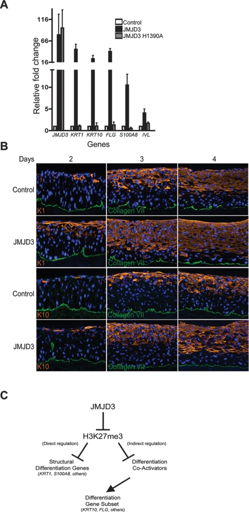
The recent discovery of H3K27me3 demethylases suggests that H3K27me3 may dynamically regulate gene expression, but this potential role in mammalian tissue homeostasis remains uncharacterized. In the epidermis, a tissue that balances stem cell self-renewal with differentiation, H3K27me3, occupies the promoters of many differentiation genes. During calcium-induced differentiation, H3K27me3 was erased at these promoters in concert with loss of PcG protein occupancy and increased binding by the H3K27me3 demethylase, JMJD3. Within epidermal tissue, JMJD3 depletion blocked differentiation, while active JMJD3 dominantly induced it. These results indicate that epigenetic derepression by JMJD3 controls mammalian epidermal differentiation.


Figures

Figure 1.
Figure 1.
Global analysis of H3K27me3 enrichment at genomic promoters during epidermal differentiation. (A) Gene Ontology (GO) analysis of promoters enriched for H3K27me3 marks. Black bars represent the observed percentage of H3K27me3 enriched sites in a certain GO category; gray bars represent the expected percentage. P-values were obtained using hypergeometric distribution. (B) Classification of epidermal differentiation genes. The left diagram depicts the number of H3K27me3 enriched epidermal differentiation genes. The right diagram shows the number of H3K27me3 enriched differentiation genes that lose methylation during differentiation. (C) H3K27me3 enrichment ratios on genomic tiling arrays comparing individual differentiation gene promoters for all probes in cells cultured in growth medium (−calcium) or differentiation medium (+calcium). The arrow below the IVL graph denotes an alternate transcription start site.
Figure 2.
Figure 2.
JMJD3 binds to the promoters of epidermal differentiation genes. (A) ChIP using H3K27me3, SUZ12, or JMJD3 antibody on the promoters of three differentiation genes, KRT1, S100A8, and IVL, in the absence or presence of calcium. SFRP4 was used as a positive control for H3K27me3 binding. (B) QPCR analysis on the mRNA levels of differentiation genes in the absence or presence of calcium. (C) Western blot depicting levels of JMJD3, SUZ12, H3K27me3, and H3K9me3 in keratinocytes grown in the absence or presence of calcium. Actin was used as a loading control.
Figure 3.
Figure 3.
JMJD3 is required for epidermal differentiation. (A) Epidermal cells receiving siRNA against JMJD3 (JMJD3i) or nonfunctional control siRNA (CTL) were used to regenerate epidermal tissue on human dermis in organotypic culture. Tissue sections were stained for the presence of keratin 1 (K1) and keratin 10 (K10). Type VII collagen (green) was used as a costain and marks the boundary between the epidermis and dermis. Note the loss of differentiation-specific keratins with JMJD3 depletion. (B) Quantitation of tissue mRNA levels by QPCR analysis, normalized to GAPDH.
Figure 4.
Figure 4.
JMJD3 can induce premature epidermal differentiation. (A) Keratinocytes were transduced with retroviral expression constructs for empty vector control, JMJD3, and the JMJD3 H1390A demethylase-defective point mutant and grown in low calcium conditions. RNA was isolated from the samples 5 d post-transduction and QPCR was used to determine levels of derepression of epidermal differentiation genes. (B) JMJD3-expressing and control regenerated organotypic tissue was harvested at days 2, 3, and 4 to determine the impact of JMJD3 expression on differentiation. Tissue sections were stained for keratin 1 and keratin 10. Note earlier expression of differentiation proteins and their localization nearer to the epidermal basement membrane in tissue engineered to express JMJD3. (C) Model of epigenetic control of epidermal differentiation.

References

    1. Agger K., Cloos P.A., Christensen J., Pasini D., Rose S., Rappsilber J., Issaeva I., Canaani E., Salcini A.E., Helin K. UTX and JMJD3 are histone H3K27 demethylases involved in HOX gene regulation and development. Nature. 2007;449:731–734. - PubMed
    1. Barski A., Cuddapah S., Cui K., Roh T.Y., Schones D.E., Wang Z., Wei G., Chepelev I., Zhao K. High-resolution profiling of histone methylations in the human genome. Cell. 2007;129:823–837. - PubMed
    1. Blanpain C., Fuchs E. Epidermal stem cells of the skin. Annu. Rev. Cell Dev. Biol. 2006;22:339–373. - PMC - PubMed
    1. Boyce S.T., Ham R.G. Calcium-regulated differentiation of normal human epidermal keratinocytes in chemically defined clonal culture and serum-free serial culture. J. Invest. Dermatol. 1983;81:33s–40s. - PubMed
    1. Boyer L.A., Plath K., Zeitlinger J., Brambrink T., Medeiros L.A., Lee T.I., Levine S.S., Wernig M., Tajonar A., Ray M.K., et al. Polycomb complexes repress developmental regulators in murine embryonic stem cells. Nature. 2006;441:349–353. - PubMed

Publication types