. 2008 Jul;21(3):248-54.
doi: 10.1080/08998280.2008.11928404.

Autologous fat grafts placed around temporomandibular joint total joint prostheses to prevent heterotopic bone formation

Affiliations

Autologous fat grafts placed around temporomandibular joint total joint prostheses to prevent heterotopic bone formation

Larry M Wolford et al. Proc (Bayl Univ Med Cent). 2008 Jul.

Abstract

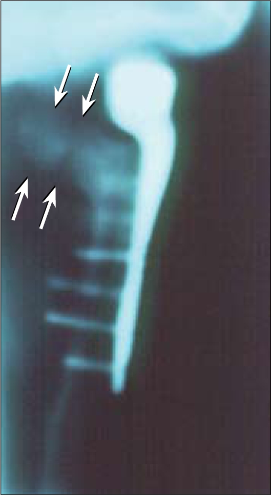
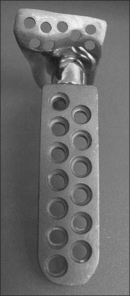
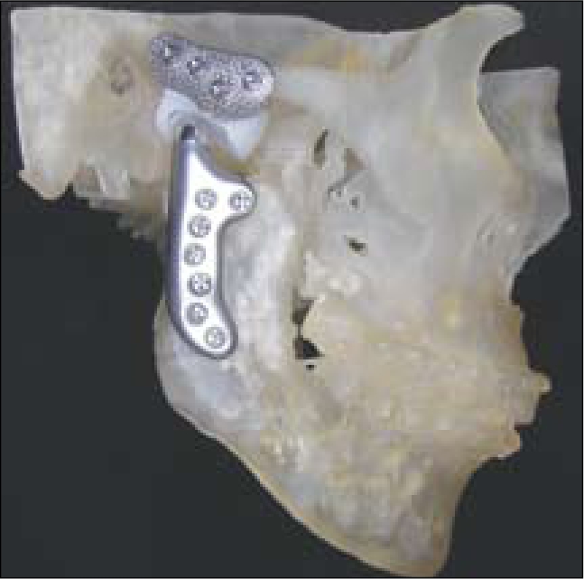
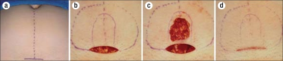
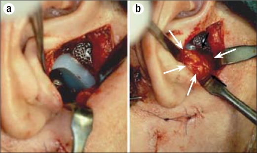
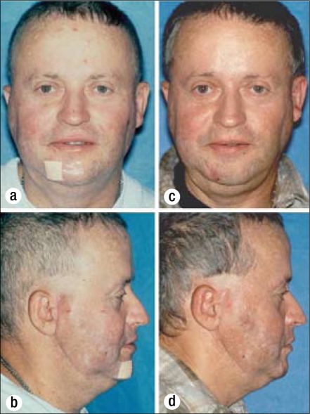
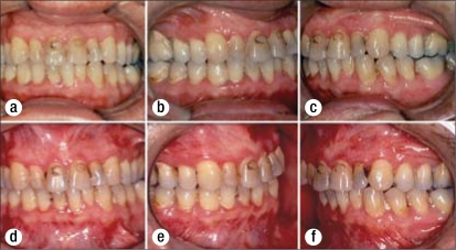
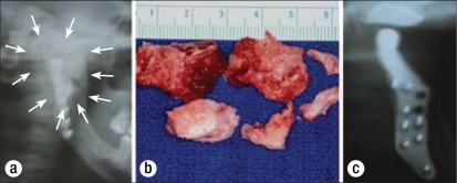
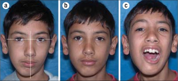
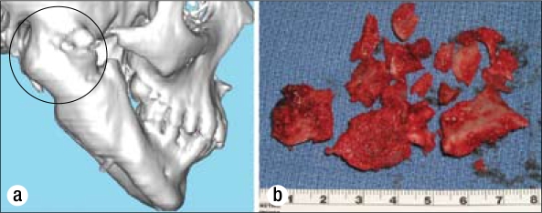
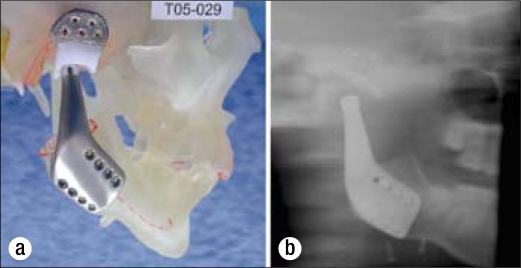
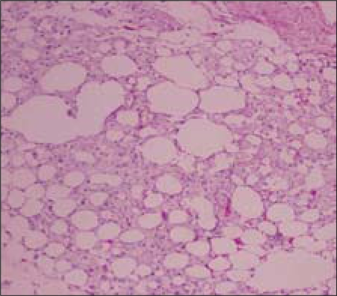
This study evaluated 1) the efficacy of packing autologous fat grafts around temporomandibular joint (TMJ) total joint prosthetic reconstructions to prevent fibrosis and heterotopic bone formation and 2) the effects on postsurgical joint mobility and jaw function. One hundred fifteen patients (5 males and 110 females) underwent TMJ reconstruction with total joint prostheses and simultaneous fat grafts (88 bilateral and 27 unilateral) for a total of 203 joints. The abdominal fat grafts were packed around the articulating portion of the joint prostheses after the fossa and mandibular components were stabilized. Patients were divided into two groups: group 1 (n = 76 joints) received Christensen total joint prostheses, and group 2 (n = 127 joints) received TMJ Concepts total joint prostheses. Clinical and radiographic assessments were performed before surgery, immediately after surgery, and at long-term follow-up. In group 1, maximal incisal opening (MIO) increased 3.5 mm, lateral excursions (LE) decreased 0.2 mm, and jaw function improved 1.9 levels. In group 2, MIO increased 6.8 mm, LE decreased 1.4 mm, and jaw function improved 2.4 levels. The improvement for MIO and patient perception of jaw function in both groups was statistically significant; no significant difference was found for LE. There was no radiographic or clinical evidence of heterotopic calcifications or limitation of mobility secondary to fibrosis in either group. Twenty-five Christensen prostheses (33%) were removed because of device failure and/or metal hypersensitivity; no fibrosis or heterotopic bone formation was seen at surgical removal. Four TMJ Concepts prostheses (3%) were removed because of metal hypersensitivity. In all instances, removal of the prostheses was unrelated to the autologous fat grafting. Ten patients (8.7%) developed complications involving the fat donor site: two patients (1.8%) developed abdominal cysts requiring surgery, and eight patients (6.9%) developed seroma formation requiring aspiration. Autologous fat transplantation is a useful adjunct to prosthetic TMJ reconstruction to minimize the occurrence of excessive joint fibrosis and heterotopic calcification, consequently providing improved range of motion and jaw function.


Figures

Figure 1
Figure 1
A coronal tomogram of a prosthetically reconstructed TMJ joint demonstrates heterotopic bone formation (arrows) between the mandibular ramous and medial side of the fossa. No fat graft was placed around the prosthesis at surgery.
Figure 2
Figure 2
The Christensen prosthesis is an off-the-shelf device with three selections for the man-dibular component and over 40 selections for the fossa component. The best-fitting components are selected to fit the anatomy. These devices have metal-on-metal articulations.
Figure 3
Figure 3
The TMJ Concepts total joint prosthesis is a patient-fitted device, constructed on a three-dimensional model and designed for each patient's specific anatomical requirements. The devices have metal-on-polyethylene articulations.
Figure 4
Figure 4
Fat graft harvesting technique used in this study. (a) The fat graft is harvested from the abdomen, usually through a 4- to 5-cm incision generally made in the suprapubic area. (b) The outer dashed line is the extent of undermining of the skin and beneath the fat pad. The inner solid line denotes the fat graft to be harvested. (c) The abdominal fat graft harvesting is complete. (d) 3-0 polyglactin sutures are used to close the deep fat layers so no depression in the harvest area will be evident. The skin is closed with subcuticular suturing.
Figure 5
Figure 5
Surgical technique used in this study. (a) The fossa component prosthesis is placed through an endaural or preauricular incision. The mandibular component is placed through a submandibular incision. (b) The abdominal fat graft (arrows) is packed into the joint space to prevent heterotopic bone formation and fibrosis.
Figure 6
Figure 6
Case 1. (a, b) This 45-year-old man was referred after 14 previous failed right TMJ surgeries; the most recent involved right TMJ reconstruction with a total joint prosthesis (Osteomed system) without a fat graft. He had severe TMJ and myofascial pain, headaches, and difficulty eating. (c, d) The patient 2 years after right TMJ debridement, removal of heterotopic bone and the Osteomed prosthesis, TMJ reconstruction with a TMJ Concepts patient-fitted total joint prosthesis, and fat grafting.
Figure 7
Figure 7
Case 1. (a, b, c) Presurgery, the patient had a class I occlusion on the left side and a class II occlusion on the right side. (d, e, f) The occlusion remained stable 2 years after surgery.
Figure 8
Figure 8
Case 1. (a) The presurgical panographic x-ray showed massive heterotopic bone formation (outlined by arrows) around the Osteomed prosthesis. (b) The heterotopic bone was removed in sections. (c) A 10-year postsurgical radiograph shows the effectiveness of the fat graft in preventing heterotopic bone development.
Figure 9
Figure 9
Case 2. (a) This 12-year-old boy had right TMJ ankylosis, and two attempts at correction by rib grafting (without fat grafts) had failed. With only 3 mm of incisal opening, he was developing significant dental problems, marked facial asymmetry, and sleep apnea. (b, c) The patient 2 years after right-side TMJ reconstruction and mandibular advancement with a TMJ Concepts total joint prosthesis and fat graft. He had improved facial balance and good jaw function (35-mm opening) without pain.
Figure 10
Figure 10
Case 2. (a) A 3-D computed tomography scan demonstrates the magnitude of the heterotopic bone and joint ankylosis. (b) The heterotopic bone was removed in sections.
Figure 11
Figure 11
Case 2. (a) A TMJ Concepts total joint prosthesis was custom made to reconstruct the TMJ and advance and vertically lengthen the right mandibular ramus. A fat graft was packed around the prosthesis to prevent heterotopic bone from redeveloping. (b) The tomogram shows no heterotopic bone formation around the prosthesis 2 years after surgery.
Figure 12
Figure 12
Histological examination of a fat graft biopsy taken 4 years after implantation around a TMJ Concepts total joint prosthesis shows viable fat still present without evidence of inflammation, heterotopic bone formation, or significant fibrosis.

References

    1. Neuber H. Fettransplantation. Chir Kong Verhandl. 1893;1:66.
    1. Blair VP. Operative treatment of ankylosis of the mandible. Trans South Surg Assoc. 1913;28:435.
    1. Murphy JB. Arthroplasty for intra-articular bony and fibrous ankylosis of the temporomandibular articulation. JAMA. 1914;62:1783.
    1. Tomas BJ. Heterotopic bone formation after total hip arthroplasty. Orthop Clin North Am. 1992;23(2):347–358. - PubMed
    1. Fingeroth RJ, Ahmed AQ. Single dose 6 Gy prophylaxis for heterotopic ossification after total hip arthroplasty. Clin Orthop Relat Res. 1995;317:131–140. - PubMed

LinkOut - more resources