

Proteomic analysis of skin invasion by blood fluke larvae
- PMID: 18629379
- PMCID: PMC2467291
- DOI: 10.1371/journal.pntd.0000262
Proteomic analysis of skin invasion by blood fluke larvae
Abstract
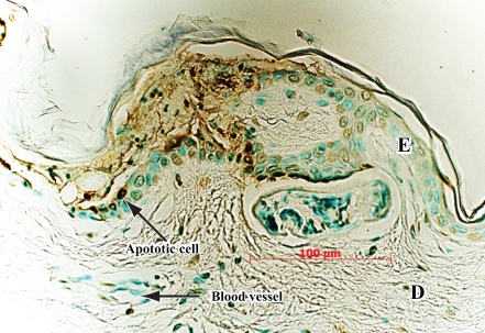
Background: During invasion of human skin by schistosome blood fluke larvae (cercariae), a multicellular organism breaches the epidermis, basement membrane, and dermal barriers of skin. To better understand the pathobiology of this initial event in schistosome infection, a proteome analysis of human skin was carried out following invasion by cercariae of Schistosoma mansoni.
Methodology and results: Human skin samples were exposed to cercariae for one-half hour to two hours. Controls were exposed to water used to collect cercariae in an identical manner, and punctured to simulate cercarial tunnels. Fluid from both control and experimental samples was analyzed by LC/MS/MS using a linear ion trap in "triple play" mode. The coexistence of proteins released by cercariae and host skin proteins from epidermis and basement membrane confirmed that cercarial tunnels in skin were sampled. Among the abundant proteins secreted by cercariae was the cercarial protease that has been implicated in degradation of host proteins, secreted proteins proposed to mediate immune invasion by larvae, and proteins implicated in protection of parasites against oxidative stress. Components of the schistosome surface tegument, previously identified with immune serum, were also released. Both lysis and apoptosis of epidermal cells took place during cercarial invasion of the epidermis. Components of lysed epidermal cells, including desmosome proteins which link cells in the stratum granulosum and stratum spinosum, were identified. While macrophage-derived proteins were present, no mast cell or lymphocyte cytokines were identified. There were, however, abundant immunoglobulins, complement factors, and serine protease inhibitors in skin. Control skin samples incubated with water for the same period as experimental samples ensured that invasion-related proteins and host protein fragments were not due to nonspecific degeneration of the skin samples.
Conclusions: This analysis identified secreted proteins from invasive larvae that are released during invasion of human skin. Analysis of specific host proteins in skin invaded by cercariae served to highlight both the histolytic events facilitating cercarial invasion, and the host defenses that attempt to arrest or retard invasion. Proteins abundant in psoriatic skin or UV and heat-stressed skin were not abundant in skin invaded by cercariae, suggesting that results did not reflect general stress in the surgically removed skin specimen. Abundant immunoglobulins, complement factors, and serine protease inhibitors in skin form a biochemical barrier that complements the structural barrier of the epidermis, basement membrane, and dermis. The fragmentation of some of these host proteins suggests that breaching of host defenses by cercariae includes specific degradation of immunoglobulins and complement, and either degradation of, or overwhelming the host protease inhibitor repertoire.
Conflict of interest statement
The authors have declared that no competing interests exist.
Figures

Comment in
-
Tracking the odysseys of juvenile schistosomes to understand host interactions.PLoS Negl Trop Dis. 2008 Jul 16;2(7):e257. doi: 10.1371/journal.pntd.0000257. PLoS Negl Trop Dis. 2008. PMID: 18628997 Free PMC article. No abstract available.
References
-
- Elias PM. Stratum corneum architecture, metabolic activity and interactivity with subjacent cell layers. Exp Dermatol. 1996;5:191–201. - PubMed
-
- Dorsey CH, Cousin CE, Lewis FA, Stirewalt MA. Ultrastructure of the Schistosoma mansoni cercaria. Micron. 2002;33:279–323. - PubMed
-
- Stirewalt MA. Schistosoma mansoni: cercaria to schistosomule. Adv Parasitol. 1974;12:115–182. - PubMed
-
- Haas W, Schmitt R. Characterization of chemical stimuli for the penetration of Schistosoma mansoni cercariae. I. Effective substances, host specificity. Z Parasitenkd. 1982;66:293–307. - PubMed
-
- Haas W, Diekhoff D, Koch K, Schmalfuss G, Loy C. Schistosoma mansoni cercariae: stimulation of acetabular gland secretion is adapted to the chemical composition of mammalian skin. J Parasitol. 1997;83:1079–1085. - PubMed
