Review
. 2008 Oct;237(10):2643-56.
doi: 10.1002/dvdy.21599.

Palatal seam disintegration: to die or not to die? that is no longer the question

Affiliations
Review

Palatal seam disintegration: to die or not to die? that is no longer the question

Ali Nawshad. Dev Dyn. 2008 Oct.

Abstract

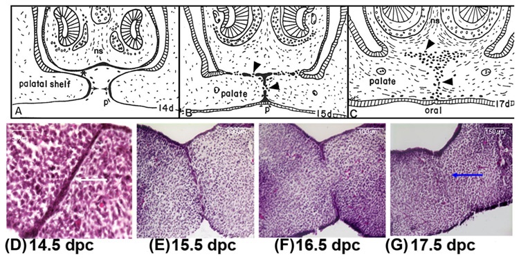
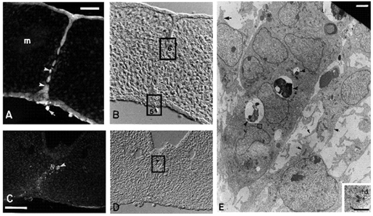
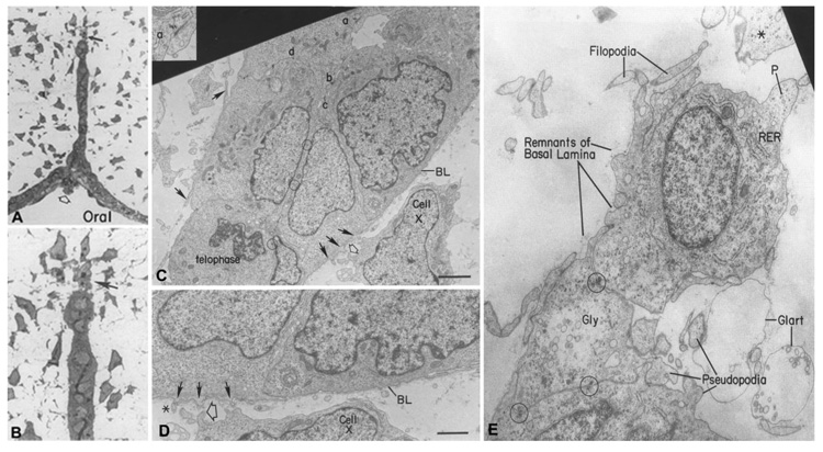
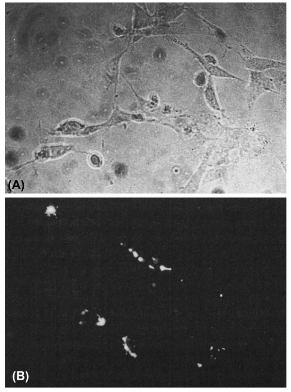
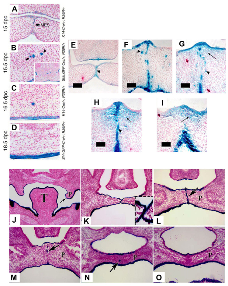
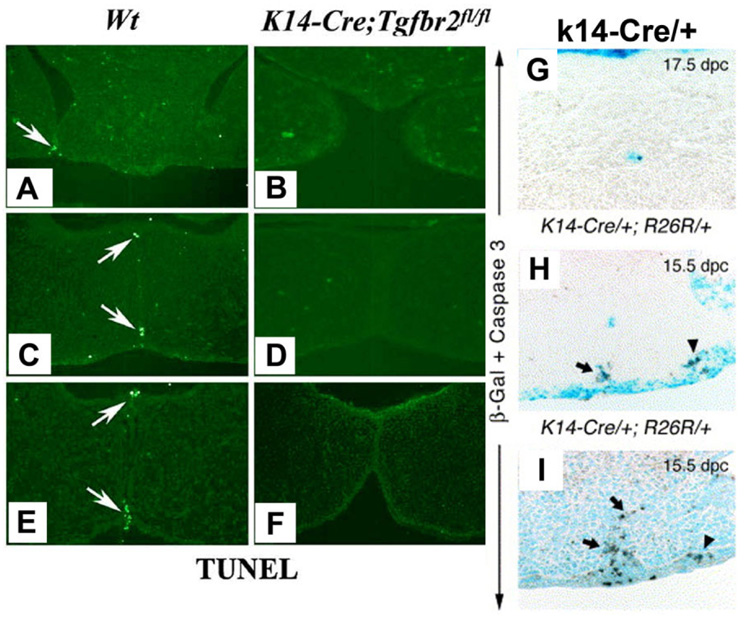
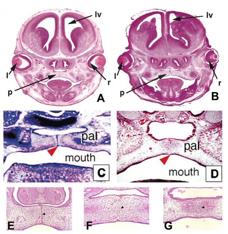
Formation of the medial epithelial seam (MES) by palatal shelf fusion is a crucial step of palate development. Complete disintegration of the MES is the final essential phase of palatal confluency with surrounding mesenchymal cells. In general, the mechanisms of palatal seam disintegration are not overwhelmingly complex, but given the large number of interacting constituents; their complicated circuitry involving feedforward, feedback, and crosstalk; and the fact that the kinetics of interaction matter, this otherwise simple mechanism can be quite difficult to interpret. As a result of this complexity, apparently simple but highly important questions remain unanswered. One such question pertains to the fate of the palatal seam. Such questions may be answered by detailed and extensive quantitative experimentation of basic biological studies (cellular, structural) and the newest molecular biological determinants (genetic/dye cell lineage, gene activity, kinase/enzyme activity), as well as animal model (knockouts, transgenic) approaches. System biology and cellular kinetics play a crucial role in cellular MES function; omissions of such critical contributors may lead to inaccurate understanding of the fate of MES. Excellent progress has been made relevant to elucidation of the mechanism(s) of palatal seam disintegration. Current understanding of palatal seam disintegration suggests epithelial-mesenchymal transition and/or programmed cell death as two most common mechanisms of MES disintegration. In this review, I discuss those two mechanisms and the differences between them.


Figures

Fig. 1
Fig. 1
Camera lucida drawings of paraffin sections of the developing rodent palate. The anterior palate (shown) of the mouse, fuses with the nasal septum (ns), but the posterior palate does not because there is no nasal septum posteriorly. A: Between 13 and 14 days post coitum (dpc), palatal shelves move horizontally (arrows) across the mouth above the tongue. At this time, the periderm (outer layer of the oral epithelium) sloughs off the epithelium along the medial palatal edge (p) and ventral nasal septum (*). B: The palatal processes do not fuse in vivo until periderm sloughs; therefore, there are few, if any, peridermal cells left to be trapped in vivo in the MEE and nasal septum seams. These epithelial seams transform to mesenchyme (arrowheads, B and C) between 15 and 17 dpc. C: The palate shelves fuse together and with the nasal septum by transforming the adherent epithelial seams to mesenchyme to become confluent. The dark cells (arrowheads) show diagrammatically the relative contribution to mesenchyme made by these epithelial seams. D–G: Hematoxylin and eosin-stained sections of rodent palates were fixed at different stages of palatogenesis in vitro showing chronological disintegration of the palatal seam (white arrow, D) to reach complete confluence (blue arrow, G) of the mesenchyme by 17.5 dpc. A–C from Griffith and Hay (2003), © Development, The Company of Biologist Ltd.
Fig. 2
Fig. 2
Demonstration of epithelial–mesenchymal transition (EMT) by tracing palatal medial edge seam (MES) cell with carboxydichlorofluorescein diacetate succinimidyl ester (CCFSE). A–D: One day after in vitro labeling, CCFSE labeling is present in the cells of the midline seam (A, B) and in the mesenchyme-like cells deriving from epithelium in the region of the seam (C, D). These labeled mesenchyme-like cells are indistinguishable from others in the mesoderm after attainment of palatal confluence, as shown in D (photographed with Nomarski optics). The CCFSE, originally diffuse in distribution, is condensing into one or more fluorescent spots per cell (arrowheads, A and C), which represent dye packaged within isolation bodies. Mesenchymal cells situated outside the midline seam (m, A) have no isolation bodies showing that the CCFSE absorbed through the periderm was confined to the epithelial cells and did not pass through the basal plasmalemma into the mesenchyme. Periderm staining is brighter than that of the basal epithelial cells. A sloughed periderm cell is labeled (arrow, A). Palate C was cultured in a medium (Abbott) that promoted faster development and it already has sloughed most of the CCFSE-containing surface epithelial cells. In the region shown here, the nasal septum epithelium does not fuse with the palate. E: This epithelial island is a remnant of the disappearing seam (as at the square labeled 6). Isolation bodies are present (arrowheads, E). Mesenchymal cells are distinguished morphologically by their shape and well-developed pseudopodia and filopodia (arrows, E). The epithelial cells in the island are joined by desmosomes, one of which (square, E) is enlarged in the inset. Scale bars = 25 mm in A, 50 mm in C, 2 µm in E; 0.5 µm in inset. A–E from Griffith and Hay (2003), © Development, The Company of Biologist Ltd.
Fig. 3
Fig. 3
Role of epithelial–mesenchymal transition (EMT) in palate epithelial seam disintegration. A, B: Light micrograph of a palate seam breaking up into mesenchyme (A), magnified further in (B) to show the transforming cell (arrow) at the tip of the breaking seam. Electron micrograph of the two-cell-thick midline epithelial seam in vivo near the nasal surface. C: Enlargement of a portion of the epithelial seam at C (a, inset) shows a desmosome, many of which appear in the major figures C (b–d). Circles indicate location of additional desmosomes between opposed epithelial cell layers. The seam shows the typical excellent health of the MEE in vivo. A telophase is present indicating mitosis is still occurring. Cell X is the same mesenchymal cell in C and D. The basal lamina (BL) is still mainly intact, but filopodia (closed arrows) are being extended through or along it. Open arrows in C and D are on the same cell process of this seam in surrounding mesenchyme. Asterisk in D is a pre-existing mesenchymal cell process in close contact with epithelial filopodium. Anterior palate (rodent). E: Electron micrograph of an elongating cell breaking away from the tip of a disappearing epithelial seam. The basal lamina is almost completely gone and the cell at the tip of the seam is extending numerous filopodia and pseudopodia typical of mesenchymal cells. Circles identify desmosomes still present in the seam. These persist until the cell undergoing EMT breaks away and the one linking the top cell to the bottom seam indicates its origin from the epithelium. Gly, glycogen; Glart, glutaraldehyde artifact; P, leading pseudopodium. Scale bars = 3 µm in C, 1.5 µm in D, 100 nm in E. From Fitchett and Hay (1989), © Developmental Biology, Academic Press Inc.
Fig. 4
Fig. 4
Labeled Palatal cells in culture maintain mesenchymal morphology. Cells isolated from the midline position of a palatal shelf after the completion of palatal fusion and placed in organ culture retain the marker for cell lineage and have a fibroblastic morphology. A: Phase-contrast microscopy of palatal mesenchyme cells in culture. B: DiI (l, l′-dioctadecyl-3, 3, 3′, 3′-tetramethylindo-carbo-cyanine perchlorate) fluorescence of the same field of cultured cells. From Shuler, CF (1995), © Crit Rev Oral Biol Med, International and American Associations for Dental Research.
Fig. 5
Fig. 5
Fate Mapping with different genetic labeling method during palatal seam disintegration. A–D: Frontal sections from 15 days post coitum (dpc) K14-Cre/+; R26R/+ (A), 15.5 dpc ShhGFPCre/+; R26R/+ (B), 16.5 dpc K14-Cre/+; R26R/+ (C), and 18.5 dpc ShhGFPCre/+; R26R/+ (D) embryos at the anterior segments of the palate stained for β-galactosidase. MES, midline epithelial seam (arrows in panels A). Inset in B is a low-magnification view of the micrograph. At 18.5 dpc, the palate is virtually cleared from lacZ-positive epithelial islands (D). Mesenchymal cells are totally devoid of β-galactosidase activity. A, B, C, and D are from Vaziri Sani et al. (2005), © Developmental Biology, Elsevier Inc. β-gal staining pattern in (K14-Cre; R26R) embryos demonstrates the occurrence of EMT during and after seam degeneration. E: β-gal staining in the anterior region of the palate at early 14.5 dpc, where the two shelves have just made contact. Note that the MEE cells were strongly labeled with β-gal (arrowhead), but no signal was present in mesenchymal cells. F: β-gal staining in the middle region of the palate at early 14.5 dpc, where seam degeneration has just been initiated. Some epithelial-like β-gal-positive cells have dissociated from the midline and migrated into the mesenchymal region (arrowhead). G, H: β-gal staining in the middle region of the palate at late 14.5 dpc, when seam degeneration is advanced. Both clump-like blue cells (arrow-head in G, H) and typical mesenchymal-looking blue cells (arrow in G, H) were observed in the mesenchymal region of the palate. I: β-gal staining in the fully fused palate at 15.5 dpc, showing that a high portion of the mesenchymal cells were β-gal positive (arrow). Scale bars = 100 µm in E, 50µm F–I. E, F, G, H, I are from Jin and Ding (2006), © Development, The Company of Biologist Ltd. β-Galactosidase staining of frontal sections from K14-Cre; R26R embryos. J: At 13.5 dpc, the secondary palate shelf projects toward the midline, palatal epithelium cells are β-galactosidase positive. K: At 14 dpc, the opposite secondary palate shelves contact each other, and the medial edge epithelial (MEE) cells are all β-galactosidase positive (insert). L: At 14.5 dpc, the palatal shelves have fused, and the midline epithelial seam (MES) is β-galactosidase positive (arrow), no β-galactosidase positive cells can be found in the palatal mesenchyme. M: At E15.5, most of the MES (arrow) has disappeared. N: At 16.5 dpc, few β-galactosidase–positive cells remain in the midline (arrow) and no β-galactosidase positive cells can be found in the palatal mesenchyme. O: At 17.5 dpc, no β-galactosidase positive cells can be found in the palatal mesenchyme. P, palatal shelf; T, tongue. Figs. J, K, L, M, N, and O are from Xu et al. (2006), © Developmental Biology, Elsevier Inc.
Fig. 6
Fig. 6
Detection of PCD in palatal seam disintegration. A–F: The fate of the medial edge epithelia (MEE) is changed in the K14-Cre; Tgfbr2fl/fl mutant palate. At 14.5 days post coitum (dpc), wild-type MEE cells show positive TUNEL staining, a marker for cell death (arrow) from anterior to posterior part of the palate. A, C, E: No cell death can be detected in the K14-Cre; Tgfbr2fl/fl mutant palate. B, D, F: At 15.5 dpc, palatal fusion process has reached the end, most of the wild-type MEE cells have disappeared. G, H, I: Immunohistochemistry with anti-activated caspase-3. Palatal sections palate from a 17.5 dpc K14-Cre/+; R26R/+ embryo showing activated-caspase-3 immunostaining (brown color) in a lacZ positive epithelial island (G). 15.5 dpc K14-Cre/+; R26R/+ embryo showing anti-activated caspase-3 immunostaining in the regressing lacZ-positive MES (arrow) and in the lateral epithelium (arrowhead; H). Palatal section from a 15.5 dpc K14-Cre/+ embryo (no R26R allele) showing cells positive for activated caspase-3, showing pre-PCD in both the regressing MES (arrow) and the epithelium at the junction of fusion between the maxillary and intermaxillary processes (arrowhead; I). A–F are from Xu et al. (2006), © Developmental Biology, Elsevier Inc. and G–I are from Vaziri Sani et al. (2005), © Developmental Biology, Elsevier Inc.
Fig. 7
Fig. 7
Palate development in Apaf-1 mutant mice. Cranial sections from wild-type and Apaf-1 mutant mice cut coronally and stained with hematoxylin and eosin are shown at ×50 magnification. A, B: Wild-type at 15.5 days post coitum (dpc; A) and Apaf-1 mutant mice at gestational ages equivalent to wild-type embryos (B). In A and B, “l”, “de”, “tv”, “v”, “lv”, “p”, “ge”, and “r” show the positions of the lens, diencephalon, telencephalic vesicle, third ventricle, lateral ventricle, palate, ganglionic eminence, and retina, respectively. A and B are from Honapour et al. (2000), © Developmental Biology, Academic Press. C: Transversal section through the caudal third of the palate of an 14.5 dpc homozygous embryo. The palatal shelves meet in the midline (arrowhead) but do not fuse. pal, secondary palate. D: Transversal section through the caudal third of the palate of an 14.5 dpc wild-type embryo, showing complete fusion of the palatal shelves in the midline (arrowhead). pal, secondary palate; C and D are from Cecconi et al. (1998), © Cell, Cell Press. E: The MEE seam (arrowhead) forms normally in Apaf-1 mutant embryos at 14.5 dpc. F: The MEE seam undergoes degeneration in Apaf-1 mutant embryos at 15.5 dpc to establish the mesenchyme confluence cross the midline (arrowheads). G: At 16.5 dpc, both wild-type and Apaf-1 mutant embryos form a continuous palate with no sign of seam cells in the midline area (arrowhead). E, F, and G are from Jin and Ding (2006) © Development, The Company of Biologist Ltd.

References

    1. Abdelilah-Seyfried S, Cox DN, Jan YN. Bazooka is a permissive factor for the invasive behavior of discs large tumor cells in Drosophila ovarian follicular epithelia. Development. 2003;130:1927–1935. - PubMed
    1. Abraham MC, Shaham S. Death without caspases, caspases without death. Trends Cell Biol. 2004;14:184–193. - PubMed
    1. Ahmed S, Liu CC, Nawshad A. Mechanisms of palatal epithelial seam disintegration by transforming growth factor (TGF) beta3. Dev Biol. 2007;309:193–207. - PMC - PubMed
    1. Arnoult D, Parone P, Martinou JC, Antonsson B, Estaquier J, Ameisen JC. Mitochondrial release of apoptosis-inducing factor occurs downstream of cytochrome c release in response to several proapoptotic stimuli. J Cell Biol. 2002;159:923–929. - PMC - PubMed
    1. Barasch J. Genes and proteins involved in mesenchymal to epithelial transition. Curr Opin Nephrol Hypertens. 2001;10:429–436. - PubMed