Review
. 2008 Dec;237(12):3464-76.
doi: 10.1002/dvdy.21598.

Morphogenesis of the node and notochord: the cellular basis for the establishment and maintenance of left-right asymmetry in the mouse

Affiliations
Review

Morphogenesis of the node and notochord: the cellular basis for the establishment and maintenance of left-right asymmetry in the mouse

Jeffrey D Lee et al. Dev Dyn. 2008 Dec.

Abstract

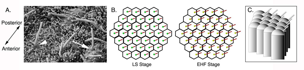
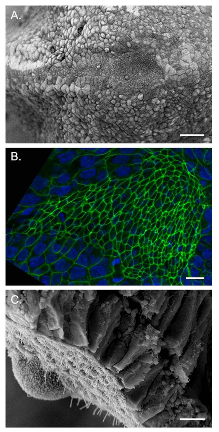
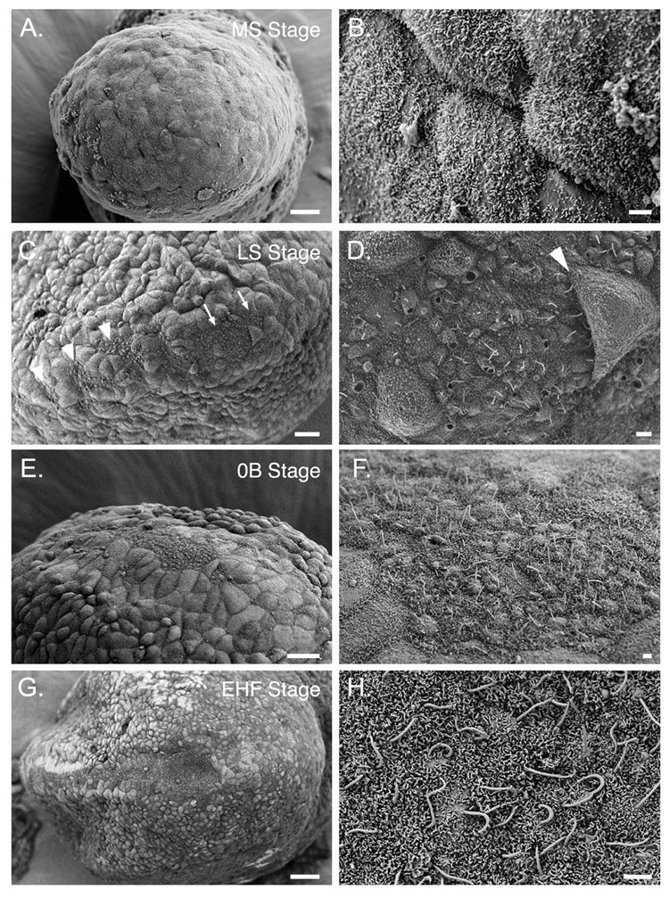
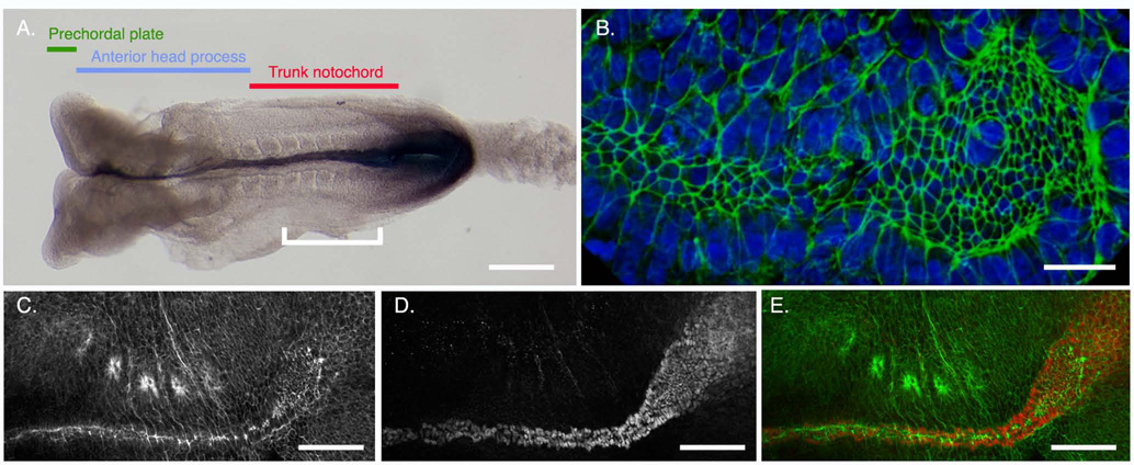
Establishment of left-right asymmetry in the mouse embryo depends on leftward laminar fluid flow in the node, which initiates a signaling cascade that is confined to the left side of the embryo. Leftward fluid flow depends on two cellular processes: motility of the cilia that generate the flow and morphogenesis of the node, the structure where the cilia reside. Here, we provide an overview of the current understanding and unresolved questions about the regulation of ciliary motility and node structure. Analysis of mouse mutants has shown that the motile cilia must have a specific structure and length, and that they must point posteriorly to generate the necessary leftward fluid flow. However, the precise structure of the motile cilia is not clear and the mechanisms that position cilia on node cells have not been defined. The mouse node is a teardrop-shaped pit at the distal tip of the early embryo, but the morphogenetic events that create the mature node from cells derived from the primitive streak are only beginning to be characterized. Recent live imaging experiments support earlier scanning electron microscopy (SEM) studies and show that node assembly is a multi-step process in which clusters of node precursors appear on the embryo surface as overlying endoderm cells are removed. We present additional SEM and confocal microscopy studies that help define the transition stages during node morphogenesis. After the initiation of left-sided signaling, the notochordal plate, which is contiguous with the node, generates a barrier at the embryonic midline that restricts the cascade of gene expression to the left side of the embryo. The field is now poised to dissect the genetic and cellular mechanisms that create and organize the specialized cells of the node and midline that are essential for left-right asymmetry.


Figures

Figure 1
Figure 1
A simplified schematic of the core left-right signaling pathway in the e8.5 (6 somite) mouse embryo. The embryo is viewed from the ventral side to highlight the ventrally located node. Nodal (blue), a TGFβ family ligand, is expressed around the periphery of the mouse node (outlined in black). Leftward fluid flow across the node requires the correct placement and rotation of nodal cilia (green), and leads to expression of Nodal in the left lateral plate mesoderm (LPM). Nodal signaling in the left LPM induces the expression of upregulates its own expression in a positive feedback loop and also induces expression lefty2 and Pitx2. Lefty2 (red) antagonizes the activity of Nodal and limits its range of activity. Nodal made in the left LPM also activates the expression of lefty1 (purple) in the left prospective floor plate, dorsal to the notochord (black line extending anteriorly toward the midline). Lefty1 antagonizes Nodal and prevents the spread of left signaling to the right LPM. The homeodomain transcription factor Pitx2 (yellow) controls later left-sided morphogenetic events. Lack of nodal and Pitx2 expression in the LPM leads to right-sided isomerism in the thorax (e.g. right pulmonary isomerism); bilateral expression of nodal and Pitx2 in the LPM leads to left pulmonary isomerism.
Figure 2
Figure 2
Cilia position on cells of the node. A. Scanning EM of the node pit at 0B/EHF stage (e8.0). The anterior-posterior axis is labeled. At this stage, some cilia emerge from the center of the cell (arrowhead) and some cilia have shifted to a more posterior position (arrow), while Scale bar = 1 µm. B. Schematics of ventral views of the node pit showing cilia position in cells at LS and EHF stages. Posterior is to the right. At LS stage cilia emerge from the center of each cell; by EHF stage most cilia emerge from the posterior. Nodal flow begins after EHF stage and is well established by LHF stage (e8.25) (Okada et. al., 1999). C. Schematic of lateral view of node pit cells at EHF stage. Posterior is to the right. Cilia emerge from the posterior of each cell; because the apical surface of each cell is domed, the cilia project posteriorly.
Figure 3
Figure 3
Morphology of the mature node. A. Scanning electron micrograph (SEM) of EHF (e8.0) stage embryo, ventral view, anterior to the left. The node is visible as a teardrop-shaped pit of cells with small apical surfaces, surrounded by larger squamous crown cells, which are contiguous with the endoderm germ layer. B. Confocal micrograph of an EHF stage embryo showing the ventral node viewed en face, anterior to the left. Phalloidin (green) labels cortical F-actin rings at the apical surface of the polarized node pit cells; DAPI (blue) labels nuclei. Note the small apical surfaces of the cells of the node and axial midline. C. SEM of a LB stage node fractured in the transverse plane, ventral side down. Ciliated, apically constricted ventral node cells are exposed to the surface, and lie beneath the columnar epithelium of the dorsal node. Scale bars: A. 40µm B. 10µm C. 5µm.
Figure 4
Figure 4
Comparison of the organs of asymmetry among vertebrate species. Transverse view, dorsal is up. Ectoderm is red, paraxial mesoderm is yellow, endoderm/hypoblast is teal, and the organ of asymmetry is violet, with green cilia. The mouse ventral node and rabbit posterior notochordal plate are positioned beneath the ectoderm and are laterally contiguous with the endoderm/hypoblast. Mesoderm fills the space between the ectoderm and endoderm germ layers lateral to the node. The ventral pit of the mouse node is covered by Reichardt's membrane, creating an enclosed space. The teleost Kupffer's vesicle is an enclosed sphere in zebrafish (shown here) and, in medakafish, a hemisphere; cilia are concentrated on the dorsal anterior surface. The Xenopus gastrocoel roof plate is contiguous with the lateral endoderm; cilia project into the gastrocoel cavity. Panels are not size-matched.
Figure 5
Figure 5
SEM of the stages of node morphogenesis. All panels show ventral views of the distal tip of the embryo, the position of the node. Anterior is to the left. Staging is according to Downs and Davies (1993). Panels B, D, F and H are higher magnifications of panels A, C, E and G, respectively. (A, B): MS (mid-streak, e7.25) stage. Visceral endoderm distal region of the embryo. (C, D). LS stage (~e7.5). Clusters of cells with small apical surfaces and cilia begin to appear in groups near the presumptive node (arrows, C) and the axial midline (arrowheads, C). Some endoderm cells lie over the node field (arrowhead, D). Most cilia are located in the center of node cells at this stage. (E, F): 0B stage (e7.5–e7.75). The node region is free of overlying endoderm but has not adopted a concave shape. Cilia are longer and continue to project outward. (G, H) EHF stage (~e7.75?). The pit of the node is concave; cilia have elongated and project posteriorly. The panel in G is a lower magnification view of the embryo shown in Fig. 3A. These images are from embryos in the C3H/HeJ inbred strain. Scale bars: A, C, E, G: 20 µm; B, D, F, H: 2 µm.
Figure 6
Figure 6
The intermediate stage of node morphogenesis, when ciliated cells emerge from beneath overlying endoderm. (A, B) Confocal images of embryos labeled with anti-Arl13b (green) to highlight cilia (Caspary et al., 2007), phalloidin (red) to show cell boundaries, and DAPI to label nuclei (blue). A. LS stage embryo, ventral view, anterior to the left; 3D rendering showing scattered clusters of ventral node cells. The notochordal plate (bracket) is visible to the left (anterior). Cilia are mostly located at the center of each cell’s apical surface. B. yz-projection of confocal z-stack, LS stage embryo; anterior is up. Cilia (arrows) are visible on some cells still covered by endoderm. Several cilia are visible on cells that have already emerged onto the ventral surface (arrowheads). C. Scanning EM of a LS stage embryo (a higher magnification of Fig. 5D), showing cilia (arrowhead) visible beneath an overlying endoderm cell. Scale bars: A: 30µm; B: 10 µm; C: 2 µm.
Figure 7
Figure 7
Steps in node morphogenesis. Camera-lucida-style renderings depict the cell boundaries, traced from SEMs of embryos shown in Fig. 5 A, C and G. A. MS stage. B. LS stage. C. EHF stage. Blue represents endoderm, purple represents ventral node and notochordal plate. Black lines depict approximate plane of section in (D–F). D–F. Cartoons showing a model of node morphogenesis, representing transverse sections through MS, LS and EHF stage nodes showing the arrangement of cell layers. Red = epiblast, purple = ventral node, yellow = paraxial mesoderm, blue = endoderm. (A, D.) Ventral node begins to differentiate prior to emergence onto the ventral surface of the embryo: cells make cilia and begin to epithelialize. (B, E) Emergence occurs gradually during 0B to LB stages, as endoderm is cleared by an unknown process from the distal tip of the embryo. (C, F). Formation of the distinctive pit shape occurs after emergence onto the ventral surface. A–C: anterior up; D–F: epiblast up, endoderm below.
Figure 8
Figure 8
Morphogenesis of the notochordal plate. A. Regions of the axial midline of e8.5 embryo with 8 somites, dorsal view, anterior to the left, hybridized to reveal expression of Brachyury (T), which is expressed in the primitive streak, node and notochord. Prechordal plate (green) arises from the early gastrula organizer. Anterior head process (blue) arises from the mid-gastrula organizer. Trunk notochord (red) is derived from the node. White bracket is the region shown in (C–F). B. EHF stage embryo, ventral view of the node and axial midline; anterior to the left. Phalloidin (green) labels cell boundaries. Cells with small apical surfaces form a continuous population from the wider node (right) tapering to the notochordal plate (left). The notochordal plate is ~4 cells wide. (C–E). 4-somite embryo, ventral view, anterior to the left. C. Phalloidin staining shows the apically constricted node and notochord cells. The notochord has begun to form a rod along the AP axis; this creates a narrowed line of F-actin staining along the midline. Four somites are visible along the left (upper) flank of the embryo. The curve between the node and notochord is an artifact of the sample preparation. D. Anti-Brachyury immunofluorescence shows nuclei of the notochordal plate and node. E. Merge of (C, D). Scale bars: A: 150 µm; B: 20 µm; C–D: 100 µm.

References

    1. Abdelkhalek HB, Beckers A, Schuster-Gossler K, Pavlova MN, Burkhardt H, Lickert H, Rossant J, Reinhardt R, Schalkwyk LC, Muller I, Herrmann BG, Ceolin M, Rivera-Pomar R, Gossler A. The mouse homeobox gene Not is required for caudal notochord development and affected by the truncate mutation. Genes Dev. 2004;18:1725–1736. - PMC - PubMed
    1. Amack JD, Wang X, Yost HJ. Two T-box genes play independent and cooperative roles to regulate morphogenesis of ciliated Kupffer's vesicle in zebrafish. Dev Biol. 2007;310:196–210. - PubMed
    1. Amack JD, Yost HJ. The T box transcription factor no tail in ciliated cells controls zebrafish left-right asymmetry. Curr Biol. 2004;14:685–690. - PubMed
    1. Ang SL, Jin O, Rhinn M, Daigle N, Stevenson L, Rossant J. A targeted mouse Otx2 mutation leads to severe defects in gastrulation and formation of axial mesoderm and to deletion of rostral brain. Development. 1996;122:243–252. - PubMed
    1. Ang SL, Rossant J. HNF-3 beta is essential for node and notochord formation in mouse development. Cell. 1994;78:561–574. - PubMed

Publication types

LinkOut - more resources