Review
. 2008 Oct;237(10):2657-66.
doi: 10.1002/dvdy.21613.

Reprogramming multipotent tumor cells with the embryonic neural crest microenvironment

Affiliations
Review

Reprogramming multipotent tumor cells with the embryonic neural crest microenvironment

Jennifer C Kasemeier-Kulesa et al. Dev Dyn. 2008 Oct.

Abstract

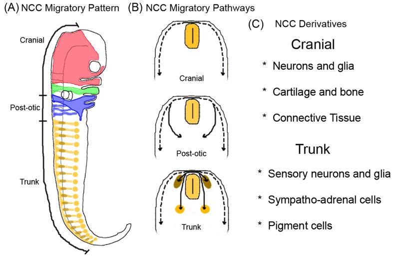
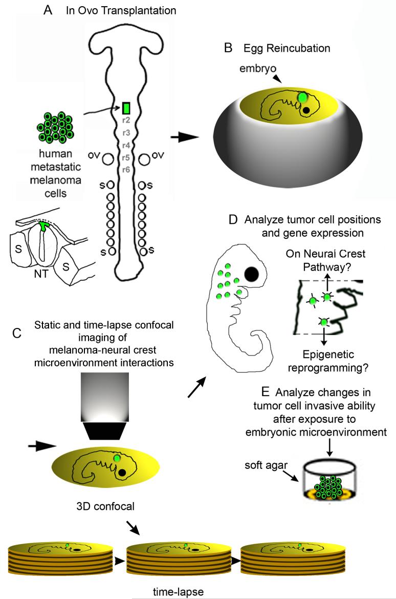
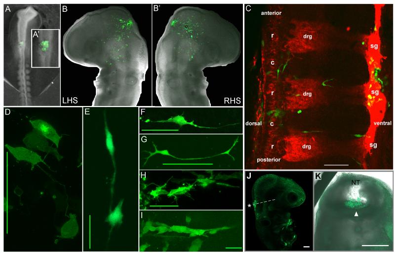
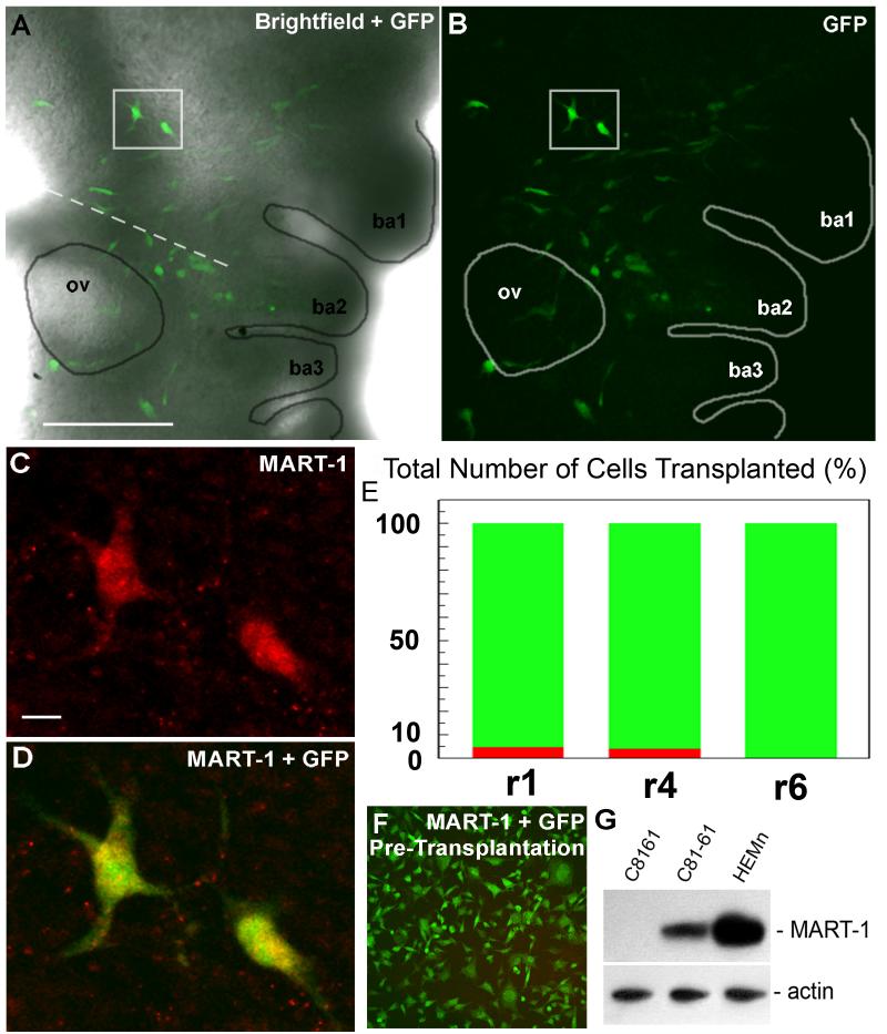
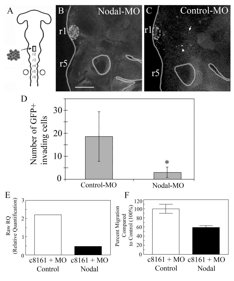
The embryonic microenvironment is an important source of signals that program multipotent cells to adopt a particular fate and migratory path, yet its potential to reprogram and restrict multipotent tumor cell fate and invasion is unrealized. Aggressive tumor cells share many characteristics with multipotent, invasive embryonic progenitors, contributing to the paradigm of tumor cell plasticity. In the vertebrate embryo, multiple cell types originate from a highly invasive cell population called the neural crest. The neural crest and the embryonic microenvironments they migrate through represent an excellent model system to study cell diversification during embryogenesis and phenotype determination. Recent exciting studies of tumor cells transplanted into various embryo models, including the neural crest rich chick microenvironment, have revealed the potential to control and revert the metastatic phenotype, suggesting further work may help to identify new targets for therapeutic intervention derived from a convergence of tumorigenic and embryonic signals. In this mini-review, we summarize markers that are common to the neural crest and highly aggressive human melanoma cells. We highlight advances in our understanding of tumor cell behaviors and plasticity studied within the chick neural crest rich microenvironment. In so doing, we honor the tremendous contributions of Professor Elizabeth D. Hay toward this important interface of developmental and cancer biology.


Figures

Figure 1
Figure 1
The neural crest cell stereotypical migratory pathways and derivatives. (A) Neural crest cells form migratory streams throughout the embryo, patterning the craniofacial area (red and green streams), the post-otic region (blue streams) that leads to the cardiovascular and enteric neural crest contribution, and the trunk migratory streams (yellow) that shape the peripheral nervous system and pigment cell contributions. (B) Neural crest cells migrate along distinct pathways that range from dorsolateral to medioventral. (C) Neural crest cells give rise to a wide variety of derivatives, a subset of which are shown.
Figure 2
Figure 2
Schematic outline of tumor cell transplantation experiments and imaging analysis. (A) In ovo transplantation of human metastatic melanoma cells into the chick neural tube and (B) egg re-incubation. (C) After egg re-incubation, the invading tumor cell positions are analyzed with static and time-lapse confocal analysis for (D) positioning along host neural crest cell migratory pathways, changes in gene expression, and invasive ability. (E) Invading tumor cells within the chick embryo are extracted by FACS and evaluated in soft agar for invasive ability. The schematic is modified from Hendrix et al., 2007
Figure 3
Figure 3
Transplanted metastatic melanoma cells invade chick neural crest cell migratory pathways and destinations. (A-A’) Adult GFP-labeled human metastatic melanoma cells (C8161) were transplanted into specific neural tube locations in host chick embryos (6-9 somite stage). Eggs were resealed and re-incubated for 48hrs. (BB’) Transplanted melanoma cells invade host tissue and spread out into the chick craniofacial (RHS is right-hand side) and (C) trunk, including the dorsal root ganglia (drg) and sympathetic ganglia (sg). (D-I) Transplanted tumor cell morphologies resemble host neural crest cell shapes. (J-K) Non-aggressive melanoma cells transplanted into close contact with the neural tube (A-A’) into similar regions of the chick neural tube at the same 6-9 somite stage remain at the transplant site. Some tumor cells may appear at the ventral lumen of the neural tube due to morphogenesis of the neural tube volume and failure of the cells to egress. Data presented in (A-C, F-K) are reproduced from Kulesa et al., 2006; otherwise are unpublished from KCKK and PMK. Scalebars are (C) 10um, (D,F,H,I) 50um, (E) 100um, (G) 200um, (J-K) 100um.
Figure 4
Figure 4
Transplanted tumor cells express melanocyte-like markers. (A- E) Expression analysis of MART-1 shows a small subset of transplanted tumor cells with expression. (F-G) The GFP-labeled tumor cells do not express MART-1 prior to transplantation. The source of the material is from Kulesa et al., 2006. All scalebars are 100um.
Figure 5
Figure 5
C8161 melanoma cells transfected with an anti-Nodal GFP morpholino were transplanted into the cranial region of host unlabeled chick embryos at Hamburger and Hamilton Stages 8-9. (B) A confocal image of a typical chick embryo at +48hrs of reincubation including transplanted melanoma (dotted circled clump). (C,D,E) Many melanoma cells transfected with a control GFP morpholino invade the host chick embryo (see the arrow and arrowhead). Graph of the cell counts of GFP+ invading cells in both the control-MO (n=10) and Nodal-MO (n=14) (p<0.001). (E) Relative quantification (RawRQ) of Nodal expression by aggressive C8161 human metastatic melanoma cells treated with either a control or Nodal morpholino (MO) as measured by real-time reverse transcriptase polymerase chain reaction. (F) Percent migration (measured over 6 hours in a modified Boyden chamber containing a gelatin-coated porous membrane) of C8161 cells treated with either a control or Nodal morpholino (MO), with the control treated cells normalized to 100% (n=4, average ± S.E.). These data are unpublished. The scalebar is 150um (B).

References

    1. Anderson RB, Newgreen DF, Young HM. Neural crest and the development of the enteric nervous system. Adv. Exp. Med. Biol. 2006;589:181–96. - PubMed
    1. Barrallo-Gimeno A, Nieto MA. The Snail genes as inducers of cell movement and survival: implications in development and cancer. Dev. 2005;132:3151–3161. - PubMed
    1. Belmadani A, Tran PB, Ren D, Assimacopoulos S, Grove EA, Miller RJ. The chemokine stromal cell-derived factor-1 regulates the migration of sensory neuron progenitors. J Neurosci. 2005;25:3995–4003. - PMC - PubMed
    1. Bielenberg DR, Hida Y, Shimizu A, Kaipainen A, Kreuter M, Kim CC, Klagsbrun M. Semaphorin 3F, a chemorepulsant for endothelial cells, induces a poorly vascularized, encapsulated, nonmetastatic tumor phenotype. J Clin Invest. 2004;114(9):1260–71. - PMC - PubMed
    1. Bilozur ME, Hay ED. Neural crest migration in 3D extracellular matrix utilizes laminin, fibronectin, or collagen. Dev Biol. 1988;125:19–33. - PubMed

Publication types

LinkOut - more resources