. 2008 Jul 16:4:26.
doi: 10.1186/1744-8069-4-26.

PKA and ERK, but not PKC, in the amygdala contribute to pain-related synaptic plasticity and behavior

Affiliations

PKA and ERK, but not PKC, in the amygdala contribute to pain-related synaptic plasticity and behavior

Yu Fu et al. Mol Pain. .

Abstract

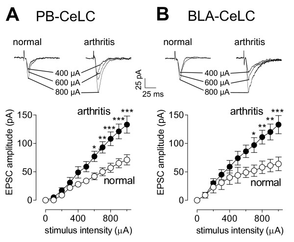
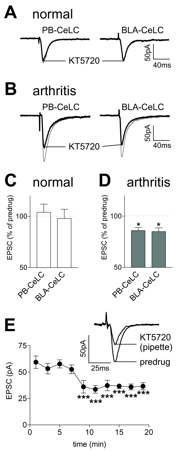
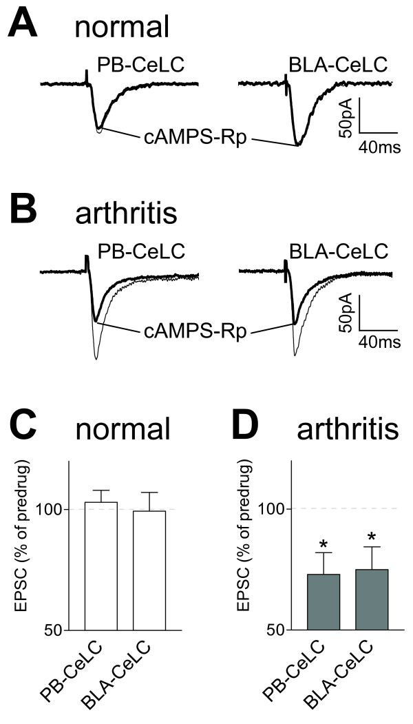
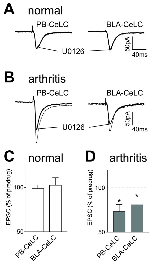
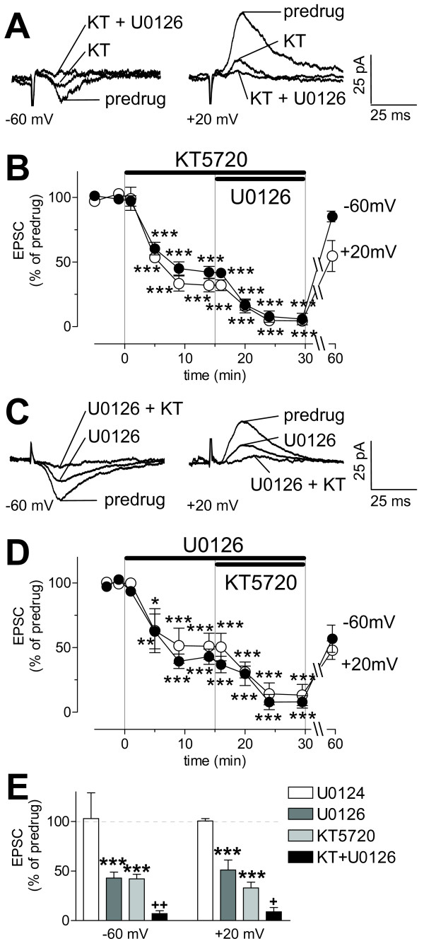
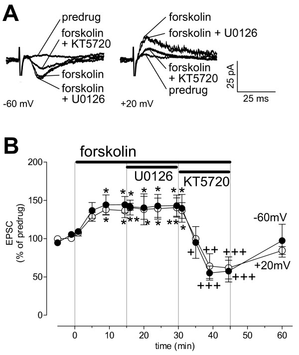
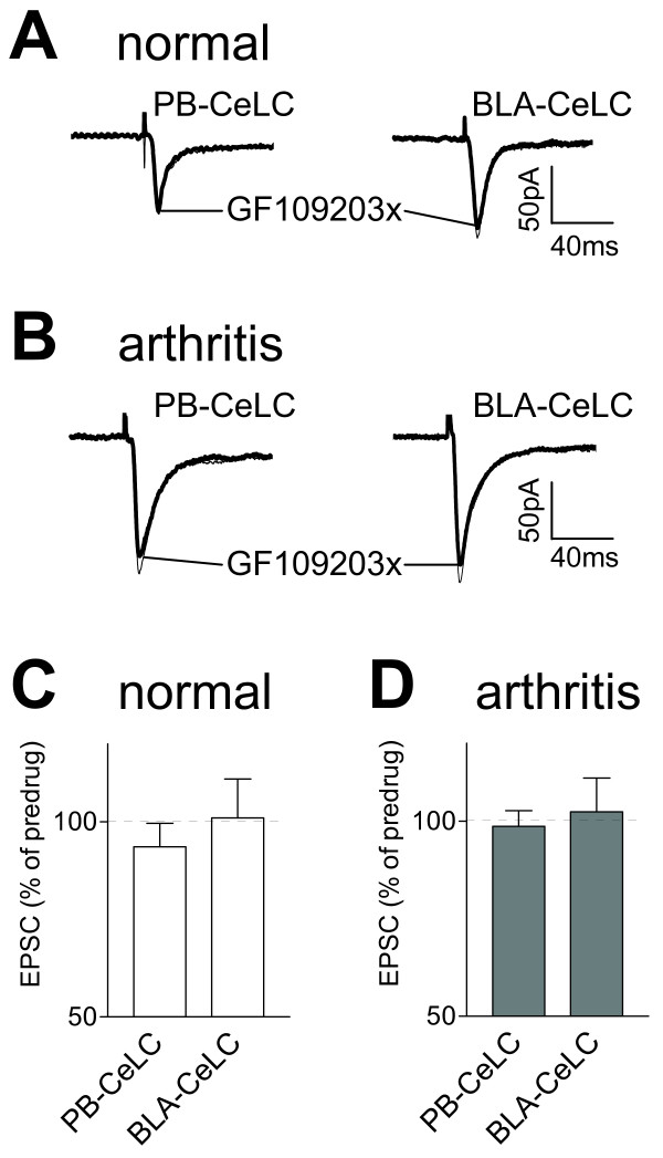
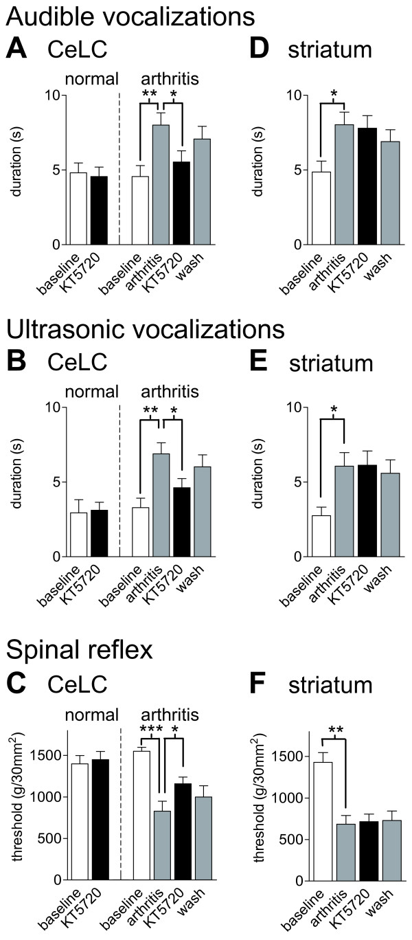
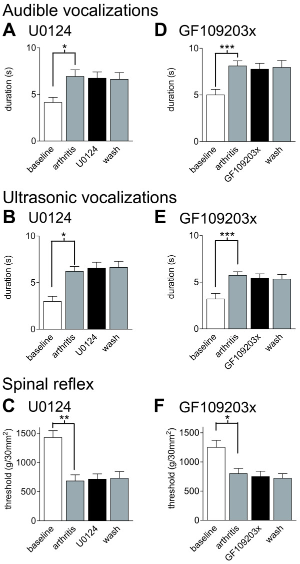
The laterocapsular division of the central nucleus of the amygdala (CeLC) has emerged as an important site of pain-related plasticity and pain modulation. Glutamate and neuropeptide receptors in the CeLC contribute to synaptic and behavioral changes in the arthritis pain model, but the intracellular signaling pathways remain to be determined. This study addressed the role of PKA, PKC, and ERK in the CeLC. Adult male Sprague-Dawley rats were used in all experiments. Whole-cell patch-clamp recordings of CeLC neurons were made in brain slices from normal rats and from rats with a kaolin/carrageenan-induced monoarthritis in the knee (6 h postinduction). Membrane-permeable inhibitors of PKA (KT5720, 1 microM; cAMPS-Rp, 10 microM) and ERK (U0126, 1 microM) activation inhibited synaptic plasticity in slices from arthritic rats but had no effect on normal transmission in control slices. A PKC inhibitor (GF109203x, 1 microM) and an inactive structural analogue of U0126 (U0124, 1 microM) had no effect. The NMDA receptor-mediated synaptic component was inhibited by KT5720 or U0126; their combined application had additive effects. U0126 did not inhibit synaptic facilitation by forskolin-induced PKA-activation. Administration of KT5720 (100 microM, concentration in microdialysis probe) or U0126 (100 microM) into the CeLC, but not striatum (placement control), inhibited audible and ultrasonic vocalizations and spinal reflexes of arthritic rats but had no effect in normal animals. GF109203x (100 microM) and U0124 (100 microM) did not affect pain behavior. The data suggest that in the amygdala PKA and ERK, but not PKC, contribute to pain-related synaptic facilitation and behavior by increasing NMDA receptor function through independent signaling pathways.


Figures

Figure 1
Figure 1
Synaptic plasticity in CeLC neurons in the arthritis pain model. Input-output functions of CeLC neurons were measured in slices from arthritic animals (6 h postinduction) and in slices from normal animals. Monosynaptic excitatory postsynaptic currents (EPSCs) were evoked in CeLC neurons by electrical stimulation of the PB-CeLC (A) and BLA-CeLC (B) synapses with increasing intensities. Input-output curves were generated by plotting peak EPSC amplitude (pA) as a function of afferent fiber volley stimulus intensity (μA). Input-output functions of neurons from arthritic animals (n = 19) were significantly different from those of control neurons (n = 16) at the PB-CeLC (P < 0.0001, F 1,363 = 66.65) and BLA-CeLC (P < 0.0001, F 1,363 = 43.30, two-way ANOVA) synapses. Individual traces (mean of 8–10 trials) show monosynaptic EPSCs recorded in one CeLC neuron from a normal rat and another CeLC neuron from an arthritic rat. Whole-cell voltage-clamp recordings were made at -60 mV. * P < 0.05, ** P < 0.01, *** P < 0.001 (Bonferroni post-tests).
Figure 2
Figure 2
A PKA inhibitor (KT5720) inhibits synaptic plasticity but not normal synaptic transmission. Monosynaptic EPSCs were evoked at the PB-CeLC and BLA-CeLC synapses in slices from normal (A, C) and arthritic (6 h postinduction; B, D, E) rats. KT5720 (1 μM, 15 min) inhibited synaptic transmission in neurons from arthritic rats 6 h postinduction (D, n = 7; P < 0.05, paired t-test) but not in control neurons from normal rats (C, n = 7). (A, B) Original recordings of EPSCs (average of 8–10 EPSCs) evoked at the two synapses. (C, D) Averaged EPSC amplitudes (mean ± SE) in the presence of KT5720 normalized to predrug (ACSF) control values (set to 100%). (E) Time course of the inhibitory effect of direct intracellular application of KT5720 (1 μM) through the patch pipette. Each symbol shows averaged EPSC amplitudes (mean ± SE; n = 4) at different times after whole-cell configuration was obtained (t = 0). The inhibitory effect was significant (P < 0.001, compared to the first EPSC after patch formation; Dunnett's Multiple Comparison Test). Insets show EPSCs (average of 8–10 trials) evoked at the PB-CeLC synapse at 1 min (predrug) and at 15 min (KT5720) after patch formation. Whole-cell voltage-clamp recordings were made at -60 mV. * P < 0.05, *** P < 0.001.
Figure 3
Figure 3
A cAMP antagonist (cAMPS-Rp) inhibits synaptic plasticity but not normal synaptic transmission. Monosynaptic EPSCs were evoked at the PB-CeLC and BLA-CeLC synapses in slices from normal (A, C) and arthritic (6 h postinduction; B, D) rats. cAMPS-Rp (10 μM, 15 min) inhibited synaptic transmission in neurons from arthritic rats 6 h postinduction (D, n = 6; P < 0.05, paired t-test) but not in control neurons from normal rats (C, n = 4). (A, B) Original recordings of EPSCs (average of 8–10 EPSCs) evoked at the two synapses. (C, D) Averaged EPSC amplitudes (mean ± SE) in the presence of cAMPS-Rp normalized to predrug control values (set to 100%). Whole-cell voltage-clamp recordings were made at -60 mV. * P < 0.05.
Figure 4
Figure 4
An ERK inhibitor (U0126) inhibits synaptic plasticity but not normal synaptic transmission. Monosynaptic EPSCs were evoked at the PB-CeLC and BLA-CeLC synapses in slices from normal (A, C) and arthritic (6 h postinduction; B, D) rats. U0126 (1 μM, 15 min) inhibited synaptic transmission in neurons from arthritic rats 6 h postinduction (D, n = 6; P < 0.05, paired t-test) but not in control neurons (C, n = 6). (A, B) Original recordings of EPSCs (average of 8–10 trials) evoked at the two synapses. (C, D) Averaged EPSC amplitudes (mean ± SE) in the presence of U0126 normalized to predrug control values (set to 100%). Whole-cell voltage-clamp recordings were made at -60 mV. * P < 0.05.
Figure 5
Figure 5
Additive effect of PKA and ERK inhibitors on NMDA receptor-mediated synaptic plasticity. (A, B) KT5720 (KT, 1 μM, 15 min; n = 3) inhibited the pharmacologically (with NBQX, 20 μM; bicuculline, 30 μM) isolated NMDA receptor-mediated synaptic component in slices from arthritic rats (P < 0.001, repeated-measures ANOVA with Bonferroni post-tests). Co-application of KT5720 and U0126 (1 μM, n = 3) further decreased the synaptic response (P < 0.001, Bonferroni post-tests). (C, D) U0126 (1 μM, 15 min) applied alone (n = 3) or together with KT5720 (n = 3) inhibited the NMDA component in the arthritis pain model (P < 0.05–0.001, Bonferroni post-tests). (E) Coapplication of KT5720 and U0126 (n = 6) had a significantly greater effect than each compound alone (P < 0.05–0.01, one-way ANOVA with Bonferroni post-tests). A negative structural analogue of U0126 (U0124, 1 μM, n = 3) had no effect. Bar histograms show averaged EPSC amplitudes (mean ± SE) normalized to predrug control values (set to 100%). (A, C) Monosynaptic EPSCs (average of 8–10 traces) recorded at -60 mV and +20 mV. (B, D) Time course of drug effects at -60 mV and +20 mV. Each symbol shows averaged EPSC amplitudes (mean ± SE) normalized to predrug control (set to 100%). * P < 0.05, ** P < 0.01, *** P < 0.001 (compared to predrug values); + P < 0.05, ++ P < 0.01 (compared to each drug alone).
Figure 6
Figure 6
Synaptic facilitation by forskolin is not impaired by an ERK inhibitor (U0126). PKA activation by forskolin (5 μM, 15 min) increased transmission at the PB-CeLC synapse in slices from normal animals and induced an NMDA receptor-mediated component in the presence of NBQX (20 μM) and bicuculline (30 μM). U0126 (1 μM, 15 min) had no effect but KT5720 (1 μM, 1 min) inhibited the forskolin-induced facilitation. (A) Monosynaptic EPSCs (average of 8–10 traces) recorded in an individual CeLC neuron held at -60 mV and +20 mV. (B) Time course of drug effects at -60 mV and +20 mV (n = 4). Each symbol shows averaged EPSC amplitudes (mean ± SE) normalized to predrug control values (set to 100%). * P < 0.05, ** P < 0.01 (compared to predrug values), + P < 0.05, ++ P < 0.01, +++ P < 0.001 (compared to forskolin without KT5720; repeated-measures ANOVA with Bonferroni post-tests).
Figure 7
Figure 7
A PKC inhibitor (GF109203X) has no effect on synaptic transmission. Monosynaptic EPSCs were evoked at the PB-CeLC and BLA-CeLC synapses in slices from normal (A, C) and arthritic (6 h postinduction; B, D) rats. GF109203X (1 μM, 15 min) had no significant effect (P > 0.05, paired t-test) in neurons from arthritic rats (n = 6) and in control neurons from normal rats (n = 5). (A, B) Original recordings of EPSCs evoked at the two synapses (average of 8–10 EPSCs). (C, D) Averaged EPSC amplitudes (mean ± SE) in the presence of GF109203X normalized to predrug control values (set to 100%).
Figure 8
Figure 8
A PKA inhibitor decreases pain-related behavior in arthritic but not in normal animals. KT5720 (100 μM, concentration in microdialysis probe, 15 min) administered into the CeLC had no significant effect on audible (A) and ultrasonic (B) vocalizations and on hindlimb withdrawal reflexes (C) in normal animals ("normal"; n = 5). In arthritic animals ("arthritis" in A-C; n = 6) KT5720 significantly inhibited vocalizations and increased mechanical thresholds. In the arthritis group, behaviors were measured before (baseline) and 6 h after arthritis induction and during (15 min) and after (30 min washout) drug application. (D-F) Administration of KT5720 (100 μM) into the striatum as placement control had no effect on audible and ultrasonic vocalizations and on spinal reflexes of arthritic rats (n = 4). Vocalizations were evoked by brief (15 s) noxious (2000 g/30 mm2) stimulation of the knee with a calibrated forceps. Duration of vocalizations was measured as the arithmetic sum of the duration of each individual vocalization event during a 1 min period beginning with the onset of the stimulus (see Methods for details). Bar histograms and error bars show mean ± SE. * P < 0.05, ** P < 0.01, *** P < 0.001 (repeated-measures ANOVA with Bonferroni post-tests).
Figure 9
Figure 9
An ERK inhibitor decreases pain-related behavior in arthritic but not in normal animals. U0126 (100 μM, concentration in microdialysis probe, 15 min) administered into the CeLC had no significant effect on audible (A) and ultrasonic (B) vocalizations and on hindlimb withdrawal reflexes (C) in normal animals ("normal"; n = 3). In arthritic animals ("arthritis" in A-C) U0126 significantly inhibited vocalizations (n = 9) and increased mechanical thresholds (n = 5). In the arthritis group, behaviors were measured before (baseline) and 6 h after arthritis induction and during (15 min) and after (30 min washout) drug application. (D-F) Administration of U0126 (100 μM) into the striatum as placement control had no effect (n = 4). Vocalizations and withdrawal reflexes were measured as in Figure 8 (see Methods for details). Bar histograms and error bars represent mean ± SE. * P < 0.05, ** P < 0.01, *** P < 0.001 (repeated-measures ANOVA with Bonferroni post-tests).
Figure 10
Figure 10
An inactive structural U0126 analogue (U0124) and a selective PKC inhibitor (GF109203X) have no effect on pain behavior. (A-C) Application of U0124 (100 μM, 15 min) into the CeLC had no effect on the significantly increased audible and ultrasonic vocalizations and on the decreased hindlimb withdrawal thresholds of arthritic rats (n = 3). (D-F) GF109203X (100 μM, 15 min) administered into the CeLC had no effect on the significantly increased vocalizations (n = 9) and on decreased withdrawal reflexes (n = 5) of arthritic rats. Vocalizations and withdrawal reflexes were measured as in Figure 8 (see Methods for details). Bar histograms and error bars represent mean ± SE. * P < 0.05, ** P < 0.01, *** P < 0.001 (repeated-measures ANOVA with Bonferroni post-tests).
Figure 11
Figure 11
Histologic verification of drug application sites. Standard diagrams [adapted from [54]] show coronal sections through the right brain hemisphere at different levels posterior to bregma (-1.88 mm and -2.12 mm). Symbols show the positions of the tips of the microdialysis probe (length of exposed membrane = 1 mm) in the CeLC (A-C) and striatum (D, placement control) in the behavioral experiments. (A) Application sites of KT5720, n = 11. (B) U0126, n = 12, filled circles; U0124, n = 3, open circles. (C) GF109203X, n = 9. (D) KT5720, n = 4, filled circles; U0126, n = 4, open circles. CeM, CeL, CeLC: medial, lateral and laterocapsular divisions of the central nucleus of the amygdala. Calibration bar (1 mm) applies to each diagram in A-D.

References

    1. Neugebauer V, Li W, Bird GC, Han JS. The amygdala and persistent pain. Neuroscientist. 2004;10:221–234. - PubMed
    1. Carrasquillo Y, Gereau RW. Activation of the extracellular signal-regulated kinase in the amygdala modulates pain perception. J Neurosci. 2007;27:1543–1551. - PMC - PubMed
    1. Ikeda R, Takahashi Y, Inoue K, Kato F. NMDA receptor-independent synaptic plasticity in the central amygdala in the rat model of neuropathic pain. Pain. 2007;127:161–172. - PubMed
    1. Pedersen LH, Scheel-Kruger J, Blackburn-Munro G. Amygdala GABA-A receptor involvement in mediating sensory-discriminative and affective-motivational pain responses in a rat model of peripheral nerve injury. Pain. 2007;127:17–26. - PubMed
    1. Heinricher MM, McGaraughty S. Pain-modulating neurons and behavioral state. In: Lydic R and Baghdoyan HA, editor. Handbook of Behavioral State Control. New York, CRC Press; 1999. pp. 487–503.

Publication types

MeSH terms