. 2008 Jul 18;29(1):21-32.
doi: 10.1016/j.immuni.2008.05.013.

Induction of immunological tolerance by apoptotic cells requires caspase-dependent oxidation of high-mobility group box-1 protein

Affiliations

Induction of immunological tolerance by apoptotic cells requires caspase-dependent oxidation of high-mobility group box-1 protein

Hirotaka Kazama et al. Immunity. .

Abstract

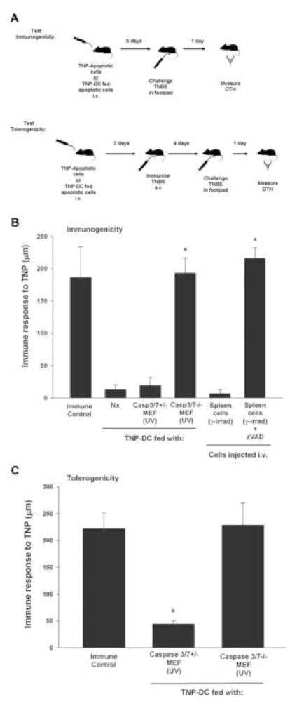
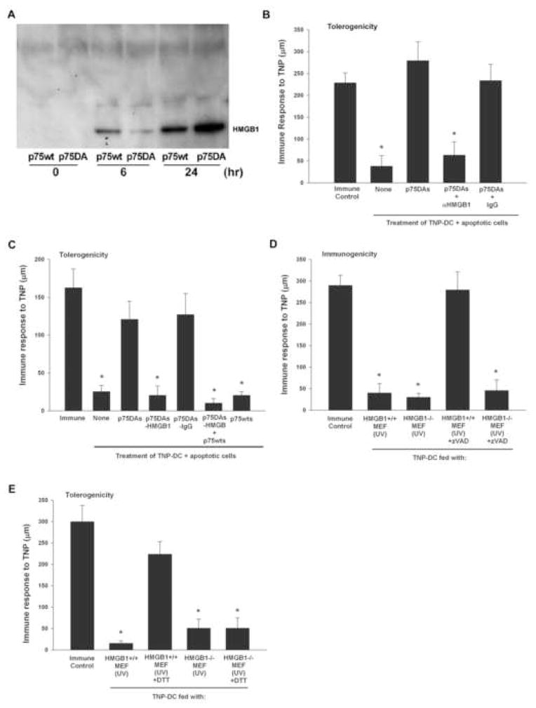
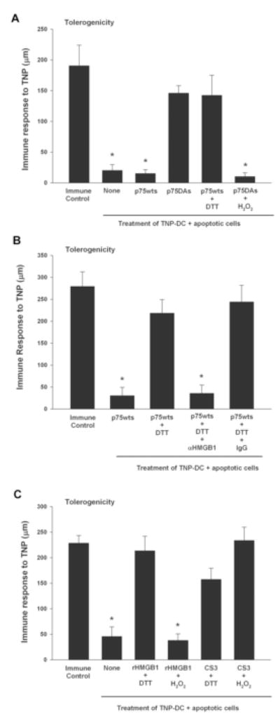
The mammalian immune system discriminates between modes of cell death; necrosis often results in inflammation and adaptive immunity, whereas apoptosis tends to be anti-inflammatory and promote immune tolerance. We have examined apoptosis for the features responsible for tolerance; specifically, we looked at the roles of caspases and mitochondria. Our results show that caspase activation targeted the mitochondria to produce reactive oxygen species (ROS), which were critical to tolerance induction by apoptotic cells. ROS oxidized the potential danger signal high-mobility group box-1 protein (HMGB1) released from dying cells and thereby neutralized its stimulatory activity. Apoptotic cells failed to induce tolerance and instead stimulated immune responses by scavenging or by mutating a mitochondrial caspase target protein when ROS activity was prohibited. Similarly, blocking sites of oxidation in HMGB1 prevented tolerance induction by apoptotic cells. These results suggest that caspase-orchestrated mitochondrial events determine the impact of apoptotic cells on the immune response.


Figures

Figure 1
Figure 1
Figure 1A. Experimental systems. Immunogenicity and tolerogenicity of apoptotic cells were measured using a well defined in vivo model. Immunogenicity test: TNP-coupled apoptotic cells (107) or TNP-DC fed apoptotic cells (106) were injected i.v. into mice. Five days later mice were injected with TNBS in the right footpad and PBS in the left footpad. DTH was measured with a micrometer 24 hrs later. Tolerogenicity test: TNP-coupled apoptotic cells (107) or TNP-DC fed apoptotic cells (106) were injected i.v. into mice. Mice were immunized 2 days later by s.c injection of TNBS. Four (4) days following immunization mice were injected with TNBS in the right footpad and PBS in the left footpad. DTH was measured with a micrometer 24 hrs later. Figure 1B. Caspase activation and immunity. Caspase 3/7 deficient MEF (caspase3/7−/−) or heterozygous knockout MEF (caspase3/7+/−) were UV irradiated and then incubated with TNP-DC overnight. DC (106) were then injected i.v. into naïve C57Bl/6 mice. Nx denotes nothing. Spleen cells were γ-irradiated and then incubated for 1 hr with or without 10mM zVAD-fmk. They were then coupled with TNP and injected i.v. (107) into naïve C57Bl/6 mice. Five (5) days following DC injection mice were injected with TNBS in the right footpad and PBS in the left footpad. With the exception of the Immune Control, all groups in this experiment were not immunized s.c with TNBS. Measurements (μm ± standard error) were taken 24 h later and represent the difference between right footpad (antigen challenge) and left footpad (PBS challenge). * denotes significantly different from unimmunized mice. Figure 1C. Effector caspases and immune tolerance. Caspase 3/7 deficient MEF (caspase3/7−/−) or heterozygous knockout MEF (caspase3/7+/−) were UV irradiated and then incubated with TNP-DC overnight. DC (106) were then injected i.v. into naïve C57Bl/6 mice. All mice in this experiment were immunized 2 days later by s.c injection of TNBS. Four (4) days following immunization mice were injected with TNBS in the right footpad and PBS in the left footpad. Measurements (μm ± standard error) were taken 24 h later and represent the difference between right footpad (antigen challenge) and left footpad (PBS challenge). * denotes significantly different from Immune Control mice.
Figure 2
Figure 2
ROS and tolerance by apoptotic cells. A. C57Bl/6 mice were injected i.v. with TNP-coupled apoptotic cells (107) (spleen cells, γ-irrad) that were untreated or treated with 100μM BHA. B. C57Bl/6 mice were injected i.v. with TNP-coupled apoptotic cells (spleen cells, γ-irrad) (107), apoptotic cells treated with H2O2 (spleen cells, γ-irrad), necrotic cells (F/T) (107 equivalence), or necrotic cells (F/T) treated with DTT. In both experiments (A and B) recipient mice were immunized s.c. with TNBS 2 days later. Four days following immunization, mice were challenged with TNBS in the right footpad and 0.033ml PBS in the left footpad. Measurements (μm ± standard error) were taken 24 h later, and represent the difference between right footpad (antigen challenge) and left footpad (PBS challenge). Immune Control groups were injected with 0.1 ml 10mM TNBS s.c. C. C57Bl/6 mice were injected i.v. with Act-mOVA spleen cells (107) (Act-mOVA cells, γ-irrad). Two days later mice were immunized s.c. with 100μg OVA emulsified 1:1 with CFA. Seven days later mice were injected with 100μg heat aggregated OVA in the right footpad and PBS in the left footpad. DTH was measured with a micrometer 24 hrs later. * denotes significantly different from immune control mice.
Figure 3
Figure 3. Mitochondrial changes and immune tolerance
A. Cells expressing wild type (p75wt) mutated mitochondrial protein p75 (p75DA), or p75wt cells in the presence of zVAD-fmk were UV irradiated. Cells were harvested at indicated time points and ROS production was measured by flow cytometry using DHE. Values are reported as % cells making ROS. B. p75wt and p75DA cells were examined at indicated time points after the induction of apoptosis by UV irradiation for PS expression using annexin V and propidium iodide (PI) staining. C. p75wt and p75DA cells were labeled with PKH26 and DC were labeled with CFSE. Cells were incubated for 2 hrs at 37oC and analyzed by flow cytometry. D. Apoptosis was induced in p75wt and p75DA cells by UV irradiation and these cells were fed to TNP-DC overnight. DC were harvested and injected i.v. (106 per mouse) into recipient mice. Five days later mice were challenged with 0.033ml 10 mM TNBS in the right footpad and 0.033ml PBS in the left footpad. Measurements (μm ± standard error) were taken 24 h later, and represent the difference between right footpad (antigen challenge) and left footpad (PBS challenge). Immune Control groups were injected with TNBS s.c. on the same day as TNP-DC injection. E. Apoptosis was induced in p75wt and p75DA cells by UV irradiation and these cells were fed to TNP-DC overnight. Some p75wt cells were treated with 10mM zVAD-fmk prior to DC culture. DC were harvested and injected i.v. (106 per mouse) into recipient mice that were immunized s.c. with TNBS 2 days later. Four days following immunization, mice were challenged with TNBS in the right footpad and PBS in the left footpad. Measurements (μm ± standard error) were taken 24 h later, and represent the difference between right footpad (antigen challenge) and left footpad (PBS challenge). Immune Control groups were injected with TNBS s.c. F. Apoptosis was induced in p75wt and p75DA cells by UV irradiation and these cells were fed alone or together to TNP-DC overnight. Four days following immunization, mice were challenged with TNBS in the right footpad and PBS in the left footpad. Measurements (μm ± standard error) were taken 24 h later, and represent the difference between right footpad (antigen challenge) and left footpad (PBS challenge). Immune Control groups were injected with TNBS s.c. D. * denotes significantly different from unimmunized mice (Background, Bkg). E. * denotes significantly different from immune control mice. F. * denotes significantly different from immune control mice.
Figure 4
Figure 4. HMGB1 release blocks tolerance
A. Cells expressing wild type p75 (p75wt) or mutated mitochondrial protein p75 (p75DA) were UV irradiated. Cells were cultured for 6 or 24 hrs and the supernatants harvested. HMGB1 was separated on a reducing SDS-PAGE gel and detected by western blotting with a monoclonal anti-HMGB1. B. Supernatant from p75DA (p75DAs) was added to TNP-DC that had been fed apoptotic spleen cells for 6 hrs. In some cases polyclonal rabbit anti-HMGB1 (αHMGB1) or control rabbit IgG (IgG) was added to the supernatant culture. After overnight culture DC were harvested and injected i.v. into recipient mice that were immunized s.c with TNBS 2 days later. Four days following immunization, mice were challenged with 0.033ml 10 mM TNBS in the right footpad and 0.033ml PBS in the left footpad. Measurements (μm ± standard error) were taken 24 h later, and represent the difference between right footpad (antigen challenge) and left footpad (PBS challenge). Immune Control group was injected with TNBS s.c. C. Supernatants from wild type p75 (p75wt) or mutated mitochondrial protein p75 (p75DA) cells were collected 24 hrs following UV irradiation. Supernatants from p75DA (p75DAs) were treated in various ways before adding to TNP-DC that had been fed apoptotic spleen cells for 6 hrs. Groups in this experiment were: Immune Control, no treatment of DC (none), P75DA supernatant treatment only (p75DAs); HMGB1 depleted p75DAs (-HMGB1); mock depleted with rabbit IgG (-IgG); p75DAs depleted of HMGB1 with p75wts added back; p75wts only. After overnight culture DC were harvested and injected i.v. into recipient mice that were immunized s.c. with TNBS 2 days later. Four days following immunization, mice were challenged with TNBS in the right footpad and PBS in the left footpad. Measurements (μm ± standard error) were taken 24 h later, and represent the difference between right footpad (antigen challenge) and left footpad (PBS challenge). Immune Control groups were injected with TNBS s.c. * denotes significantly different from Immune Control mice. D. HMGB1 deficient MEF (HMGB1−/−) or wild type control MEF (HMGB1+/+) were UV irradiated and then incubated with TNP-DC overnight. DC (1x10⁶) were then injected i.v. into naïve C57Bl/6 mice. Five (5) days following DC injection mice were injected with TNBS in the right footpad and PBS in the left footpad. With the exception of the Immune Control, all groups in this experiment were not immunized s.c with TNBS. Measurements (μm ± standard error) were taken 24 h later and represent the difference between right footpad (antigen challenge) and left footpad (PBS challenge). * denotes significantly different from Immune Control mice. E. HMGB1 deficient MEF (HMGB1−/−) or wild type control MEF (HMGB1+/+) were UV irradiated and then incubated with TNP-DC overnight. DC (1x10⁶) were then injected i.v. into naïve C57Bl/6 mice. All mice in this experiment were immunized 2 days later by s.c injection of TNBS. Four (4) days following immunization mice were injected with TNBS in the right footpad and PBS in the left footpad. Measurements (μm ± standard error) were taken 24 h later and represent the difference between right footpad (antigen challenge) and left footpad (PBS challenge). * denotes significantly different from Immune Control mice.
Figure 5
Figure 5. HMGB1 function and ROS
A. Supernatants from wild type p75 (p75wts) or mutated mitochondrial protein p75 (p75DAs) cells were collected 24 hrs following UV irradiation. Supernatants were treated in various ways before adding to TNP-DC that had been fed apoptotic spleen cells for 6 hrs. Groups in this experiment were: Immune Control, no treatment of DC (none); p75wts only; p75DAs only; p75wts treated with DTT; p75DAs treated with H2O2. B. Supernatants from wild type p75 (p75wts) or mutated mitochondrial protein p75 (p75DAs) cells were collected 24 hrs following UV irradiation. Some supernatant was treated with DTT for 30 min and anti-HMGB1 or control IgG was added. Groups in this experiment were Control, p75wts only; p75wts treated with DTT; p75wts treated with DTT with anti-HMGB1 added; p75wts treated with DTT with IgG added. C. Recombinant HMGB1 (rHMGB1) or rHMGB1-CS3 mutant (CS3; 500ng/ml each) was added to TNP-DC that had been fed apoptotic spleen cells for 6 hrs. Groups in this experiment were: Immune Control; no treatment of DC (none); rHMGB1 +DTT; rHMGB1 + H2O2; HMGB1-CS3 treated with DTT (CS3 + DTT); HMGB1-CS3 treated with H2O2 (CS3 + H2O2). For A-C treated DC were cultured overnight and injected i.v. into recipient mice that were immunized s.c. with TNBS 2 days later. Four days following immunization, mice were challenged with TNBS in the right footpad and PBS in the left footpad. Measurements (μm ± standard error) were taken 24 h later, and represent the difference between right footpad (antigen challenge) and left footpad (PBS challenge). Immune Control groups were injected with TNBS s.c. * denotes significantly different from Immune Control mice.
Figure 6
Figure 6. HMGB1 function and Cys modification
A. Cells expressing wild type p75 (p75wt) or mutated mitochondrial protein p75 (p75DA) were UV irradiated. Supernatants were collected for 0–6 hr or 6–24 hrs. HMGB1 was separated on a non-reducing SDS-PAGE gel and detected by western blotting with a polyclonal anti-HMGB1. B. Recombinant HMGB1 (rHMGB1), rHMGB1-C23S mutant (500ng/ml) rHMGB1-C45S (500ng/ml) mutant, or rHMGB1-C106S (500ng/ml) mutant was treated with DTT or H2O2 and added to TNP-DC that had been fed apoptotic spleen cells for 6 hrs. After overnight culture DC were harvested and injected i.v. into recipient mice that were immunized s.c. with TNBS 2 days later. Four days following immunization, mice were challenged with TNBS in the right footpad and PBS in the left footpad. Measurements (μm ± standard error) were taken 24 h later, and represent the difference between right footpad (antigen challenge) and left footpad (PBS challenge). Immune Control groups were injected with TNBS s.c. * denotes significantly different from Immune Control mice.

Comment in

  • ROS eliminate danger.
    Peter ME. Peter ME. Immunity. 2008 Jul 18;29(1):1-2. doi: 10.1016/j.immuni.2008.06.006. Immunity. 2008. PMID: 18631448

References

    1. Albert ML, Jegathesan M, Darnell RB. Dendritic cell maturation is required for the cross-tolerization of CD8+ T cells. Nat Immunol. 2001;2:1010–1017. - PubMed
    1. Andersson U, Erlandsson-Harris H, Yang H, Tracey KJ. HMGB1 as a DNA-binding cytokine. J Leukoc Biol. 2002;72:1084–1091. - PubMed
    1. Andersson U, Wang H, Palmblad K, Aveberger AC, Bloom O, Erlandsson-Harris H, Janson A, Kokkola R, Zhang M, Yang H, Tracey KJ. High mobility group 1 protein (HMG-1) stimulates proinflammatory cytokine synthesis in human monocytes. J Exp Med. 2000;192:565–570. - PMC - PubMed
    1. Asano K, Miwa M, Miwa K, Hanayama R, Nagase H, Nagata S, Tanaka M. Masking of phosphatidylserine inhibits apoptotic cell engulfment and induces autoantibody production in mice. J Exp Med. 2004;200:459–467. - PMC - PubMed
    1. Battisto JR, Bloom BR. Dual immunological unresponsiveness induced by cell membrane coupled hapten or antigen. Nature. 1966;212:156–157. - PubMed

Publication types