. 2008 Jan;4(1):6-13.
doi: 10.1016/j.jalz.2007.10.015. Epub 2007 Dec 21.

Elevated microsomal prostaglandin-E synthase-1 in Alzheimer's disease

Affiliations

Elevated microsomal prostaglandin-E synthase-1 in Alzheimer's disease

Uzma A Chaudhry et al. Alzheimers Dement. 2008 Jan.

Abstract

Background: The proinflammatory prostaglandin E(2) (PGE(2)) fluctuates over time in the cerebrospinal fluid of patients with Alzheimer's disease (AD), but the cerebral distribution and expression patterns of microsomal prostaglandin-E synthase (mPGES)-1 have not been compared with those of normal human brains.

Methods: Middle frontal gyrus tissue from AD and age-matched control brains was analyzed by Western blot, immunofluorescence, and immunohistochemistry with mPGES-1-specific antibodies.

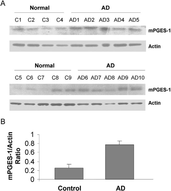
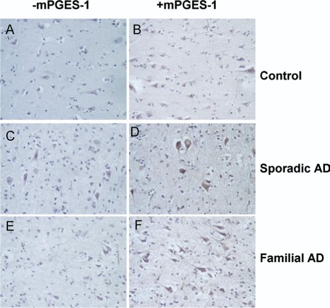
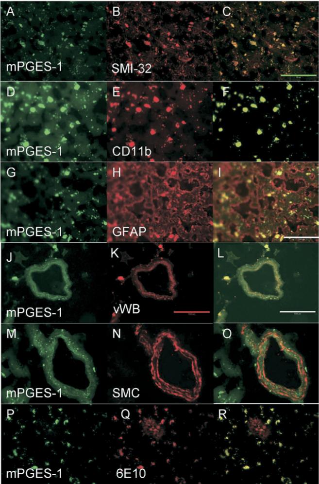
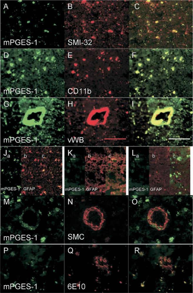
Results: Western blotting revealed that mPGES-1 expression was significantly elevated in AD tissue. Furthermore, immunofluorescence of mPGES-1 was observed in neurons, microglia, and endothelial cells of control and AD tissue. Although mPGES-1 was consistently present in astrocytes of control tissue, it was present in only some astrocytes of AD tissue. Immunohistochemical staining suggested that mPGES-1 was elevated in pyramidal neurons of AD tissue when compared with controls.

Conclusions: The results suggest that mPGES-1 is normally expressed constitutively in human neurons, microglia, astrocytes, and endothelial cells but is up-regulated in AD.


Figures

Fig 1
Fig 1
Differential expression of mPGES-1 in human brain MFG. (A) Western blot analysis showed that mPGES-1 protein was expressed in the MFG of AD (n = 10) and age-matched control (n = 9) brains. (B) After normalization to actin, the mean expression of mPGES-1 in the AD brains was shown to be significantly greater than that of the control brains; **P < .001.
Fig 2
Fig 2
Immunohistochemical study of mPGES-1 in AD. (A) Negative control normal brain; (B) a normal brain positively immunostained with anti–mPGES-2. (C) Negative control sporadic AD brain tissue; (D) mPGES-1–stained sporadic AD brain tissue. (E) Negative control familial AD brain tissue; (F) positively immunostained familial AD brain tissue. Neurons displayed visible detectable immunostaining. Original magnification, 40×.
Fig 3
Fig 3
Microsomal PGES-1 (green) colocalization with various cell types (red) in normal brain. Yellow areas show overlap of mPGES-1 with the cell type, indicating colocalization. mPGES-1 colocalized with neurons labeled by SMI-32 antibody (A–C), with microglia labeled by CD11b antibody (D–F), with astrocytes labeled by GFAP antibody (G–I), and with endothelial cells labeled by vWB antibody (J–L). mPGES-1 did not colocalize with smooth muscle cells labeled by SMC antibody (M–O). Aβ1−42, which was stained with 6E10 antibody, colocalized with mPGES-1 in microglial-shaped cells (P–R). The plaque itself (red area seen in R) did not remarkably colocalize with mPGES-1. Green scale bar = 50 μm; red/white scale bar = 100 μm.
Fig 4
Fig 4
Microsomal PGES-1 (green) colocalization with various cell types (red) in AD brain. Staining was similar to that of control brain. mPGES-1 colocalized with neurons labeled by SMI-32 antibody (A–C), microglia labeled by CD11b antibody (D–F), and endothelial cells labeled by vWB antibody (G–I). mPGES-1 colocalized with astrocytes (labeled by anti-GFAP) in some areas (Jc, Kc) but not in others (Lc). mPGES-1 did not colocalize with smooth muscle cells labeled by SMC antibody (M–O). Aβ1−42, which was stained with 6E10 antibody, colocalized with mPGES-1 in microglial-shaped cells (P–R). The plaque itself (red area seen in R) did not colocalize with mPGES-1. Scale bar = 100 μm.

References

    1. Combrinck M, Williams J, De Berardinis MA, Warden D, Puopolo M, Smith AD, et al. Levels of CSF prostaglandin E2, cognitive decline, and survival in Alzheimer's disease. J Neurol Neurosurg Psychiatry. 2006;77:85–8. - PMC - PubMed
    1. Breitner JC, Welsh KA, Helms MJ, Gaskell PC, Gau BA, Roses AD, et al. Delayed onset of Alzheimer's disease with nonsteroidal anti-inflammatory and histamine H2 blocking drugs. Neurobiol Aging. 1995;16:523–30. - PubMed
    1. Zandi PP, Breitner JC. Do NSAIDs prevent Alzheimer's disease? and, if so, why? the epidemiological evidence. Neurobiol Aging. 2001;22:811–7. - PubMed
    1. Szekely CA, Thorne JE, Zandi PP, Ek M, Messias E, Breitner JC, et al. Nonsteroidal anti-inflammatory drugs for the prevention of Alzheimer's disease: a systematic review. Neuroepidemiology. 2004;23:159–69. - PubMed
    1. McGeer PL, Schulzer M, McGeer EG. Arthritis and anti-inflammatory agents as possible protective factors for Alzheimer's disease: a review of 17 epidemiologic studies. Neurology. 1996;47:425–32. - PubMed

Publication types

Substances