. 2008 Jul 22;105(29):10238-43.
doi: 10.1073/pnas.0802759105. Epub 2008 Jul 16.

Single treatment with RNAi against prion protein rescues early neuronal dysfunction and prolongs survival in mice with prion disease

Affiliations

Single treatment with RNAi against prion protein rescues early neuronal dysfunction and prolongs survival in mice with prion disease

Melanie D White et al. Proc Natl Acad Sci U S A. .

Abstract

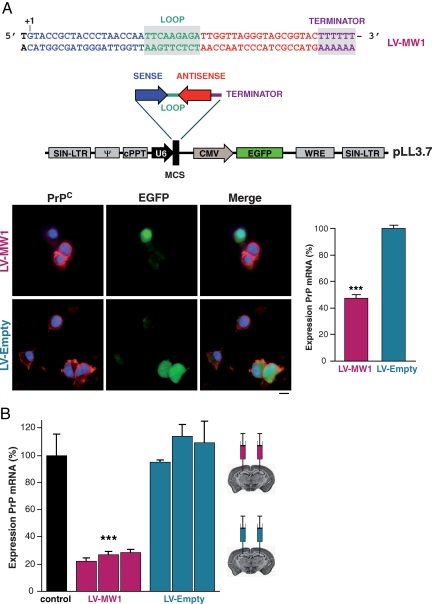
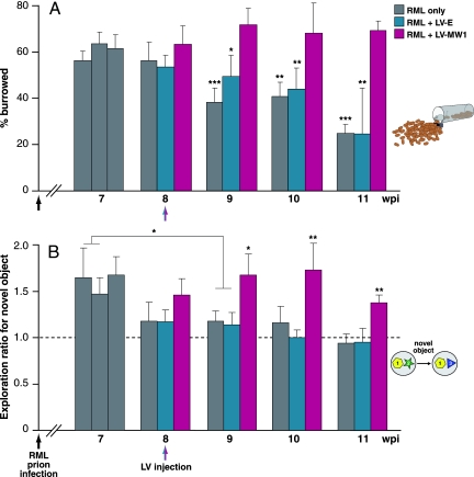
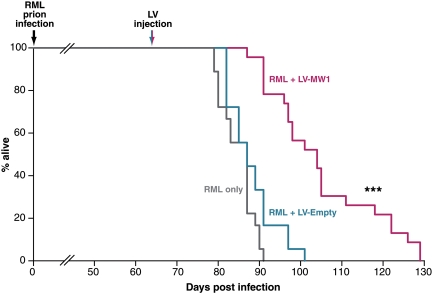
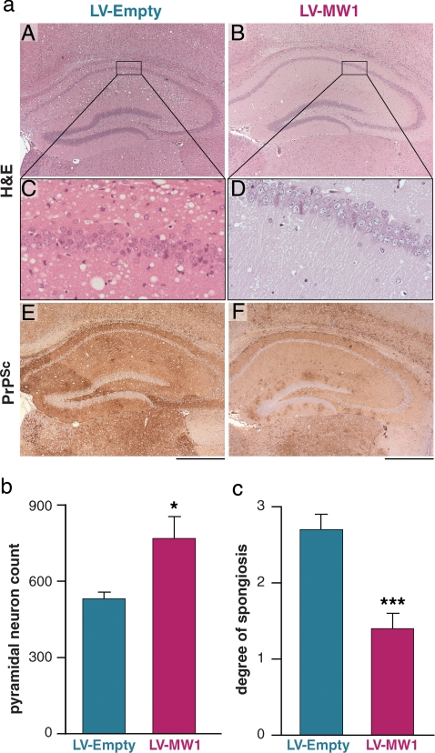
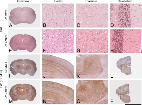
Prion diseases are fatal neurodegenerative conditions for which there is no effective treatment. Prion propagation involves the conversion of cellular prion protein, PrP(C), to its conformational isomer, PrP(Sc), which accumulates in disease. Here, we show effective therapeutic knockdown of PrP(C) expression using RNAi in mice with established prion disease. A single administration of lentivirus expressing a shRNA targeting PrP into each hippocampus of mice with established prion disease significantly prolonged survival time. Treated animals lived 19% and 24% longer than mice given an "empty" lentivirus, or not treated, respectively. Lentivirally mediated RNAi of PrP also prevented the onset of behavioral deficits associated with early prion disease, reduced spongiform degeneration, and protected against neuronal loss. In contrast, mice receiving empty virus or no treatment developed early cognitive impairment and showed severe spongiosis and neuronal loss. The focal use of RNAi therapeutically in prion disease further supports strategies depleting PrP(C), which we previously established to be a valid target for prion-based treatments. This approach can now be used to define the temporal, quantitative, and regional requirements for PrP knockdown for effective treatment of prion disease and to explore mechanisms involved in predegenerative neuronal dysfunction and its rescue.


Conflict of interest statement

Conflict of interest statement: J.C. is a director and shareholder of D-Gen Limited, an academic spin-out company working in the field of prion disease diagnosis, decontamination, and therapeutics. D-Gen markets the ICSM35 and ICS18 antibodies used in this study.

Figures

Fig. 1.
Fig. 1.
Lentiviruses expressing anti-PrP shRNAs reduce PrP protein and mRNA levels in vitro and in vivo. (A) Oligonucleotide insert encoding shRNA targeting PrP was cloned into the pLL3.7 lentivector, which also expresses EGFP reporter protein to generate virus LV-MW1. A control lentivirus LV-Empty lacking the shRNA insert was also produced. Transduction of N2a cells with LV-MW1 markedly reduced PrPC expression detected with anti-PrP antibody ICSM18 (red) in transduced cells (green), but LV-Empty had no effect. Cell nuclei were stained with DAPI (blue). RT-PCR showed LV-RNAi-mediated reduction of PrP mRNA by LV-MW1 but not LV-Empty (P = 0.0014; Student's t test, two tails). Error bars represent SEM. Three replicates were performed for each sample. All data shown are 4 days after transduction. (B) PrP mRNA levels were reduced in the hippocampi of uninfected FVB mice 2 weeks after treatment with LV-MW1 (P < 0.0001; Student's t test, two tails) but not with LV-Empty (n = 3 in each case) compared with total levels in uninjected control mice. Error bars and replicates as above.
Fig. 2.
Fig. 2.
Lentivirally mediated RNAi of PrP prevents loss of burrowing behavior and memory deficits in prion-infected mice. (A) Mice were infected with RML prions at 1 week of age (black arrows) and tested for their ability to actively displace (burrow) food pellets from a tube over 24 h from 7 weeks after inoculation. At 8 wpi, mice were treated with LV-MW1 or LV-Empty or with no virus (red and blue arrows). LV-MW1-treated mice continued to burrow actively, whereas LV-Empty and untreated mice lost burrowing behavior (P = 0.011 and P = 0.0001, respectively, at 9 wpi and P < 0.005 for LV-MW1 compared with LV-Empty-treated and untreated mice at all time points after 9 wpi). (B) Mice were also tested for object recognition memory. LV-MW1-treated mice retained the capacity to distinguish novel objects, whereas this was lost in LV-Empty-treated mice and untreated animals (P = 0.021 and 0.027, respectively, for exploration at 7 wpi compared with 9 wpi in these groups). In contrast, mice treated with LV-MW1 retained object-recognition memory (P = 0.016 and 0.014, compared with LV-Empty-treated and untreated mice, respectively, at 9 wpi), and this was sustained for the course of the experiment (P < 0.005 for all groups at 10 and 11 wpi). Dashed line (exploratory ratio = 1) indicates random exploration of both objects, denoting no memory. n = 22 for LV-MW1 and n = 18 for LV-Empty and RML-only-treated groups.
Fig. 3.
Fig. 3.
Treatment with anti-PrP shRNA expressing lentivirus prolongs survival in mice with established prion disease. Mice were infected with RML prions at 1 week of age and were treated with bilateral hippocampal injections of either LV-MW1 (n = 22) or LV-Empty (n = 18) at 8 wpi or with no virus (n = 18). RML-infected mice treated with no virus or with LV-Empty died within 91 and 101 days postinfection (dpi), respectively; mice treated with LV-MW1 survived longer, living up to 129 dpi. (P < 0.0001 Student's t test, two tails, compared with both LV-Empty-treated and untreated mice).
Fig. 4.
Fig. 4.
Lentivirally mediated RNAi of PrP expression protects against prion neurodegeneration in vivo. (a) LV-MW1 treatment protected against neuronal loss and reduced spongiform degeneration in the CA1–3 region (B and D) compared with LV-Empty-treated mice where neuronal loss is evident and spongiosis is marked (A and C). (The right hippocampus is shown for all mice.) PrPSc deposition was reduced in LV-MW1-treated mice (F) but was extensive in LV-Empty-treated animals (E). (Scale bar, 500 μm.) (b) Systematic counting of pyramidal cell neurons in CA1 confirmed neuroprotective effects of LV-MW1 (P = 0.029, Student's t test, two tails) and (c) semiquantitative scoring of spongiosis showed significantly less spongiosis in LV-MW1- treated mice (P = 0.0007, Student's t test, two tails) (n = 6 in each case).
Fig. 5.
Fig. 5.
Focal delivery of lentivirally mediated RNAi of PrP expression to hippocampus reduces spongiosis and PrPSc deposition in other brain regions. Representative sections from various brain regions from prion-infected mice treated with bilateral hippocampal injections of LV-MW1 8 weeks after infection (A–D and I–L) and LV-Empty (E–H and M–P). Sections are stained with H&E to show spongiosis (top two rows) and ICSM35 for PrPSc deposition (bottom two rows). LV-MW1 treatment reduces prion spongiosis and PrPSc accumulation in thalamus and cortex and in the hippocampus. [Scale bar, 5 mm (A, E, I, M), 4 mm (L and P), 1.3 mm (J, K, N, O), and 170 μm (B–D, F–H).]

References

    1. Bueler H, et al. Mice devoid of PrP are resistant to scrapie. Cell. 1993;73:1339–1347. - PubMed
    1. Sailer A, et al. No propagation of prions in mice devoid of PrP. Cell. 1994;77:967–968. - PubMed
    1. Manson JC, et al. PrP gene dosage determines the timing but not the final intensity or distribution of lesions in scrapie pathology. Neurodegeneration. 1994;3:331–340. - PubMed
    1. Mallucci G, et al. Depleting neuronal PrP in prion infection prevents disease and reverses spongiosis. Science. 2003;302:871–874. - PubMed
    1. Mallucci GR, et al. Targeting cellular prion protein reverses early cognitive deficits and neurophysiological dysfunction in prion-infected mice. Neuron. 2007;53:325–335. - PubMed

Publication types

MeSH terms