. 2008 Jul 22;105(29):9959-64.
doi: 10.1073/pnas.0804336105. Epub 2008 Jul 16.

A modified cysteinyl-labeling assay reveals reversible oxidation of protein tyrosine phosphatases in angiomyolipoma cells

Affiliations

A modified cysteinyl-labeling assay reveals reversible oxidation of protein tyrosine phosphatases in angiomyolipoma cells

Benoit Boivin et al. Proc Natl Acad Sci U S A. .

Abstract

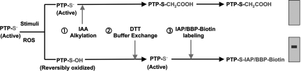
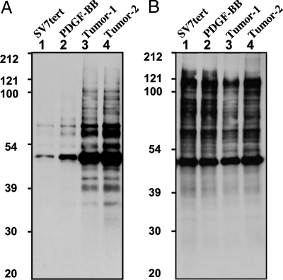
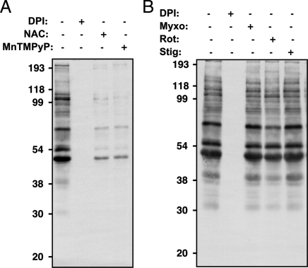
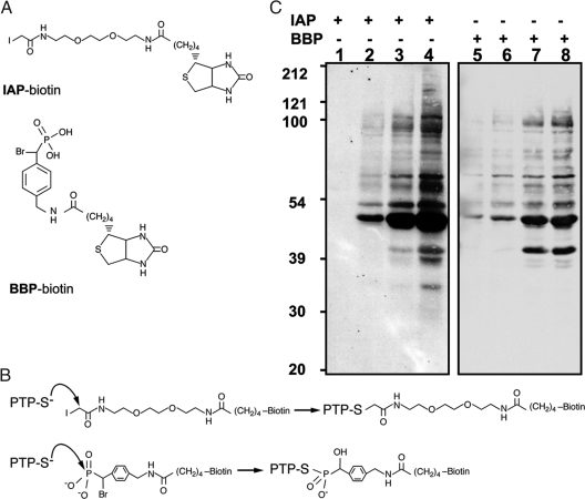
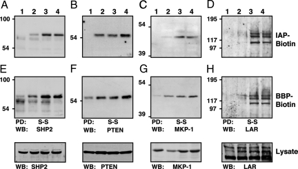
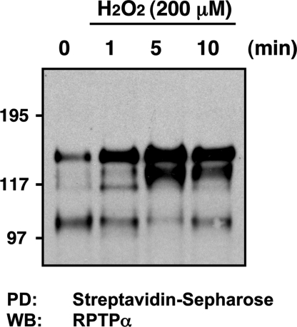
The production of reactive oxygen species (ROS) exerts an additional tier of control over tyrosine phosphorylation-dependent signal transduction by transiently inhibiting the catalytic activity of specific protein tyrosine phosphatases (PTPs). Hence, the ability to detect reversible oxidation of PTPs in vivo is critical to understanding the complex biological role of ROS in the control of cellular signaling. Here, we describe an assay for identifying those PTPs that are reversibly oxidized in vivo, which utilizes the unique chemistry of the invariant catalytic Cys residue in labeling the active site with biotinylated small molecules under mildly acidic conditions. We have applied this cysteinyl-labeling assay to the study of platelet-derived growth factor (PDGF) receptor signaling in an angiomyolipoma cell model. Doing so has allowed us to detect reversible oxidation of several proteins in response to sustained PDGF stimulation. As in other cell systems, we have observed the reversible oxidation of the classical PTP SHP2 and the tumor suppressor phosphatase PTEN in response to PDGF stimulation. Furthermore, we detected reversible oxidation of members of two other subclasses of PTPs, the receptor PTP LAR and the dual-specificity phosphatase MKP1. These data demonstrate the broad selectivity of the assay, allowing us to detect representatives of all of the major subgroups of the PTP superfamily. We anticipate that this cysteinyl-labeling enrichment strategy can be applied broadly to study reversible oxidation as a mechanism of harnessing PTP catalytic activity in a variety of signaling pathways.


Conflict of interest statement

The authors declare no conflict of interest.

Figures

Fig. 1.
Fig. 1.
Schematic outline of the cysteinyl-labeling assay. An active PTP is indicated with the active-site cysteinyl residue as a thiolate anion, representing the cell at a resting state. (1) After a physiological stimulus, the cells were lysed in a degassed buffer at pH 5.5 containing IAA. The low-pKa Cys residue at the active site of those PTPs that remained in a reduced state are alkylated, terminally inactivating this pool of PTPs. Conversely, the Cys residues that were oxidized by second-messenger ROS molecules were protected from irreversible alkylation. (2) IAA was then removed from the lysate by buffer exchange using size-exclusion spin columns, and the oxidized Cys residues were reduced back to the thiolate ion with DTT. (3) The oxidized PTPs were maintained in pH 5.5 buffers and incubated with either of two biotinylated, active-site-directed compounds, a sulfhydryl-reactive IAP probe or a BBP activity-based probe. Purification by streptavidin pull-down and immunoblotting permits identification of ROS-targeted PTPs.
Fig. 2.
Fig. 2.
Detection of reversible PTP oxidation by using a sulfhydryl-reactive IAP probe. Serum-deprived (16 h) angiomyolipoma cell lines (lanes: 1, SV7tert; 2, SV7tert-PDGF; 3, SV7tertPDGF-Tumor1; 4, SV7tertPDGF-Tumor2) were subjected to the cysteinyl-labeling assay by using biotinylated IAP at pH 5.5 (A) or pH 8 (B). Biotinylated proteins were purified on streptavidin–Sepharose beads, resolved by SDS/PAGE, and visualized by using streptavidin–HRP.
Fig. 3.
Fig. 3.
Reversible PTP oxidation in PDGF-BB-derived tumor cells. Tumor-derived PDGF-transformed SV7tert cells were serum-deprived (48 h) and treated for a 48-h period with (A) 25 μM DPI, 100 μM NAC, or 5 μM MnTMPyP; or (B) 25 μM DPI, 10 μM myxothiazol, 100 μM rotenone, or 10 μM stigmatellin, then subjected to the cysteinyl-labeling assay with the biotinylated IAP probe. Biotinylated proteins were purified on streptavidin–Sepharose beads, resolved by SDS/PAGE, and visualized by using streptavidin–HRP.
Fig. 4.
Fig. 4.
Comparison of IAP- and BBP- based probes for detection of reversibly oxidized PTPs in PDGF-BB-transformed angiomyolipoma cells. (A) Structures of biotinylated IAP and BBP probes. (B) Mechanisms of PTP labeling by the IAP and BBP probes. (C) Serum-deprived (16 h) angiomyolipoma cell lines (lanes: 1 and 5, SV7tert; 2 and 6, SV7tert-PDGF; 3 and 7, SV7tertPDGF-Tumor1; 4 and 8, SV7tertPDGF-Tumor2) were subjected to the cysteinyl-labeling assay using either biotinylated IAP (lanes 1–4) or BBP probes (lanes 5–8). Biotinylated proteins were purified on streptavidin–Sepharose beads, resolved by SDS/PAGE and visualized by using streptavidin–HRP.
Fig. 5.
Fig. 5.
Identification of reversibly oxidized PTPs in PDGF-BB-transformed angiomyolipoma cells by using IAP- and BBP-based-probes. Serum-deprived (16 h) angiomyolipoma cell lines (lanes: 1, SV7tert; 2, SV7tert-PDGF; 3, SV7tertPDGF-Tumor1; 4, SV7tertPDGF-Tumor2) were subjected to the cysteinyl-labeling assay using either biotinylated IAP (A–D) or BBP probes (E and F). Biotinylated proteins were purified on streptavidin–Sepharose beads, resolved by SDS/PAGE, and immunoblotted against SHP2 (A and E), PTEN (B and F), MKP-1 (C and G), or LAR (D and H). Reversible PTP oxidation was restricted to PDGF-BB-transformed angiomyolipoma cells using both probes. WB, Western blotting.
Fig. 6.
Fig. 6.
RPTPα reversible oxidation is detected by using the cysteinyl-labeling assay. Serum-deprived (16 h) angiomyolipoma SV7tert cells were exposed to 200 μM H2O2 for 1, 5, or 10 min and subjected to the cysteinyl-labeling assay using a biotinylated IAP probe. Biotinylated proteins were purified on streptavidin–Sepharose beads, resolved by SDS/PAGE, and immunoblotted by using anti-RPTPα antiserum.

References

    1. Tonks NK. Protein tyrosine phosphatases: From genes, to function, to disease. Nat Rev Mol Cell Biol. 2006;7:833–846. - PubMed
    1. Finkel T. Oxidant signals and oxidative stress. Curr Opin Cell Biol. 2003;15:247–254. - PubMed
    1. Rhee SG, et al. Intracellular messenger function of hydrogen peroxide and its regulation by peroxiredoxins. Curr Opin Cell Biol. 2005;17:183–189. - PubMed
    1. Barford D, Flint AJ, Tonks NK. Crystal structure of human protein tyrosine phosphatase 1B. Science. 1994;263:1397–1404. - PubMed
    1. Denu JM, Dixon JE. Protein tyrosine phosphatases: Mechanisms of catalysis and regulation. Curr Opin Chem Biol. 1998;2:633–641. - PubMed

Publication types

MeSH terms