. 2008 Jul 22;105(29):10101-6.
doi: 10.1073/pnas.0804286105. Epub 2008 Jul 15.

CXCR4-CCR5: a couple modulating T cell functions

Affiliations

CXCR4-CCR5: a couple modulating T cell functions

Rita Lucia Contento et al. Proc Natl Acad Sci U S A. .

Abstract

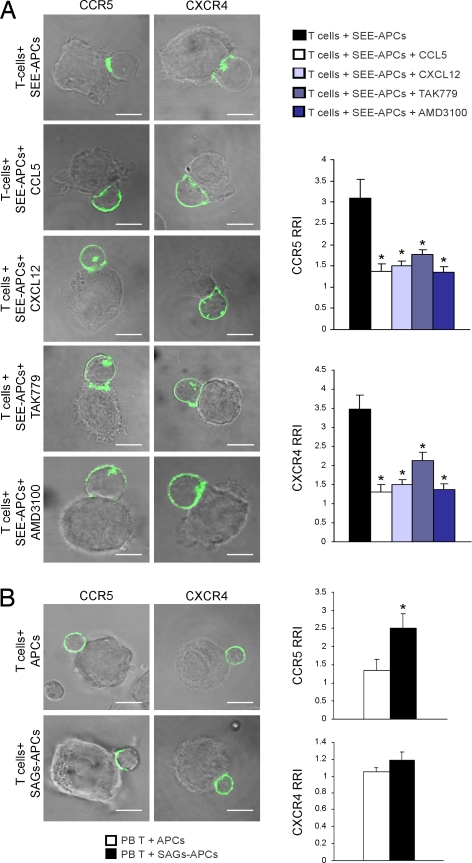
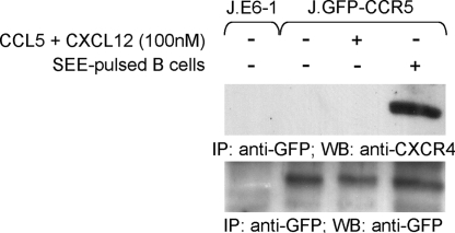
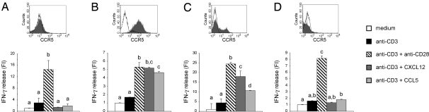
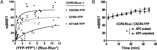
Chemokines and their receptors direct leukocyte migration among blood, lymph and tissues. Evidence has recently accumulated that, besides their chemotactic functions, chemokine receptors are highly versatile players that fine tune immune responses. During human T cell activation by antigen-presenting cells, the chemokine receptors CCR5 and CXCR4 are recruited into the immunological synapse, where they deliver costimulatory signals. However, the molecular mechanisms allowing signaling versatility of chemokine receptors are unknown. Here, we describe the functional interaction between CXCR4 and CCR5 to exert specific biological functions and modulate T lymphocyte responses. We demonstrate that simultaneous expression and cooperation between CCR5 and CXCR4 are required for chemokine-induced T cell costimulation at the immunological synapse. In addition, we provide evidence for a physical association of the two receptors in a signaling complex that activates distinct T cell functions. We suggest that cooperation between receptors represents one key strategy for the functional plasticity of chemokines.


Conflict of interest statement

The authors declare no conflict of interest.

Figures

Fig. 1.
Fig. 1.
CXCR4 and CCR5 are co-recruited into the IS. (A) Jurkat cells expressing GFP-tagged CCR5 or CXCR4 were incubated with SEE-loaded B cells for 10 min in the presence of AMD3100, TAK779, CXCL12 or CCL5. (B) PB T cells expressing GFP-tagged CCR5 or CXCR4 were incubated with superantigens (SAGs)-loaded B cells for 10 min. (A and B) Cells were then fixed and analyzed by confocal microscopy. Bar, 10 μm. Quantitative analyses of CCR5 and CXCR4 accumulation at the IS are shown. The relative recruitment index (RRI) (5) represents mean (± SE) of 30 cells out of four independent experiments. *, P < 0.05 compared with control T cells.
Fig. 2.
Fig. 2.
Physical association between CXCR4 and CCR5. GFP-CCR5 Jurkat T cells were stimulated as indicated, and CCR5 was immunoprecipitated with anti-GFP. Immunoprecipitated proteins were blotted with anti-CXCR4. Blotting with anti-GFP resolved in the same gel is shown, and Jurkat cells lacking GFP-CCR5 were included as control. Results are representative of at least three experiments.
Fig. 3.
Fig. 3.
CXCL12-induced costimulation requires CCR5 expression. IFN-γ production in resting (A), activated (B), CCR5-transfected resting (C), or CCR5Δ32 activated (D) human PB T cells stimulated with beads coated with anti-CD3 mAb in the presence or absence of anti-CD28 mAb, CXCL12, or CCL5. For each cell type, the expression of CCR5, as analyzed by flow cytometry, is indicated (the empty histograms represent the isotype control). Results are representative of at least three experiments. Bars with different letters are significantly different from each other (Student-Newman-Keuls test, P < 0.05). Mean amounts of IFN-γ produced by single living cells upon anti-CD3 stimulation were: 1.2 fg/ml (A), 18 fg/ml (B), 1.1 fg/ml (C) and 15 fg/ml (D). The percentage of living cells after 48 h of stimulation was: ≈100% in A and B, 10% in C, and 20% in D. FI = Fold of induction over unstimulated cells.
Fig. 4.
Fig. 4.
Constitutive association between CXCR4 and CCR5. (A) BRET saturation curves obtained by measuring BRET in Jurkat T cells expressing fixed quantities of BRET donor (CCR5-Rluc) and increasing amounts of BRET acceptors (indicated C-terminally YFP-tagged GPCR constructs). Relative amounts of BRET acceptor are expressed as the ratio between the fluorescence of the acceptor over the luciferase activity of the donor. YFP° corresponds to background fluorescence in cells expressing the BRET donor alone. BRET-ratio values were from 18 individual transfections grouped as a function of the amount of BRET acceptor. (B) The transfer of energy between CCR5-Rluc and CXCR4-YFP was initiated by the addition of coelenterazine h, and the BRET ratio was monitored in real time in live Jurkat cells incubated with SEE-pulsed APCs (closed squares) or unpulsed APCs (closed triangles). The data shown represent the mean ± SD. of triplicates in an experiment representative of two independent experiments at constant (YFP-YFP°)/(Rluc-RLuc°) values (between 0.5 and 2).
Fig. 5.
Fig. 5.
Co-modulation of CXCR4 and CCR5. CCR5+CXCR4+ Jurkat T cells (A and B) or CCR5+CXCR4 A7 cells (C) were stimulated with CCL5 or CXCL12 for 90 min, and plasma membrane expression of CCR5 (A and C) and CXCR4 (B) was analyzed at different time points. The graphs show the mean fluorescence intensity (MFI) for treated cells as a proportion of the MFI for untreated cells at the indicated times. Data points represent mean (± SE) of triplicates.

References

    1. Lanzavecchia A, Lezzi G, Viola A. From TCR engagement to T cell activation: A kinetic view of T cell behavior. Cell. 1999;96:1–4. - PubMed
    1. Gerard C, Rollins BJ. Chemokines and disease. Nat Immunol. 2001;2:108–115. - PubMed
    1. Viola A, Luster AD. Chemokines and Their Receptors: Drug Targets in Immunity and Inflammation. Annu Rev Pharmacol Toxicol. 2008;48:171–197. - PubMed
    1. Viola A, Contento RL, Molon B. T cells and their partners: The chemokine dating agency. Trends Immunol. 2006;27:421–427. - PubMed
    1. Molon B, Gri G, Bettella M, Gomez-Mouton C, Lanzavecchia A, et al. T cell costimulation by chemokine receptors. Nat Immunol. 2005;6:465–471. - PubMed

Publication types

MeSH terms