. 2008 Jul 15;68(14):5972-8.
doi: 10.1158/0008-5472.CAN-07-6818.

Ability of mature dendritic cells to interact with regulatory T cells is imprinted during maturation

Affiliations

Ability of mature dendritic cells to interact with regulatory T cells is imprinted during maturation

Ravikumar Muthuswamy et al. Cancer Res. .

Abstract

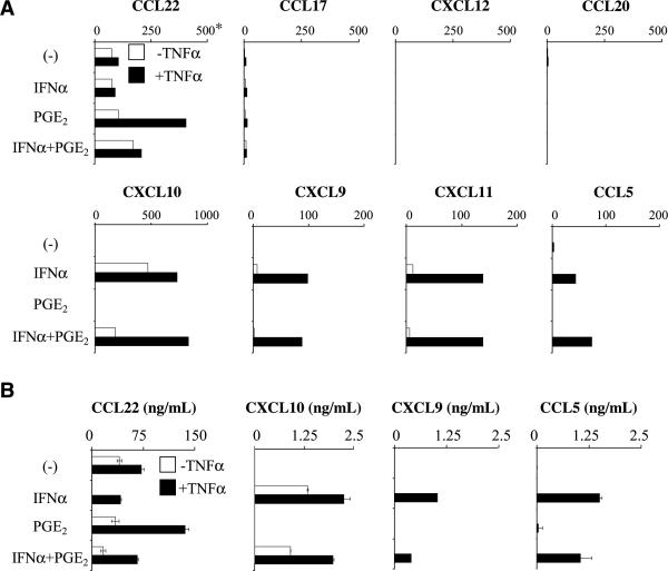
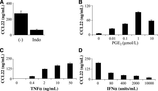
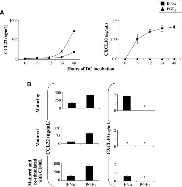
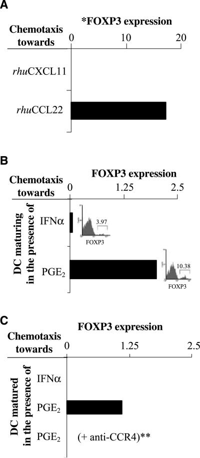
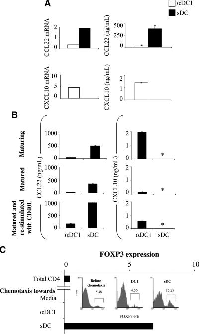
Preferential activation of regulatory T (Treg) cells limits autoimmune tissue damage during chronic immune responses but can also facilitate tumor growth. Here, we show that tissue-produced inflammatory mediators prime maturing dendritic cells (DC) for the differential ability of attracting anti-inflammatory Treg cells. Our data show that prostaglandin E(2) (PGE(2)), a factor overproduced in chronic inflammation and cancer, induces stable Treg-attracting properties in maturing DC, mediated by CCL22. The elevated production of CCL22 by PGE(2)-matured DC persists after the removal of PGE(2) and is further elevated after secondary stimulation of DC in a neutral environment. This PGE(2)-induced overproduction of CCL22 and the resulting attraction of FOXP3(+) Tregs are counteracted by IFN alpha, a mediator of acute inflammation, which also restores the ability of the PGE(2)-exposed DC to secrete the Th1-attracting chemokines: CXCL9, CXCL10, CXCL11, and CCL5. In accordance with these observations, different DCs clinically used as cancer vaccines show different Treg-recruiting abilities, with PGE(2)-matured DC, but not type 1-polarized DC, generated in the presence of type I and type II IFNs, showing high Treg-attracting activity. The current data, showing that the ability of mature DC to interact with Treg cells is predetermined at the stage of DC maturation, pave the way to preferentially target the regulatory versus proinflammatory T cells in autoimmunity and transplantation, as opposed to intracellular infections and cancer.


Figures

Figure 1
Figure 1
IFNα and PGE2 cross-regulate the production of Teff- and Treg-attracting chemokines by DC. Day 6 monocyte-derived immature DCs were exposed to PGE2 and/or IFNα either alone (□) or in the presence of TNFα (■) as a DC maturation-inducing agent. A and B, dominant effect of IFNα on the chemokine production profiles in maturing DC. A, expression of Treg-attracting (top) and Teff-attracting (bottom) chemokine genes in the differentially treated DC. *, the data are expressed as the ratios between the expression of the individual chemokine genes and HPRT1 (see Materials and Methods) and represent one of five experiments that all yielded similar results. B, secretion of CCL5, CXCL9, CXCL10, and CCL22 proteins by the differentially treated DC. PGE2-induced CCL22 production was significantly higher (P < 0.01) compared with the TNFα treatment alone.
Figure 2
Figure 2
Dose-dependent cross-regulation of CCL22 production by TNFα, PGE2, and IFNα. A, involvement of endogenous prostaglandins in the production of “baseline” levels of CCL22 by maturing DC. Please note the reduction (P < 0.05) of the baseline CCL22 production in the presence of prostaglandin synthesis inhibitor indomethacin (Indo; 50 μmol/L; added at the beginning of cultures). B, dose-dependent enhancement of CCL22 production in maturing DC by exogenous PGE2. Indomethacin (50 μmol/L) was present in all cultures to eliminate the endogenous PGE2 production. C, DCs were treated with 1 μmol/L PGE2 but with increasing doses of TNFα to reveal TNFα-dependent induction of CCL22 by PGE2. D, inhibition of CCL22 production by increasing doses of IFNα in the TNFα-treated (50 ng/mL) and PGE2-treated (1 μmol/L) DC. Data from one of two (C and D) or four (A and B) experiments that all yielded similar results.
Figure 3
Figure 3
The ability of DC to produce Teff- and Treg-attracting chemokines is imprinted during DC maturation. A, kinetics of CXCL10 and CCL22 protein secretion by DC maturing in the presence of IFNα (■) or PGE2 (▲). TNFα was added to both types of cultures. Figure represents combined data from two experiments that yielded similar results. The difference in CCL22 production between IFNα-treated and PGE2-treated DC was significant at 24 h (P < 0.05) and 48 h (P < 0.01) of stimulation. B, stability of the maturation-induced chemokine profiles in DC matured with either IFNα or PGE2 in the presence of TNFα. Chemokine concentrations in the supernatants from the DC maturation cultures (top) and in the 48-h cultures of the DC harvested, washed, and replated in the absence (middle) or presence (bottom) of CD40L. In all three situations, the IFNα- versus PGE2-treated DC showed significant differences in CCL22 production (P < 0.01, 0.05, and 0.01, respectively). *, below 0.05 ng/mL.
Figure 4
Figure 4
PGE2 imprints a stable Treg-attracting function in maturing DC: key role of CCR4 in the ability of DC to attract FOXP3+ T cells. Purified CD4+ T cells were loaded into the upper chamber of the Transwells and allowed to migrate toward recombinant CCR4 or CXCR3 ligands or the supernatants of IFNα- or PGE2-treated DC. The migrated CD4+ T cells from the bottom chambers were pooled (eight wells per group, with higher numbers needed for the intracellular staining experiments) and analyzed by Taqman for the presence of FOXP3+ cells. A, Taqman analysis of CD4+ T cells migrated in response to rhuCCL22 or to CXCL10 (see Supplementary Fig. S2 for the intracellular expression of FOXP3 protein in the individual CD4+ T cells). Similar data were obtained in two additional experiments. B, Taqman analysis of the CD4+ T cells migrated in response to the factors released during the TNFα-driven maturation of DC exposed to IFNα or PGE2. Insets, frequencies of FOXP3+ T cells in the CD4+ T-cell populations recruited by IFNα- or PGE2-treated DC supernatants. Similar data were obtained in three additional experiments. C, CD4+ T cells migrating in response to the supernatants from harvested and reseeded DC after maturation. *, all mRNA data are normalized for HPRT1. **, the CD4+ T cells were pretreated with CCR4 blocking antibody before addition to the upper chambers of the chemotaxis plate. Similar data were obtained using excess CCL22 to abrogate the CCL22 gradient.
Figure 5
Figure 5
Different clinically applied DC types display strong differences in the ability to attract FOXP3+ T cells. A, sDC (■) generated in the presence of IL-1β, TNFα, IL6, and PGE2 (35) and αDC1s (□) matured in the presence of IL-1β, TNFα, IFNα, IFNγ, and polyI:C (36) were tested for the expression of Teff- or Treg-attracting chemokine genes (left; also see Supplementary Fig. S5) and the secretion of the relevant chemokines (right; P < 0.05). Representative data from over 10 donors. B, stability of the chemokine production pattern in different types of clinically applied DC. Chemokine contents in the supernatants from DC maturation cultures (top) and in the 48-h cultures of the harvested, washed, and replated DC in the absence (middle) or presence (bottom) of CD40L. αDC1 and sDC showed significant differences in CCL22 production in all stages tested (P < 0.01). *, below 0.05 ng/mL. Similar data were obtained in two to four additional experiments. C, Taqman analysis of freshly isolated purified CD4+ T cells (before migration) and of the CD4+ T cells migrated in response to medium (negative control, spontaneous migration only), αDC1, or sDC supernatants. Data from one of five experiments that all yielded similar results. Inset, frequencies of FOXP3+ T cells in the CD4+ T-cell populations recruited by the supernatants from αDC1s or sDCs were determined by flow cytometry.

References

    1. Zou W. Regulatory T cells, tumour immunity and immunotherapy. Nat Rev Immunol. 2006;6:295–307. - PubMed
    1. Taams LS, Palmer DB, Akbar AN, Robinson DS, Brown Z, Hawrylowicz CM. Regulatory T cells in human disease and their potential for therapeutic manipulation. Immunology. 2006;118:1–9. - PMC - PubMed
    1. Curiel TJ, Coukos G, Zou L, et al. Specific recruitment of regulatory T cells in ovarian carcinoma fosters immune privilege and predicts reduced survival. Nat Med. 2004;10:942–9. - PubMed
    1. Hirahara K, Liu L, Clark RA, Yamanaka K, Fuhlbrigge RC, Kupper TS. The majority of human peripheral blood CD4+CD25highFoxp3+ regulatory T cells bear functional skin-homing receptors. J Immunol. 2006;177:4488–94. - PubMed
    1. Iellem A, Mariani M, Lang R, et al. Unique chemotactic response profile and specific expression of chemokine receptors CCR4and CCR8 by CD4(+)CD25(+) regulatory T cells. J Exp Med. 2001;194:847–53. - PMC - PubMed

Publication types

MeSH terms