. 2008 Sep 5;283(36):24889-98.
doi: 10.1074/jbc.M804236200. Epub 2008 Jul 15.

Adiponectin blocks interleukin-18-mediated endothelial cell death via APPL1-dependent AMP-activated protein kinase (AMPK) activation and IKK/NF-kappaB/PTEN suppression

Affiliations

Adiponectin blocks interleukin-18-mediated endothelial cell death via APPL1-dependent AMP-activated protein kinase (AMPK) activation and IKK/NF-kappaB/PTEN suppression

Bysani Chandrasekar et al. J Biol Chem. .

Abstract

The adipocyte-derived cytokine adiponectin is known to exert anti-inflammatory and anti-apoptotic effects. In patients with atherosclerotic cardiovascular disease, circulating levels of adiponectin correlate inversely with those of the proinflammatory, proapoptotic cytokine interleukin (IL)-18. The opposing actions of IL-18 and adiponectin on both cell survival and inflammation led us to investigate whether adiponectin signaling antagonizes IL-18-mediated endothelial cell death and to identify the underlying molecular mechanisms. Treatment with IL-18 suppressed Akt phosphorylation and its associated kinase activity, induced IkappaB kinase (IKK)-NF-kappaB-dependent PTEN activation, and promoted endothelial cell death. Pretreatment with adiponectin stimulated APPL1-dependent AMPK activation, reversed Akt inhibition in a phosphatidylinositol 3-kinase-dependent manner, blocked IKK-NF-kappaB-PTEN signaling, reduced caspase-3 activity, blocked Bax translocation, and inhibited endothelial cell death. The cytoprotective effect of adiponectin signaling was recapitulated by treatment with the pharmacological AMPK activator 5-aminoimidazole-4-carboxamide-1-beta-riboside. Collectively, these results demonstrated that adiponectin reverses IL-18-mediated endothelial cell death through an AMPK-associated mechanism, which may thus have therapeutic potential for diminishing IL-18-dependent vascular injury and inflammation.


Figures
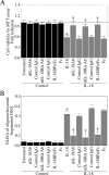
FIGURE 1.
FIGURE 1.
IL-18 induces endothelial cell death. A, IL-18 induces EC death. Quiescent EC were treated with neutralizing mouse anti-IL-18 or goat anti-IL-18Rα antibodies or IL-18BP/Fc chimera (10 μg/ml for 1 h) prior to the addition of rhIL-18 (100 ng/ml for 24 h). Normal mouse IgG1, goat IgG, and Fc served as the respective controls. Cell viability was assessed by the MTT assay, and the results were expressed as -fold reduction from untreated cells (n = 12/group). *, p < 0.001 versus untreated; †, p < 0.01 versus IL-18. B, IL-18-mediated EC death was confirmed by ELISA. EC treated as described in A were analyzed for cell death by quantifying mono- and oligonucleosomal fragmented DNA in the cytoplasmic extracts by ELISA (n = 12/group). *, p < 0.001 versus untreated; †, p < 0.001 versus IL-18 by ANOVA.
FIGURE 2.
FIGURE 2.
IL-18 suppresses Akt activity and stimulates IKK-dependent NF-κB activation. A, IL-18 suppresses Akt phosphorylation. Quiescent EC were treated with rhIL-18 (100 ng/ml for 1 h). Total and phospho-Akt levels in cell lysates were analyzed by immunoblotting using activation-specific antibodies. B, IL-18 suppresses Akt kinase activity. Quiescent EC treated as described in A were analyzed for Akt kinase activity by an immune complex kinase assay using GSK as a substrate. The specificity of IL-18 was determined by incubating EC with IL-18-neutralizing antibodies (10 μg/ml for 1 h; lane 3) prior to IL-18 addition. A representative of three independent experiments is shown. C, IL-18 induces IκB kinase activity. Quiescent EC treated as described in A were analyzed for IKK kinase activity by an in vitro kinase assay using GST-IκB fusion protein as a substrate. A representative of three independent experiments is shown. D, SC-514 blunts IL-18-mediated IKK activation. Quiescent EC were treated with the IKKβ-specific inhibitor SC-514 (100 μm in DMSO for 1 h) prior to IL-18 addition. DMSO served as a control. IKK activity was analyzed as in C. E, IL-18 activates NF-κB. Quiescent EC treated as described in A were analyzed for NF-κB p50 and p65 subunits in the nuclear protein extracts by ELISA (n = 12). *, p < 0.001 versus the respective untreated cells (n = 12). F, the purity of nuclear extract was confirmed by immunoblotting using anti-lamin A/C and tubulin antibodies (n = 3). G, IL-18-mediated NF-κB activation was confirmed by reporter assay. EC transduced with adenoviral NFκB reporter vector (Ad.NFκB-Luc) were treated with IL-18 for 12 h. Ad.MCS-Luc served as a control. Ad.β-gal served as an internal control. *, p < 0.001 versus the respective untreated cells (n = 12). H, IL-18 induces NF-κB activation in IKK-dependent manner. EC either transduced with Ad.dnIKKβ (24 h) or pretreated with SC-514 (1 h) were treated with rhIL-18 for 12 h. Ad.GFP and DMSO served as the respective controls. Nuclear extracts were analyzed for NFκBp65 by ELISA. *, p < 0.001 versus respective untreated cells; †, p < 0.01 versus IL-18 by ANOVA (n = 12/group).
FIGURE 3.
FIGURE 3.
IL-18 stimulates PTEN expression. A, IL-18 induces PTEN promoter-reporter activity in IKK-NF-κB-dependent manner. EC transiently transfected with pGL3-PTEN were transduced with adenoviral dnIKKβ, dnp65, or dnIκB-α for 24 h and then treated with IL-18 for 12 h (n = 6). Ad.GFP served as a control. pGL3-Basic served as a vector control. EC were cotransfected with pRL-TK vector (100 ng) to normalize for variations in transfection efficiency. *, p < 0.05 versus pGL3-Basic alone, **, p < 0.001 versus respective untreated cells; †, p < 0.01 versus IL-18 and IL-18+GFP (n = 12/group). B, IL-18 induces PTEN mRNA expression in an IKK-NF-κB-dependent manner. Quiescent EC treated as described in A, except for 2 h, were analyzed for PTEN mRNA expression by real-time quantitative PCR. In addition, IKKβ, p65 and IκB-α were targeted by specific siRNA for 48 h prior to IL-18 treatment. Non-targeting siRNA served as a control, and β-actin served as an internal control. *, p < 0.001 versus respective untreated cells; †, p < 0.01 versus IL-18 by ANOVA (n = 6/group). C, knockdown of IKKβ, p65, or IκB-α after 48 h was confirmed by immunoblotting (n = 3). Tubulin served as a loading control. D, IL-18 induces PTEN protein expression in an IKK-NF-κB-dependent manner. EC transduced with adenoviral dnIKKβ, dnp65, or dnIκB-α for 24 h were treated with IL-18 for 12 h. PTEN protein levels were analyzed in cleared cell lysates by immunoblotting. Tubulin served as an internal control. A representative of three independent experiments is shown. E, PTEN knockdown blunts IL-18-mediated EC death. EC were treated with PTEN-specific siRNA (100 nm for 48 h) prior to IL-18 addition. After 24 h, cell death was analyzed by ELISA. *, p < 0.001 versus respective untreated cells; †p < 0.001 versus IL-18 (n = 12/group). Knockdown of PTEN, confirmed by immunoblotting (n = 3), is shown in the right panel. Tubulin served as a loading control.
FIGURE 4.
FIGURE 4.
Adiponectin blocks IL-18-mediated EC death. A, adiponectin attenuates IL-18-mediated EC death in a dose-dependent manner. Quiescent EC were treated with adiponectin at the indicated concentrations for 1 h prior to IL-18 addition. EC death was assessed at 24 h by ELISA. *, p < 0.001 versus respective untreated cells; †, p < 0.001 versus IL-18 (n = 12/group). B, adiponectin inhibits IL-18-mediated caspase-3 activation. Quiescent EC were treated with adiponectin (30 μg/ml for 1 h) prior to IL-18 addition (8 h). Capase-3 activity was assayed essentially as described under “Experimental Procedures.” *, p < 0.001 versus untreated cells; †, p < 0.01 versus IL-18 (n = 12/group). C, adiponectin blunts Bax translocation. Quiescent EC were treated with adiponectin (30 μg/ml for 1 h) prior to IL-18 addition. Bax levels in mitochondrial and cytoplasmic fractions were analyzed by immunoblotting. A representative of three independent experiments is shown.
FIGURE 5.
FIGURE 5.
Adiponectin blocks IL-18-mediated EC death via AMPK activation. A, adiponectin-mediated AMPK activation is inhibited by compound C. Quiescent EC were treated with adiponectin (30 μg/ml) for the indicated time periods. AMPK activation was analyzed by immunoblotting using activation-specific antibodies. A representative of three independent experiments is shown. B, adiponectin stimulates AMP kinase activity. Quiescent EC treated as described in A were analyzed for AMPK activity using an in vitro assay as described under “Experimental Procedures.” *, p < 0.001 versus respective untreated cells (n = 6/group). C, adiponectin-mediated AMPK phosphorylation is attenuated by compound C. Quiescent EC were treated with the AMPK inhibitor compound C (40 μm in DMSO for 1 h) prior to adiponectin (30 μg/ml for 30 min). AMPK activation was analyzed as described A. A representative of three independent experiments is shown. D, knockdown of APPL1 blunts adiponectin-mediated AMPK activation. EC treated with APPL1 siRNA (100 nm for 48 h) were treated with adiponectin for 30 min. AMPK phosphorylation was analyzed by Immunoblotting. Scrambled siRNA served as a control. Knockdown of APPL1 was confirmed by immunoblotting (right panel). A representative of three independent experiments is shown. E, dnAMPK or kinase-dead AMPK (kdAMPK) reverses adiponectin prosurvival effects. EC transduced with adenoviral dnAMPK or kinase-dead AMPK (100 m.o.i. for 24 h) or treated with compound C (40 μm in DMSO or 1 h) were treated with adiponectin for 1 h followed by IL-18 for 24 h. Ad.GFP and DMSO served as controls. Cell death was assessed by ELISA. *, p < 0.001 versus untreated cells; §, p < 0.01 versus IL-18; †, p < 0.001 versus IL-18+DMSO or IL-18+GFP by ANOVA (n = 12/group). F, AMPKα1 knockdown reverses prosurvival effects of adiponectin. EC treated with AMPKα1 siRNA (100 m.o.i. for 48 h) were treated with adiponectin for 1 h followed by IL-18 for 24 h. Cell death was assessed by ELISA. *, p < 0.001 versus untreated cells; †, p < 0.001 versus IL-18+adiponectin+control siRNA; §, p < 0.01 versus IL-18+adiponectin (n = 12/group). Knockdown of AMPKα1 was confirmed by immunoblotting (right panel). G, APPL1 knockdown reverses prosurvival effects of adiponectin. EC treated with APPL1 siRNA (100 m.o.i. for 48 h) were treated with adiponectin for 1 h followed by IL-18 for 24 h. Cell death was assessed by ELISA. *, p < 0.001 versus untreated cells; †, p < 0.001 versus IL-18+adiponectin+control siRNA; §, p < 0.01 versus IL-18+adiponectin (n = 12/group).
FIGURE 6.
FIGURE 6.
Adiponectin reverses IL-18-mediated Akt suppression and IKK-NF-κB-dependent PTEN induction. A, adiponectin induces PI3K activation. Quiescent EC were incubated with adiponectin for 30 min. Equal amounts of cleared cell lysates were immunoprecipitated with the anti-p85 regulatory subunit antibody (Ab) of PI3K followed by an immune complex kinase assay as described under “Experimental Procedures.” The bottom panel shows an immunoblot analysis (WB) of the same samples with anti-p85 antibody. A representative of three independent experiments is shown. PI3P, phosphatidyl 3-phosphate. B, adiponectin reverses IL-18-mediated Akt suppression. Quiescent EC were treated with adiponectin for 1 h prior to IL-18 addition. Total and phospho-Akt levels were assessed by immunoblotting (n = 3). C, dnPI3K reverses adiponectin-mediated Akt activation. EC transduced with adenoviral dnPI3Kp85 (100 m.o.i. for 24 h) were treated with adiponectin followed by IL-18 addition. Total and phospho-Akt levels were assessed as described in B. D, AMPKα1 knockdown reverses adiponectin-mediated Akt activation. EC treated with AMPKα1 siRNA for 48 h were treated with adiponectin followed by IL-18 addition. Total and phospho-Akt levels were assessed as described in B. E, adiponectin reverses IL-18-induced IKK activity. Quiescent EC were treated with adiponectin for 1 h prior to IL-18 addition. IKK activity was analyzed by an in vitro kinase assay (n = 3). Tubulin served as a loading control. F, AMPKα1 knockdown reverses adiponectin-mediated IKK suppression. EC treated with AMPKα1 siRNA for 48 h were treated with adiponectin followed by IL-18 addition. IKK activity was assessed as described in E. G, adiponectin. blunts IL-18-mediated NF-κB activation. EC treated with AMPKα1 siRNA for 48 h were treated with adiponectin followed by IL-18 addition for 2 h. Nuclear protein was extracted and analyzed for NFκBp65 levels by ELISA. *, p < 0.001 versus untreated cells; †, p < 0.01 versus IL-18; §, p < 0.05 versus IL-18+adiponectin. H, adiponectin reverses IL-18-mediated PTEN induction. Quiescent EC were treated with adiponectin for 1 h prior to IL-18 addition. PTEN levels were analyzed by immunoblotting. Tubulin served as a loading control. A representative of three independent experiments is shown. I, knockdown of AMPKα1 reverses the cell survival effects of adiponectin. EC treated with IL-18 as described in D, but for 24 h, were analyzed for cell death by quantifying mono- and oligonucleosomal fragmented DNA in the cytoplasmic extracts by ELISA (n = 12/group). *, p < 0.001 versus untreated cells; †, p < 0.001 versus IL-18; §, p < 0.005 versus IL-18+Control siRNA by ANOVA.
FIGURE 7.
FIGURE 7.
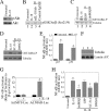
AICAR inhibits EC death. A, AICAR induces AMPK phosphorylation. Quiescent EC were treated with AICAR (1 mm for 1 h). AMPK activation was analyzed by immunoblotting using activation-specific antibodies (n = 3). B, AICAR stimulates AMP kinase activity. Quiescent EC treated with AICAR as described in A were analyzed for AMP kinase activity by an in vitro kinase assay as described under “Experimental Procedures.” *, p < 0.01 versus untreated cells; †, p < 0.05 versus IL-18 (n = 12). C, AICAR blunts IL-18-induced IKK activity. Quiescent EC were treated with AICAR for 1 h prior to IL-18 addition. IKK activity was analyzed by an in vitro kinase assay (n = 3). D, dnAMPK reverses adiponectin-mediated IKK suppression. EC transduced with dnAMPK were treated with adiponectin followed by IL-18. IKK activity was assessed as described in C (n = 3). E, AICAR reverses IL-18-mediated NF-κB activation. Quiescent EC were treated with AICAR for 1 h prior to IL-18 addition. Nuclear protein was extracted and analyzed for NFκBp65 levels by ELISA. *, p < 0.001 versus untreated cells; †, p < 0.01 versus IL-18 (n = 12). F, AICAR reverses IL-18-mediated Akt suppression. Quiescent EC were treated with AICAR for 1 h prior to IL-18 addition. Total and phospho-Akt levels were assessed by immunoblotting (n = 3). G, AICAR reverses IL-18-mediated PTEN induction. Quiescent EC were treated with AICAR for 1 h prior to IL-18 addition. PTEN levels were analyzed by immunoblotting. Tubulin served as a loading control. H, AICAR attenuates IL-18-mediated EC death. Quiescent EC were treated with AICAR prior to IL-18 addition for 24. EC death was assessed by ELISA. *, p < 0.001 versus untreated cells; †, p < 0.001 versus IL-18 (n = 12/group). I, schema showing signaling mechanisms involved in IL-18-mediated EC death and those targeted by adiponectin and AICAR.

References

    1. Landmesser, U., Hornig, B., and Drexler, H. (2004) Circulation 109 Suppl. 1, II27—II33 - PubMed
    1. Kockx, M. M., De Meyer, G. R., Muhring, J., Jacob, W., Bult, H., and Herman, A. G. (1998) Circulation 97 2307-2315 - PubMed
    1. Dimmeler, S., Hermann, C., and Zeiher, A. M. (1998) Eur. Cytokine Netw. 9 697-698 - PubMed
    1. Diamant, M., and Tushuizen, M. E. (2006) Curr. Diab. Rep. 6 279-286 - PubMed
    1. Koenig, W., and Khuseyinova, N. (2007) Arterioscler. Thromb. Vasc. Biol. 27 15-26 - PubMed

Publication types

MeSH terms