. 2008 Sep;3(5):1339-49.
doi: 10.2215/CJN.01600408. Epub 2008 Jul 16.

Renal lesions associated with IgM-secreting monoclonal proliferations: revisiting the disease spectrum

Affiliations

Renal lesions associated with IgM-secreting monoclonal proliferations: revisiting the disease spectrum

Vincent Audard et al. Clin J Am Soc Nephrol. 2008 Sep.

Abstract

Background and objectives: Since the first description of pathology of the kidney in Waldenström disease in 1970, there have been few reports on kidney complications of IgM-secreting monoclonal proliferations. Here, we aimed to revisit the spectrum of renal lesions occurring in patients with a serum monoclonal IgM.

Design, setting, participants, & measurements: Fourteen patients with a circulating monoclonal IgM and a kidney disease related to B cell proliferation were identified retrospectively. Demographic, clinical, and laboratory data were assessed for each patient at the time of kidney biopsy.

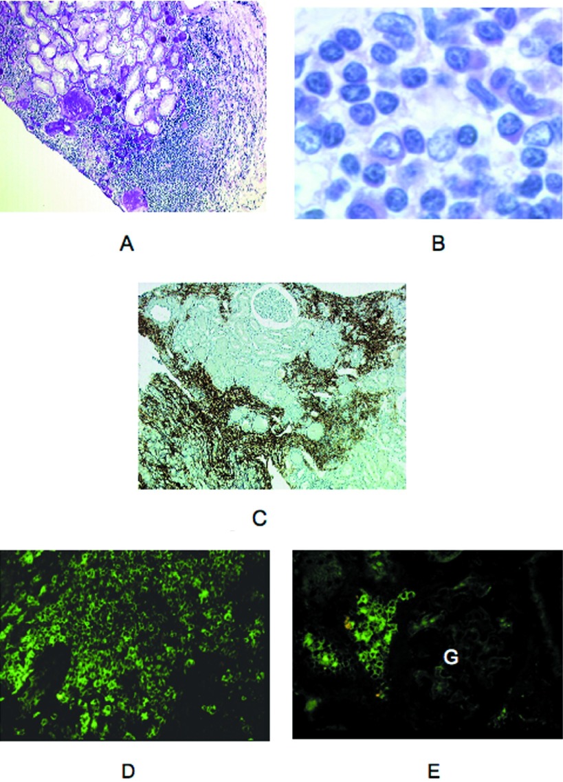
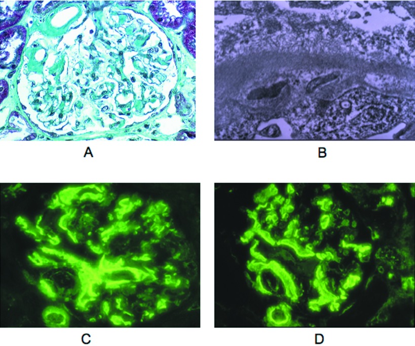
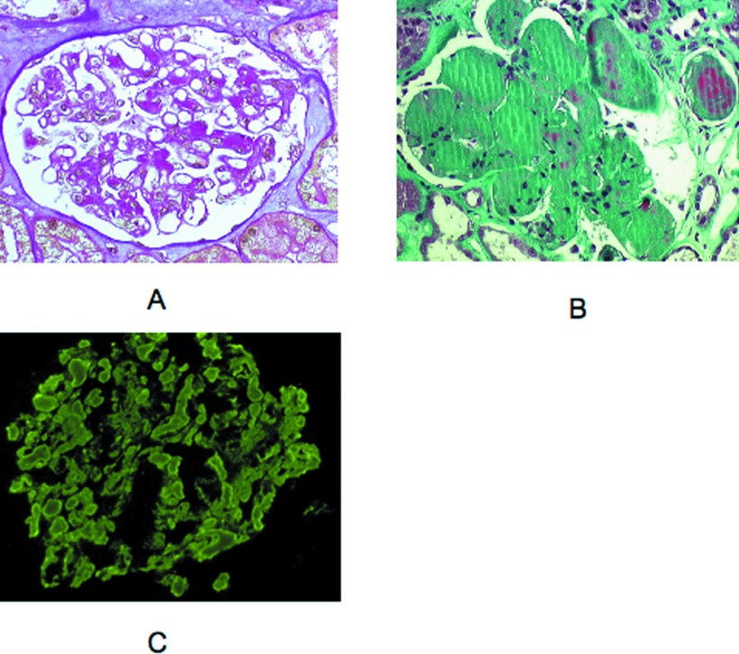
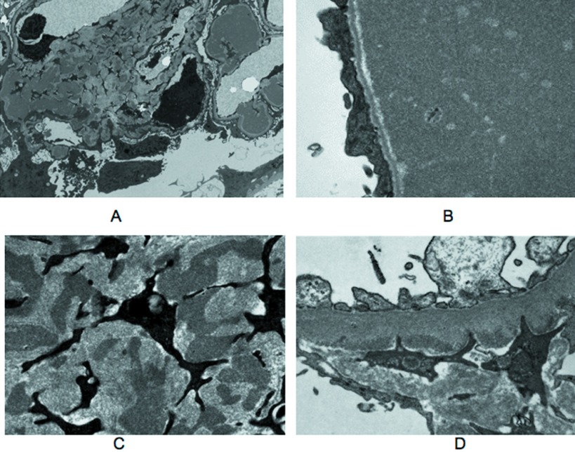
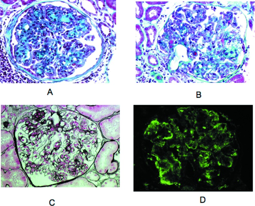
Results: Seven patients had a nephrotic syndrome. Patients without nephrotic syndrome all had impaired renal function. Mean serum creatinine was 238 micromol/L. For five patients, the diagnosis of monoclonal IgM preceded the kidney disease by 28.8 mo (range 12 to 60). Seven patients had Waldenström disease, two had a small B cell non-Hodgkin lymphoma, one had an IgM-excreting multiple myeloma, one had a marginal zone B cell lymphoma, and three had an IgM-related disorder. Renal lesions included (1) intracapillary monoclonal deposits disease with granular, electron-dense IgM thrombi occluding capillary lumens (5); (2) atypical membranoproliferative glomerulonephritis (3); (3) lambda light chain amyloidosis (2) associated with mu deposits in one patient; (4) acute tubular necrosis (1); and (5) CD20(+) lymphomatous infiltration (3). Remission of the nephrotic syndrome was attained in three of seven patients, and renal function improved after chemotherapy.

Conclusions: Although renal complications of IgM proliferations are rare, a wide spectrum of kidney lesions is observed, without correlation with the type of hematologic disorder.


Figures

Figure 1.
Figure 1.
Representative cases of lymphomatous infiltrate in the context of Waldenström macroglobulinemia (WM; light microscopy, immunofluorescence). (A) patient 3. Lymphoplasmacytic infiltrate of the interstitium in the external cortex close to an area of interstitial fibrosis with global glomerulosclerosis and tubular atrophy. Note cellular infiltration of the renal capsule (light microscopy, periodic acid-Schiff [PAS] stain). (B) Patient 1. Interstitial expansion secondary to lymphoplasmacytic infiltration (light microscopy, hematoxylin and eosin stain). (C) Patient 1. Immunocytochemical staining of patchy cortical cellular infiltrates showing numerous CD20+ cells. (D) Patient 2. By immunofluorescence (IF), cellular infiltrate stained brightly for μ chain. Some cells showed a cytoplasmic staining (FITC-conjugated anti-μ antibody). (E) Patient 2. Interstitial neoplastic cells were also stained for λ chain. Note the pericellular or cytoplasmic pattern of fluorescence. No deposits were seen in the glomerulus (G; FITC-conjugated anti-λ antibody). Magnifications: ×100 in A and C; ×1000 in B; ×400 in D and E.
Figure 2.
Figure 2.
AHL amyloidosis in patient 5 (light microscopy, electron microscopy [EM], IF). (A). Amyloid deposits were seen in the hilum, in mesangial areas, and segmentally along glomerular capillary walls (light microscopy, Masson trichrome stain). (B) Ultrastructural analysis showed focal transmembranous and subepithelial accumulation of amyloid fibrils with effacement of podocyte foot processes (EM). (C and D) Glomerular and arteriolar amyloid deposits were equally stained for μ and λ chains and were negative for γ, α, and κ chains as well as for C3, C1q, and albumin (data not shown; FITC-conjugated anti μ [C] and anti-λ antibodies [D]). Magnifications: ×400 in A, C, and D; ×20,000 in B.
Figure 3.
Figure 3.
Western blot analysis of IgM-enriched fractions from patient 5 (P) and from two control subjects (1 and 2) with (+) or without (−) PNGase treatment. A predominant μ chain subunit with normal apparent molecular weight (approximately 60 kD) and a less abundant lower sized (approximately 45 kD) component are detected in the patient 5 sample.
Figure 4.
Figure 4.
Representative cases of intracapillary monoclonal deposits disease (ICMDD; light microscopy, IF). (A) Patient 7. PAS positive intracapillary thrombi in a glomerulus. Note the absence of cellular proliferation (light microscopy, PAS stain). (B) Patient 8. Huge polychromatic capillary plugs responsible for massive occlusion of glomerular capillary lumens. A large deposit also occluded some arteriolar lumens (light microscopy, Masson trichrome stain). (C) Patient 7. Bright staining of intraglomerular thrombi for μ chain. The same fluorescence pattern was obtained with an anti-λ antibody but not with anti-κ, anti-C3, and anti-C1q antibodies (data not shown; IF, FITC-conjugated anti-μ antibody). Magnification, ×400.
Figure 5.
Figure 5.
Ultrastructural analysis of representative cases of ICMDD. (A) Patient 8. Low-power magnification electron micrograph of a glomerulus showing intracapillary deposits and irregular deposits in increased mesangial matrix. (B) Patient 8. Electron micrograph of a glomerular capillary lumen filled by electron-dense material. No organized structure was seen. Note capillary wall basement membrane stretching with sparing of the lamina densa and effacement of podocyte foot processes. (C) Patient 8. High-power magnification revealed the same granular aspect of mesangial deposits. (D) Patient 6. Electron micrograph showing scarce granular subendothelial deposits. Magnifications: ×6000 in A; ×25,000 in B and C; 20,000 in D.
Figure 6.
Figure 6.
Representative cases of membranoproliferative glomerulonephritis (MPGN) with atypical features (light microscopy, IF). (A) Patient 13. Membranoproliferative pattern was shown with accentuation of glomerular lobules and segmentally thickened capillary walls (light microscopy, Masson trichrome stain). (B) Patient 13. Some small subendothelial and intraluminal deposits stained red with the trichrome stain. Proliferation of mesangial and endothelial cells was moderate. Note segmental mesangiolysis (light microscopy, Masson trichrome stain). (C) Patient 12. Numerous infiltrating neutrophils were observed in some glomeruli (light microscopy, Silver stain). (D) Patient 13. Peripheral capillary wall deposits with granular texture were predominant in segmental areas outlining the double contours. This deposits stain for μ chain and κ chain (data not shown; IF, FITC-conjugated anti-μ antibody). Magnification, ×400.

References

    1. Solomon A, Weiss DT, Kattine AA: Nephrotoxic potential of Bence Jones proteins. N Engl J Med 324 :1845 –1851,1991 - PubMed
    1. Ronco P, Aucouturier P, Mougenot B: Monoclonal gammopathies: Multiple myeloma, amyloidosis, and related disorders. In: Diseases of the Kidney and Urinary Tract, 8th Ed., edited by Schrier RW, Philadelphia, Lippincott Williams & Wilkins,2007. , pp1941 –1985
    1. Morel-Maroger L, Basch A, Danon F, Verroust P, Richet G: Pathology of the kidney in Waldenström's macroglobulinemia: Study of sixteen cases. N Engl J Med 283 :123 –129,1970 - PubMed
    1. Da'as N, Kleinman Y, Polliack A, Amir G, Ne'eman Z, Kopolovic J, Bits H, Darmon D: Immunotactoid glomerulopathy with massive bone marrow deposits in a patient with IgM kappa monoclonal gammopathy and hypocomplementemia. Am J Kidney Dis 38 :395 –399,2001 - PubMed
    1. Dussol B, Kaplanski G, Daniel L, Brunet P, Pellissier JF, Berland Y: Simultaneous occurrence of fibrillary glomerulopathy and AL amyloid. Nephrol Dial Transplant 13 :2630 –2632,1998 - PubMed

Publication types

MeSH terms