. 2008 Oct;82(19):9329-36.
doi: 10.1128/JVI.00646-08. Epub 2008 Jul 16.

Human endogenous retrovirus K (HML-2) elements in the plasma of people with lymphoma and breast cancer

Affiliations

Human endogenous retrovirus K (HML-2) elements in the plasma of people with lymphoma and breast cancer

Rafael Contreras-Galindo et al. J Virol. 2008 Oct.

Abstract

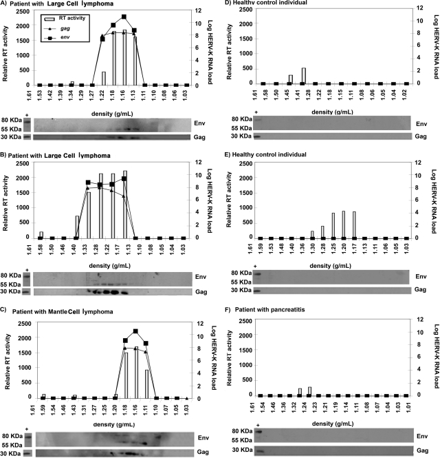
Actively replicating endogenous retroviruses entered the human genome millions of years ago and became a stable part of the inherited genetic material. They subsequently acquired multiple mutations, leading to the assumption that these viruses no longer replicate. However, certain human tumor cell lines have been shown to release endogenous retroviral particles. Here we show that RNA from human endogenous retrovirus K (HERV-K) (HML-2), a relatively recent entrant into the human genome, can be found in very high titers in the plasma of patients with lymphomas and breast cancer as measured by either reverse transcriptase PCR or nucleic acid sequence-based amplification. Further, these titers drop dramatically with cancer treatment. We also demonstrate the presence of reverse transcriptase and viral RNA in plasma fractions that contain both immature and correctly processed HERV-K (HML-2) Gag and envelope proteins. Finally, using immunoelectron microscopy, we show the presence of HERV-K (HML-2) virus-like particles in the plasma of lymphoma patients. Taken together, these findings demonstrate that elements of the endogenous retrovirus HERV-K (HML-2) can be found in the blood of modern-day humans with certain cancers.


Figures

FIG. 1.
FIG. 1.
HERV-K (HML-2) RNA titers in the plasma of patients and healthy individuals. (A) Viral RNA was isolated from plasma samples, and the HERV-K (HML-2) gag RNA load was measured by real-time RT-PCR. (B and C) The env RNA loads of type 1 (B) and type 2 (C) HERV-K (HML-2) strains were quantified by NASBA. Bars indicate the log median HERV-K (HML-2) RNA viral load. A statistically significant difference was observed between the mean HERV-K (HML-2) RNA load of patients with lymphoma (t test, P < 0.0001) or breast cancer (t test, P < 0.0001) and that of healthy individuals.
FIG. 2.
FIG. 2.
HERV-K (HML-2) plasma titers in HIV-associated lymphoma patients at time of diagnosis and remission. Plasma was available pre- and posttreatment for nine patients (four with diffuse large B-cell lymphoma, three with Hodgkin lymphoma, one with Burkitt lymphoma, and one with marginal zone lymphoma). The plasma HERV-K (HML-2) gag viral load was measured by real-time RT-PCR. The P value was calculated using the t test and comparing the mean HERV-K (HML-2) RNA loads at diagnosis and upon remission. Bars indicate the log median HERV-K (HML-2) RNA viral loads pre- and posttreatment. One patient with diffuse large B-cell lymphoma was treated with foscarnet alone, which resulted in dramatic shrinkage of the tumor. Four other patients with diffuse large B-cell lymphoma were treated with cycles of cyclophosphamide, adriamycin, vincristine, and prednisone. The one patient with Burkitt lymphoma was treated with cycles of cyclophosphamide, vincristine, prednisone, and intrathecal methotrexate. The single patient with marginal zone lymphoma was treated with rituxan plus cyclophosphamide, adriamycin, vincristine, and prednisone. The patients with Hodgkin disease were treated with cycles of adriamycin, bleomycin, velban, prednisone, and dacarbazine. In two patients, viral loads did not become undetectable following treatment: one patient with Hodgkin lymphoma completed only three of six cycles of chemotherapy, and the single patient with marginal zone lymphoma had only a partial remission. The other seven patients completed therapy and achieved complete and sustained remissions.
FIG. 3.
FIG. 3.
HERV-K RNA titers, proteins, and RT activity in fractions from plasma samples of healthy individuals and non-HIV lymphoma patients. Plasma samples from three patients with lymphoma (large-cell lymphoma [A and B] and mantle cell lymphoma [C]) and three control individuals (healthy individuals [D and E] and a patient with pancreatitis [F]) were fractionated by density in iodixanol gradients. The density of each fraction is given on the x axis. The RT activity in each fraction, represented by the light bars, is given on the y axis in relative fluorescent units. The HERV-K (HML-2) gag and env RNA titers (lines) were assessed by real-time RT-PCR. HERV-K (HML-2) viral proteins were detected by Western blotting of protein extracts from each fraction. Cell lysates from the HERV-K (HML-2) particle-producing cell line NCCIT were used as a positive control (first lane of the Western blot in each panel). The band representing processed Env is found at 55 kDa and that corresponding to the unprocessed Env at 80 kDa.
FIG. 4.
FIG. 4.
HERV-K (HML-2) particles are found in the plasma of lymphoma patients. Viral particles found in the plasma fraction at a density of 1.16 g/ml were visualized by EM. (A to C) Particles were found in all lymphoma patients. High magnification of one particle (A) shows mature morphology with a condensed core and prominent spikes. Two particles (B and C) show Env spikes, but the core is incompletely condensed, indicating that these virions are likely still not completely mature. (D to M) Immuno-EM images of particles labeled with an antibody specific to the HERV-K Env and a secondary antibody linked to 5 (D to K) or 10-nm gold particles (L and M). All viral particles detected have a size of between 90 and 100 nm, as expected for HERV-K (HML-2). Some particles (G, I, J, K, and L) showed condensed cores and spikes symmetrically distributed around the viral membrane, suggesting mature viruses. (N and O) No clustering of gold particles was observed in the negative control in which the same preparations were labeled with purified mouse IgG antibody and detected with gold-labeled (5-nm particles) secondary antibody.

References

    1. Armbruester, V., M. Sauter, E. Krautkraemer, E. Meese, A. Kleiman, B. Best, K. Roemer, and N. Mueller-Lantzsch. 2002. A novel gene from the human endogenous retrovirus K expressed in transformed cells. Clin. Cancer Res. 81800-1807. - PubMed
    1. Armbruester, V., M. Sauter, K. Roemer, B. Best, S. Hahn, A. Nty, A. Schmid, S. Philipp, A. Mueller, and N. Mueller-Lantzsch. 2004. Np9 protein of human endogenous retrovirus K interacts with ligand of numb protein X. J. Virol. 7810310-10319. - PMC - PubMed
    1. Bannert, N., and R. Kurth. 2004. Retroelements and the human genome: new perspectives on an old relation. Proc. Natl. Acad. Sci. USA 10114572-14579. - PMC - PubMed
    1. Barbulescu, M., G. Turner, M. Seaman, A. Deinard, K. Kidd, and J. Lenz. 1999. Many human endogenous retrovirus K (HERV-K) proviruses are unique to humans. Curr. Biol. 9861-868. - PubMed
    1. Beimforde, N., K. Hanke, I. Ammar, R. Kurth, and N. Bannert. 2008. Molecular cloning and functional characterization of the human endogenous retrovirus K113. Virology 371216-225. - PubMed

Publication types