. 2008 Sep;100(3):1590-601.
doi: 10.1152/jn.90548.2008. Epub 2008 Jul 16.

AMPA receptor-dependent H2O2 generation in striatal medium spiny neurons but not dopamine axons: one source of a retrograde signal that can inhibit dopamine release

Affiliations

AMPA receptor-dependent H2O2 generation in striatal medium spiny neurons but not dopamine axons: one source of a retrograde signal that can inhibit dopamine release

Marat V Avshalumov et al. J Neurophysiol. 2008 Sep.

Abstract

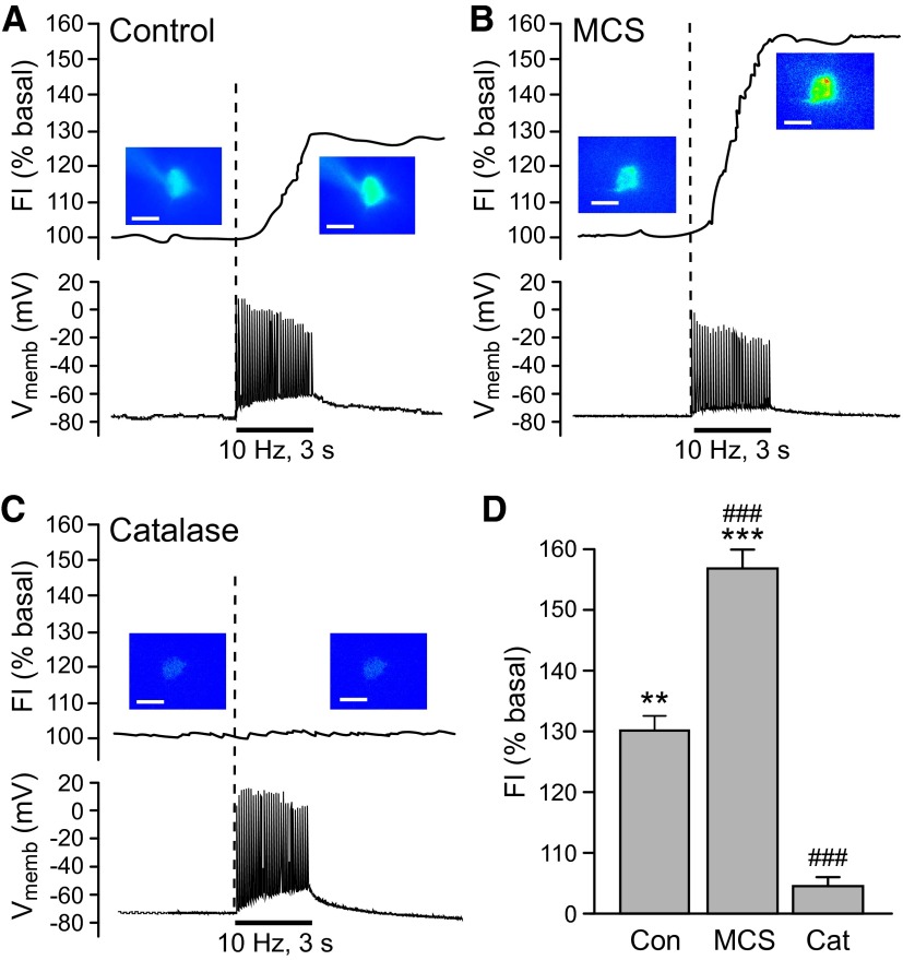
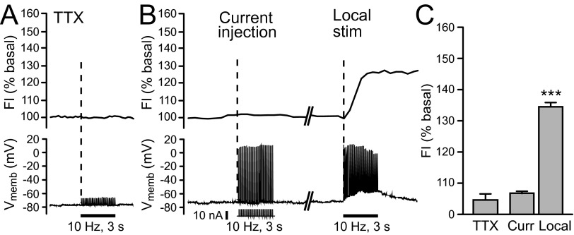
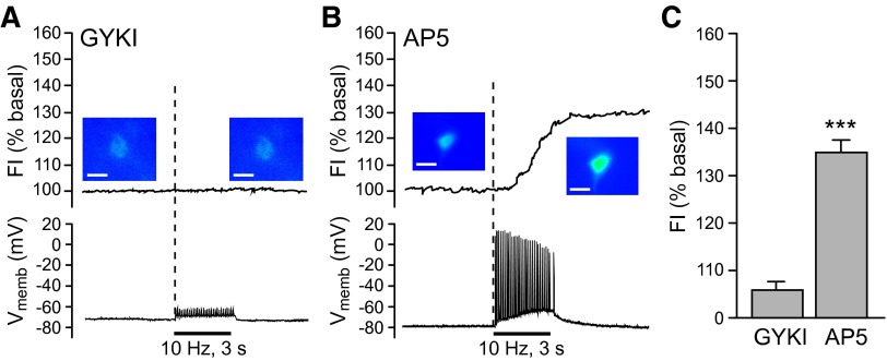
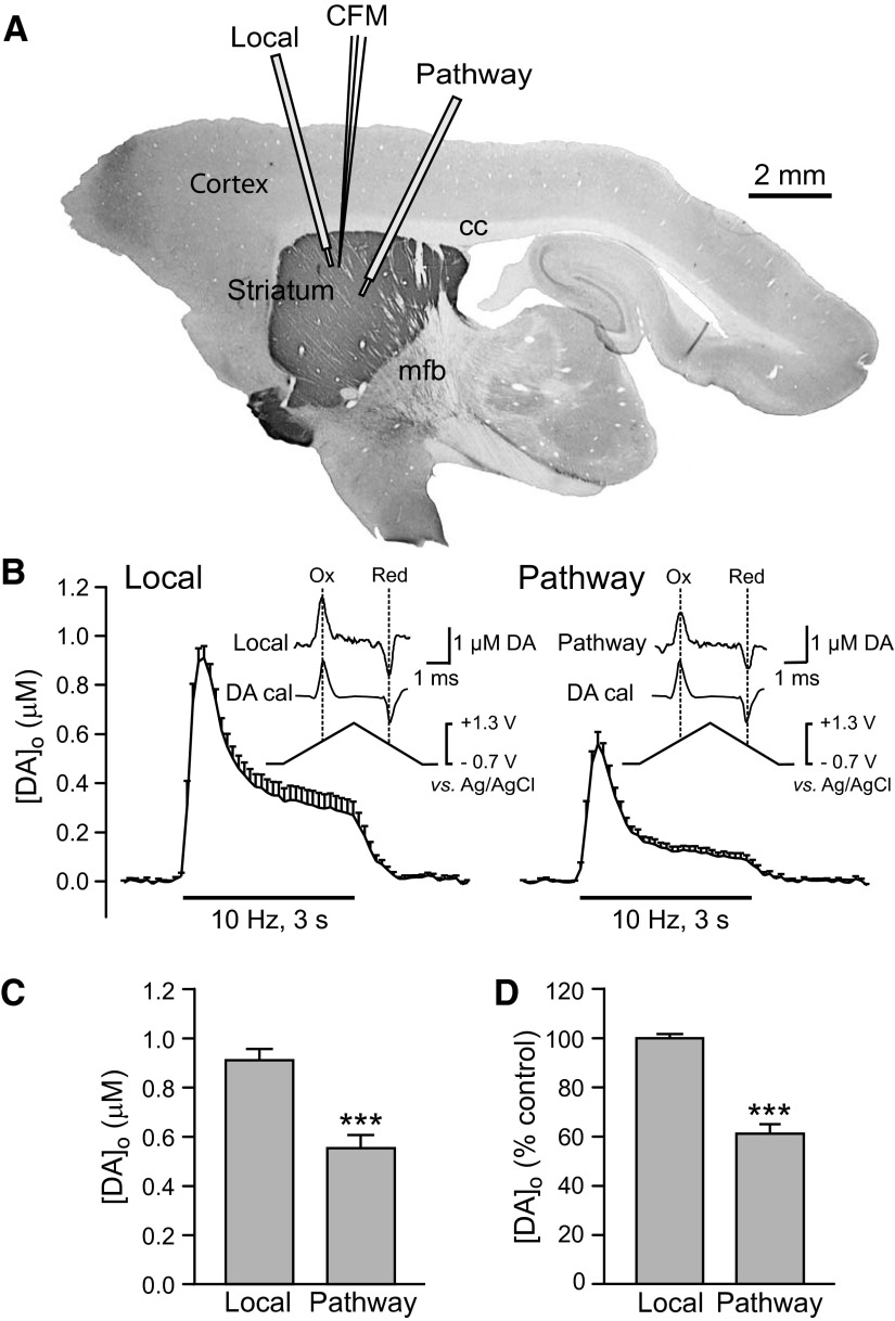
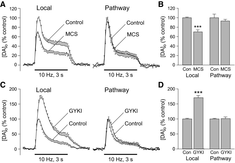
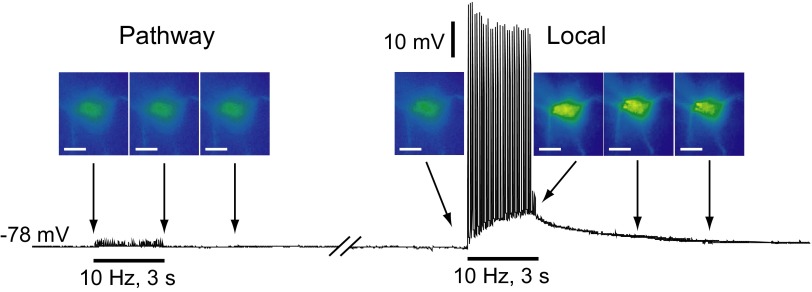
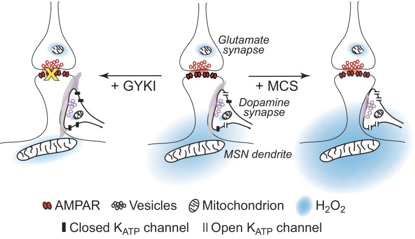
Dopamine-glutamate interactions in the striatum are critical for normal basal ganglia-mediated control of movement. Although regulation of glutamatergic transmission by dopamine is increasingly well understood, regulation of dopaminergic transmission by glutamate remains uncertain given the apparent absence of ionotropic glutamate receptors on dopaminergic axons in dorsal striatum. Indirect evidence suggests glutamatergic regulation of striatal dopamine release is mediated by a diffusible messenger, hydrogen peroxide (H2O2), generated downstream from glutamatergic AMPA receptors (AMPARs). The mechanism of H2O2-dependent inhibition of dopamine release involves activation of ATP-sensitive K+ (KATP) channels. However, the source of modulatory H2O2 is unknown. Here, we used whole cell recording, fluorescence imaging of H2O2, and voltammetric detection of evoked dopamine release in guinea pig striatal slices to examine contributions from medium spiny neurons (MSNs), the principal neurons of striatum, and dopamine axons to AMPAR-dependent H2O2 generation. Imaging studies of H2O2 generation in MSNs provide the first demonstration of AMPAR-dependent H2O2 generation in neurons in the complex brain-cell microenvironment of brain slices. Stimulation-induced increases in H2O2 in MSNs were prevented by GYKI-52466, an AMPAR antagonist, or catalase, an H2O2 metabolizing enzyme, but amplified by mercaptosuccinate (MCS), a glutathione peroxidase inhibitor. By contrast, dopamine release evoked by selective stimulation of dopamine axons was unaffected by GYKI-52466 or MCS, arguing against dopamine axons as a significant source of modulatory H2O2. Together, these findings suggest that glutamatergic regulation of dopamine release via AMPARs is mediated through retrograde signaling by diffusible H2O2 generated in striatal cells, including medium spiny neurons, rather than in dopamine axons.


Figures

FIG. 1.
FIG. 1.
Activity-dependent generation of endogenous H2O2 in striatal medium spiny neurons (MSNs). A–C: representative examples of intracellular H2O2 (top) and membrane voltage (Vmemb; bottom) monitored simultaneously in identified MSNs during local pulse-train stimulation (30 pulses, 10 Hz). The time course of stimulus-induced changes in dichlorofluorescein (DCF) fluorescence intensity (FI) is shown along with pseudocolor images of DCF FI recorded under basal conditions and at the end of stimulation (scale bar = 20 μm). A: stimulus-induced increase in DCF FI in an MSN during local stimulation; under control conditions, 7 of 11 MSNs showed a significant increase in DCF FI (P < 0.01 vs. basal). Simultaneous current-clamp recording indicated that a single action potential was generated with each stimulus pulse in all recorded MSNs. B: inhibition of glutathione (GSH) peroxidase by mercaptosuccinate (MCS, 1 mM) amplified the stimulus-evoked increases in DCF FI, consistent with selective detection of H2O2, with no effect on action potential generation in recorded MSNs. In the presence of MCS, 7 of 7 MSNs showed a significant increase in DCF FI (P < 0.001). C: the presence of the H2O2 metabolizing enzyme catalase (Cat; 500 U/mL) did not alter spike generation during stimulation but prevented the stimulus-induced increase in DCF FI in 5 of 5 MSNs. D: average stimulus-induced changes in DCF FI in H2O2 source MSNs under control conditions (Con; n = 7), in the presence of MCS (n = 7), or in the presence of catalase (n = 5; **P < 0.01 vs. basal; ***P < 0.001 vs. basal). The increase in DCF FI in MCS was nearly twofold greater than under control conditions, whereas the presence of catalase markedly attenuated the usual control response (###P < 0.001 vs. control), confirming the H2O2-dependence of the evoked increases in DCF FI.
FIG. 2.
FIG. 2.
Endogenous H2O2 generation in striatal MSNs requires coherent synaptic activation. A: intracellular H2O2 (top) and membrane voltage (Vmemb; bottom) monitored simultaneously in MSNs during local pulse-train stimulation (30 pulses, 10 Hz) in tetrodotoxin (TTX, 1 μM). In the presence of TTX, stimulus-induced action potential generation was prevented and no increase in DCF FI was seen in any recorded MSN (5 of 5). B: current injection (30 pulses, 10 Hz; pulse amplitude: 5–10 nA; pulse duration: 100 μs) alone elicited action potentials in individual MSNs, but no increase in DCF FI was detected in any MSN examined (n = 5), whereas subsequent local stimulation (30 pulses, 10 Hz) caused the usual 30% increase in DCF FI in the same cells (n = 5; P < 0.001 vs. basal). This suggests that synaptic activation of MSNs, rather than action potential generation alone, is required to generate modulatory H2O2. C: average DCF FI in the presence of TTX (n = 5) or with current injection with subsequent local stimulation, as described for panel B (n = 5; ***P < 0.001 vs. basal).
FIG. 3.
FIG. 3.
Glutamate-dependent H2O2 generation in striatal MSNs requires AMPAR activation. A and B: intracellular H2O2 (top) and membrane voltage (Vmemb; bottom) monitored simultaneously in MSNs during local pulse-train stimulation (30 pulses, 10 Hz). Time course of stimulus-induced changes in DCF FI are shown with pseudocolor images of DCF FI recorded under basal conditions and at the end of stimulation (scale bar = 20 μm). A: the usual stimulus-induced increase in DCF FI in MSNs was prevented by GYKI-52466 (50–100 μM), an AMPAR antagonist as were stimulus-evoked action potentials monitored during simultaneous current-clamp recording (n = 7; P > 0.05 vs. basal). B: blockade of NMDARs by AP5 (100 μM) had no effect on either DCF FI or action potential generation in recorded MSNs with 5 of 5 MSNs showing a significant increase in DCF FI (P < 0.001 vs. basal). C: summary of average changes in DCF-FI in MSNs in the presence of GYKI (n = 7) or AP5 (n = 5) (***P < 0.001 vs. basal).
FIG. 4.
FIG. 4.
Comparison of striatal dopamine release evoked by local stimulation and by pathway stimulation of dopaminergic axons. A: orientation of stimulation electrodes and recording electrode for local stimulation and pathway stimulation of dopaminergic axons in an angled parasagittal section of guinea pig brain stained for tyrosine-hydroxylase immunoreactivity using methods published previously (Rice et al. 1997). B: average [DA]o vs. time profiles evoked at a single site by alternating local and pathway stimulation (30 pulses, 10 Hz). Data are means ± SE. Insets: the applied triangular waveform for fast-scan cyclic voltammetry at a carbon-fiber microelectrode with representative voltammograms for evoked [DA]o and dopamine calibration (DA cal) showing characteristic oxidation (Ox) and reduction (Red) peak potentials that confirm dopamine identity. C: average peak evoked [DA]o recorded at a given site with local or pathway stimulation; pathway evoked [DA]o was significantly lower than that evoked by local stimulation. (n = 12; ***P < 0.001 vs. local). D: average peak [DA]o evoked by local or pathway stimulation normalized to locally evoked release with locally evoked peak [DA]o taken as 100% (n = 12; ***P < 0.001 vs. local).
FIG. 5.
FIG. 5.
Modulatory H2O2 is not generated in dopaminergic axons. A: average [DA]o vs. time profiles evoked at a single site by alternating local and pathway stimulation (30 pulses, 10 Hz) in the absence and presence of the GSH peroxidase inhibitor mercaptosuccinate (MCS; 1 mM; n = 6). B: summary of the effect of MCS on peak [DA]o at a given site evoked by local vs. dopamine pathway stimulation; control peak evoked [DA]o for either local or pathway stimulation was taken as 100%. Increasing endogenous H2O2 availability by inhibiting GSH peroxidase caused a significant decrease in [DA]o evoked by local stimulation but had no effect on pathway evoked [DA]o (n = 6; ***P < 0.001 vs. local control). C: average [DA]o vs. time profiles evoked at a single site by alternating local and pathway stimulation (30 pulses, 10 Hz) in the absence and presence of an AMPAR blocker, GYKI-52466 (GYKI; 50–100 μM; n = 6). D: summary of the effect of GYKI on peak [DA]o at a given site evoked by local and pathway stimulation; control peak evoked [DA]o for either local or pathway stimulation is taken as 100%. Blockade of AMPARs caused a significant increase in [DA]o evoked by local stimulation but had no effect on that evoked by selective stimulation of dopaminergic axons (n = 6; ***P < 0.001 vs. local control).
FIG. 6.
FIG. 6.
Absence of detectable H2O2 generation in MSNs during dopaminergic axon pathway stimulation. Representative example of H2O2 imaging (top) and simultaneous current-clamp (bottom) recording in an MSN during pathway and local pulse-train (30 pulses, 10 Hz) stimulation; the break in the current-clamp record indicates an interval of 2 min. Corresponding pseudocolor DCF FI images were taken at the time points indicated by the arrows; scale bars = 20 μm. Pathway stimulation of dopaminergic axons did not induce MSN firing or a change in DCF FI, whereas subsequent local stimulation induced firing of action potentials and ∼30% increase in DCF FI in these same cells (n = 7).
FIG. 7.
FIG. 7.
Model of axonal dopamine release regulation by glutamate acting via AMPARs and generation of diffusible H2O2 in striatal MSNs. Center: activation of AMPARs on MSN dendrites generates H2O2 that diffuses to adjacent dopamine axons and inhibits dopamine release via opening of KATP channels. Left: when AMPARs are blocked (+ GYKI), H2O2-dependent regulation of dopamine release via KATP channels is lost and dopamine release is enhanced. Right: when activity-dependent levels of H2O2 are amplified by inhibiting GSH peroxidase with MCS, this leads to enhanced H2O2-dependent KATP-channel activation and further suppression of dopamine release.

Similar articles

Cited by

References

    1. Albin RL, Young AB, Penney JB. The functional anatomy of basal ganglia disorders. Trends Neurosci 12: 366–375, 1989. - PubMed
    1. Avshalumov MV, Bao L, Patel JC, Rice ME. H2O2 signaling in the nigrostriatal dopamine pathway via ATP-sensitive potassium channels: issues and answers. Antioxid Redox Signal 9: 219–231, 2007. - PubMed
    1. Avshalumov MV, Chen BT, Koós T, Tepper JM, Rice ME. Endogenous hydrogen peroxide regulates the excitability of midbrain dopamine neurons via ATP-sensitive potassium channels. J Neurosci 25: 4222–4231, 2005. - PMC - PubMed
    1. Avshalumov MV, Chen BT, Marshall SP, Peña DM, Rice ME. Glutamate-dependent inhibition of dopamine release in striatum is mediated by a new diffusible messenger, H2O2. J Neurosci 23: 2744–2750, 2003. - PMC - PubMed
    1. Avshalumov MV, MacGregor DG, Sehgal LM, Rice ME. The glial antioxidant network and neuronal ascorbate: protective yet permissive for H2O2 signaling. Neuron Glia Biol 1: 365–376, 2004. - PMC - PubMed

Publication types

MeSH terms

LinkOut - more resources