Comparative Study
. 2008 Jul 16;28(29):7304-12.
doi: 10.1523/JNEUROSCI.1778-08.2008.

Interactions between medial temporal lobe, prefrontal cortex, and inferior temporal regions during visual working memory: a combined intracranial EEG and functional magnetic resonance imaging study

Affiliations
Comparative Study

Interactions between medial temporal lobe, prefrontal cortex, and inferior temporal regions during visual working memory: a combined intracranial EEG and functional magnetic resonance imaging study

Nikolai Axmacher et al. J Neurosci. .

Abstract

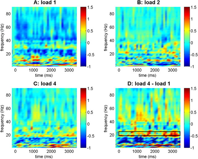
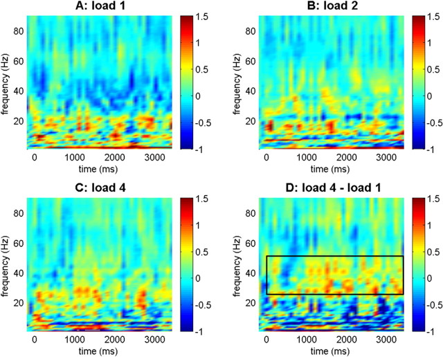
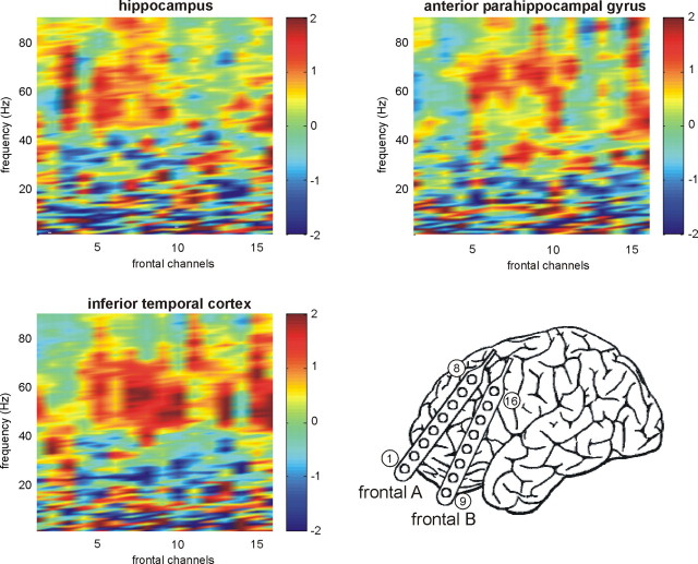
It is a fundamental question whether the medial temporal lobe (MTL) supports only long-term memory encoding, or contributes to working memory (WM) processes as well. Recent data suggest that the MTL is activated whenever multiple items or item features are being maintained in WM. This may rely on interactions between the MTL or the prefrontal cortex (PFC) and content-specific areas in the inferior temporal (IT) cortex. Here, we investigated the neural mechanism through which the MTL, PFC, and IT cortex interact during WM maintenance. First, we quantified phase synchronization of intracranial EEG data in epilepsy patients with electrodes in both regions. Second, we used directional coupling analysis to study whether oscillatory activity in the IT cortex drives the MTL or vice versa. Finally, we investigated functional connectivity in functional magnetic resonance imaging data of healthy subjects with seeds in the MTL and PFC. With increasing load, EEG phase synchronization between the IT cortex and anterior parahippocampal gyrus and within the MTL increased. Coupling was bidirectional in all load conditions, but changed toward an increased top-down (anterior parahippocampal gyrus --> IT) coupling in the high gamma range (51-75 Hz) with increasing load. Functional connectivity between the MTL seed and the visual association cortex increased with load, but activity within the MTL and the PFC correlated with fewer voxels, suggesting that more specific neural networks were engaged. These data indicate that WM for multiple items depends on an increased strength of top-down control of activity within the IT cortex by the MTL.


Figures

Figure 1.
Figure 1.
Synchronization between anterior parahippocampal gyrus and inferior temporal cortex. Baseline-normalized synchronization values are indicated for the different load conditions and comparing load 4 and load 1. Color bars indicate log10-transformed synchronization values in all plots. The box in the bottom right panel refers to the upper beta frequency range (19–25 Hz) where significant linear load effects were observed.
Figure 2.
Figure 2.
Synchronization between anterior parahippocampal gyrus and hippocampus. Baseline-normalized synchronization values are indicated for the different load conditions and comparing load 4 and load 1. Color bars indicate log10-transformed synchronization values in all plots. The box in the bottom right panel refers to the lower gamma frequency range (26–50 Hz) where significant linear load effects were observed.
Figure 3.
Figure 3.
Synchronization with frontal contacts. Results from one patient with subdural frontal electrodes (diagram) and electrodes in the hippocampus, anterior parahippocampal gyrus, and inferior temporal cortex are shown: increased synchronization during maintenance of four items compared with one item. All regions showed enhanced synchronization in the upper gamma ranges, but this effect was most pronounced for frontal–inferior temporal synchronization.
Figure 4.
Figure 4.
Directional coupling analysis. A, Effects of memory load on the direction of coupling between the anterior (ant.) parahippocampal gyrus and the IT cortex. We observed a significant load × band interaction (F(2,10) = 3.014; p < 0.05) and a trend for a load effect in the gamma2 frequency range (F(2,10) = 3.260; p < 0.05). B, Effects of memory load on the direction of coupling between anterior parahippocampal gyrus and hippocampus. Although there was a trend for a load × band interaction (F(6,66) = 2.104; p < 0.05), coupling did not show a load effect in the separate bands (all p values > 0.1). Error bars indicate SEM.
Figure 5.
Figure 5.
Overview of regions showing correlated activity with the PFC and MTL seeds. Significantly correlated voxels in two different networks defined by the seeds in the left hippocampus (yellow) and the left prefrontal cortex (blue) are shown. Color bars indicate t values (group comparison against zero).
Figure 6.
Figure 6.
Load-dependent changes in functional connectivity. A, Connectivity between the MTL seed and the ipsilateral lingual gyrus increased with memory load, whereas connectivity with regions in the contralateral inferior and middle frontal gyrus decreased. B, Connectivity between the prefrontal seed and the cingulate gyrus decreased with memory load.
Figure 7.
Figure 7.
Extension of networks defined by functional connectivity decreases with working memory load. A, Exemplary single-trial beta scatter plots: correlations between the seed in the hippocampus and a voxel in the fusiform cortex (MNI coordinates: −33, −55, −25) in one subject. B, Numerical comparison of the size (normalized number of correlated voxels) of the two networks as a function of memory load. Error bars indicate SEM.

References

    1. Aggleton JP, Shaw C, Gaffan EA. The performance of postencephalitic amnesic subjects on two behavioural tests of memory: concurrent discrimination learning and delayed matching-to-sample. Cortex. 1992;28:359–372. - PubMed
    1. Allison T, Ginter H, McCarthy G, Nobre AC, Puce A, Luby M, Spencer DD. Face recognition in human extrastriate cortex. J Neurophysiol. 1994;71:821–825. - PubMed
    1. Axmacher N, Mormann F, Fernández G, Elger CE, Fell J. Memory formation by neuronal synchronization. Brain Res Rev. 2006;52:170–182. - PubMed
    1. Axmacher N, Mormann F, Fernández G, Cohen MX, Elger CE, Fell J. Sustained neural activity patterns during working memory in the human medial temporal lobe. J Neurosci. 2007;27:7807–7816. - PMC - PubMed
    1. Axmacher N, Elger CE, Fell J. Memory formation by refinement of neural representations: the inhibition hypothesis. Behav Brain Res. 2008;189:1–8. - PubMed

Publication types